2011, IX, 349 p.
Printed book
Hardcover▶ 139,99 € | £126.00 | $189.00
▶ *149,79 € (D) | 153,99 € (A) | CHF 186.50
eBook
Available from your library or ▶ springer.com/shop
MyCopy
Printed eBook for just ▶ € | $ 24.99▶ springer.com/mycopy
Order online at springer.com▶or for the Americas call (toll free) 1-800-SPRINGER▶or email us at:
[email protected].▶For outside the Americas call +49 (0) 6221-345-4301▶or email us at: [email protected]. The first € price and the £ and $ price are net prices, subject to local VAT. Prices indicated with * include VAT for books; the €(D) includes 7% for Germany, the €(A) includes 10% for Austria. Prices indicated with ** include VAT for electronic products; 19% for Germany, 20% for Austria. All prices exclusive of carriage charges. Prices and other details are subject to change without notice. All errors and omissions excepted.
J. Tavares, R.M.N. Jorge (Eds.)
Computational Vision and Medical Image Processing
Recent Trends
Series: Computational Methods in Applied Sciences, Vol. 19
▶
Contains extended versions of papers presented to the International
Conference VIPIMAGE 2009. This conference was the second
ECCOMAS thematic conference on computational vision and medical
image-processing
▶
It covers topics related to image processing and analysis, medical
imaging and computational modeling and simulation, and considers
their multidisciplinary nature
This book contains extended versions of papers presented at the international Conference VIPIMAGE 2009 – ECCOMAS Thematic Conference on Computational Vision and Medical Image, that was held at Faculdade de Engenharia da Universidade do Porto, Portugal, from 14th to 16th of October 2009. This conference was the second ECCOMAS thematic conference on computational vision and medical image processing. It covered topics related to image processing and analysis, medical imaging and computational modelling and simulation, considering their multidisciplinary nature.
Preface
Nowadays, computational methodologies of signal processing and imaging analysis for 2D, 3D and even 4D data are commonly used for various applications in society. For example, Computational Vision systems are progressively used for surveillance tasks, traffic analysis, recognition process, inspection purposes, human-machine in-terfaces, 3D vision and deformation analysis.
One of the main characteristics of the Computational Vision domain is its inter-multidisciplinary nature. In fact, in this domain, methodologies of several other fundamental sciences, such as Informatics, Mathematics, Statistics, Psychology, Mechanics and Physics are regularly used. Besides this inter-multidisciplinary char-acteristic, one of the main rationale that promotes the continuous effort being made in this area of human knowledge is the number of applications in the med-ical area. For instance, statistmed-ical or physmed-ical procedures on medmed-ical images can be used in order to model the represented structures. This modelling can have different goals, for example: shape reconstruction, segmentation, registration, be-havioural interpretation and simulation, motion and deformation analysis, virtual reality, computer-assisted therapy or tissue characterization.
The main objective of the ECCOMAS Thematic Conferences on Computational Vision and Medical Image Processing (VIPimage) is to promote a comprehensive forum for discussion on the recent advances in the related fields and try to identify areas of potential collaboration between researchers of different sciences.
This book contains the extended versions of nineteen papers selected from works presented at the second ECCOMAS thematic conference on Computational Vision and Medical Image processing (VIPimage 2009), which was held at the Engineering Faculty of the University of Porto, Portugal. It gathers together the state-of-the-art on the subject of Computational Vision and Medical Image processing contributing to the development of these knowledge areas and showing new trends in these fields. The Editors would like to take this opportunity to thank to the European Com-munity on Computational Methods in Applied Sciences, the Portuguese Association of Theoretical, Applied and Computational Mechanics, the University of Porto, all sponsors, all members of the International Scientific Committee and to all Invited Lecturers and Authors.
Faculty of Engineering Jo˜ao Manuel R.S. Tavares University of Porto, Portugal R.M. Natal Jorge
Contents
Automatic Segmentation of the Optic Radiation Using DTI
in Healthy Subjects and Patients with Glaucoma. . . 1 Ahmed El-Rafei, Tobias Engelhorn, Simone Waerntges,
Arnd Doerfler, Joachim Hornegger, and Georg Michelson
Real Time Colour Based Player Tracking in Indoor Sports. . . 17 Catarina B. Santiago, Armando Sousa, Lu´ıs Paulo Reis,
and Maria Lu´ısa Estriga
Visualization of the Dynamics of the Female Pelvic Floor
Reflex and Steady State Function. . . 37 Christos E. Constantinou, Qiyu Peng, and Sadao Omata
Population Exposure and Impact Assessment: Benefits of Modeling Urban Land Use in Very High Spatial
and Thematic Detail. . . 75 Christoph Aubrecht, Mario K¨ostl, and Klaus Steinnocher
Dynamic Radiography Imaging as a Tool in the Design
and Validation of a Novel Intelligent Amputee Socket. . . 91 George Papaioannou, Dimitris Tsiokos, Goeran Fiedler,
Christos Mitrogiannis, Ilya Avdeev, Jake Wood, and Ray McKinney
A Discrete Level Set Approach for Texture Analysis
of Microscopic Liver Images. . . .113 Daniela Iacoviello
Deformable and Functional Models. . . .125 Demetri Terzopoulos
viii Contents
Medical-GiD: From Medical Images to Simulations, 4D MRI
Flow Analysis. . . .145 Eduardo Soudah, Julien Pennecot, Jorge S. P´erez, Maurizio
Bordone, and Eugenio O˜nate
KM and KHM Clustering Techniques for Colour Image
Quantisation. . . .161 Mariusz Frackiewicz and Henryk Palus
Caries Detection in Panoramic Dental X-ray Images. . . .175 Jo˜ao Oliveira and Hugo Proenc¸a
Noisy Medical Image Edge Detection Algorithm
Based on a Morphological Gradient Using Uninorms. . . .191 Manuel Gonz´alez-Hidalgo, Arnau Mir Torres, Daniel Ruiz
Aguilera, and Joan Torrens Sastre
Leveraging Graphics Hardware for an Automatic
Classification of Bone Tissue. . . .209 Manuel Jes´us Mart´ın-Requena and Manuel Ujald´on
A Novel Template-Based Approach to the Segmentation
of the Hippocampal Region. . . .229 M. Aiello, P. Calvini, A. Chincarini, M. Esposito, G. Gemme,
F. Isgr`o, R. Prevete, M. Santoro, and S. Squarcia
Model-Based Segmentation and Fusion of 3D Computed Tomography and 3D Ultrasound of the Eye
for Radiotherapy Planning. . . .247 M. Bach Cuadra, S. Gorthi, F.I. Karahanoglu, B. Paquier, A. Pica,
H.P. Do, A. Balmer, F. Munier, and J.-Ph. Thiran
Flow of a Blood Analogue Solution Through Microfabricated
Hyperbolic Contractions. . . .265 P.C. Sousa, I.S. Pinho, F.T. Pinho, M.S.N. Oliveira, and M.A. Alves
Molecular Imaging of Hypoxia Using Genetic Biosensors. . . .281 Pablo Iglesias and J.A. Costoya
Microscale Flow Dynamics of Red Blood Cells in Microchannels:
An Experimental and Numerical Analysis. . . .297 R. Lima, C.S. Fernandes, R. Dias, T. Ishikawa, Y. Imai,
Contents ix
Two Approaches for Automatic Nuclei Cell Counting in Low
Resolution Fluorescence Images. . . .311 Thierry Brouard and Aur´elie Chantˆome
Cerebral Aneurysms: A Patient-Specific and Image-Based
Management Pipeline. . . .327 M.C. Villa-Uriol, I. Larrabide, J.M. Pozo, M. Kim, M. De Craene,
Microscale Flow Dynamics of Red Blood Cells
in Microchannels: An Experimental
and Numerical Analysis
R. Lima, C.S. Fernandes, R. Dias, T. Ishikawa, Y. Imai, and T. Yamaguchi
Abstract The blood flow dynamics in microcirculation depends strongly on the microvascular networks composed with short irregular vessel segments which are linked by numerous bifurcations. This paper presents the application of a confo-cal micro-PTV system to track RBCs through a rectangular polydimethysiloxane (PDMS) microchannel with a bifurcation. By using a confocal micro-PTV sys-tem, we have measured the effect of bifurcation on the flow behaviour of both fluorescent particles diluted in pure water and RBCs in concentrated suspensions. After performing simulations with the commercial finite element software package POLYFLOWR
, some experimental results were compared with the numerical re-sults and the limitations of these simulations were outlined.
Keywords Blood flowMicrovascular networksBifurcationMicrochannel
R. Lima () and R. Dias
ESTiG, IPB, C. Sta. Apolonia, 5301-857 Braganca, Portugal e-mail:[email protected];[email protected]
and
CEFT, FEUP, R. Dr. Roberto Frias, 4200-465 Porto, Portugal
C.S. Fernandes
ESTiG, IPB, C. Sta. Apolonia, 5301-857 Braganca, Portugal
e-mail:[email protected]
T. Ishikawa and Y. Imai
Department of Bioengineering & Robotics, Graduate School of Engineering, Tohoku University, 6-6-01 Aoba, 980-8579 Sendai, Japan
e-mail:[email protected];[email protected]
T. Yamaguchi
Department of Biomedical Engineering, Graduate School of Engineering, Tohoku University, 6-6-01 Aoba, 980-8579 Sendai, Japan
e-mail:[email protected]
J.M.R.S. Tavares and R.M.N. Jorge (eds.),Computational Vision and Medical Image Processing: Recent Trends, Computational Methods in Applied Sciences 19,
DOI 10.1007/978-94-007-0011-6 17, cSpringer Science+Business Media B.V. 2011
298 R. Lima et al.
1 Introduction
The phenomena of blood flow in microvessels (diameters less than 300m) are crucial in maintaining healthy organs and tissues. Although these phenomena have been studied for many years it still remains incompletely understood [25]. It is therefore important to investigate the behaviour of blood flow occurring at microvessels in order to better understand the role of blood cells in the process of delivering oxygen and materials to the organs and tissues.
The complexity to control and obtain reliable measurements of the blood flow behaviour through the in vivo microvascular system has led several researchers to perform their studies by using narrow glass tubes with simple geometries. By using in vitro models it allows a more precise control over the experimental variables of interest and extracts detailed information of the flow behaviour of in-dividual blood cells. In fact, much of the understanding of the haemodynamics phenomena observed in microcirculation was obtained from studies on the both macro and microrheology properties of blood flowing through glass microtubes [3,11,13,16,17,25].
Although glass microchannels present certain similarities to in vivo microcircula-tion, it is also clear that these kind of in vitro experiments differ from microvessels in several respects, such as: elasticity of microvessels, effect of the endothelial surface layer and microvascular networks composed with short irregular vessel segments which are linked by numerous bifurcations. Thus it was not surprising that several studies on blood flow in glass microtubes and in microvessels have yielded conflict-ing results with respect to blood viscosity [20] and flow resistance [27]. Hence, the rheological properties of blood should not be only interpreted from measurements in viscometers and microtubes with simple geometries.
In vivo microvascular networks are composed of many divergent microvascular bifurcations which likely influence the blood flow behaviour in the microcirculation. In the present work, a confocal micro-PTV system is used to measure the blood flow through a symmetric PDMS bifurcation fabricated by soft lithography. Moreover, the experimental data were compared numerically by using the commercial finite element software package POLYFLOWR
. By using this combination we expect to gain understanding about several important parameters that affect the blood flow through a diverging microvessel bifurcation.
2 Materials and Methods
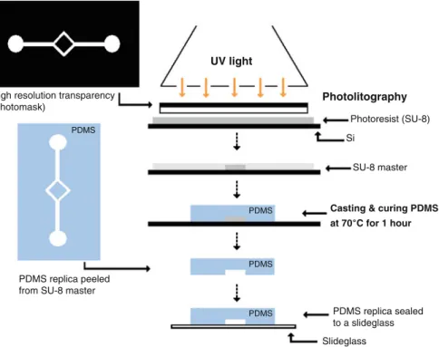
2.1 Fabrication of the Microchannels
Microscale Flow Dynamics of Red Blood Cells in Microchannels 299
UV light
Photolitography
Casting & curing PDMS at 70˚C for 1 hour
Photoresist (SU-8)
SU-8 master
PDMS replica sealed to a slideglass PDMS replica peeled
from SU-8 master High resolution transparency (photomask)
Slideglass PDMS
PDMS
PDMS PDMS
Si
Fig. 1 Main steps of the soft lithographic technique to manufacture a microchannel with a sym-metrical bifurcation geometry
300 R. Lima et al.
was formed spontaneously. The input/output ports were made by means of a 200l micro-pipette tip. This tip was inserted tightly into the connection channels where it exerts pressure on the PDMS and provides a liquid proof seal. Top tubes were also fitted tightly into the micro-pipette tip in order to deliver the working fluids from the syringe pump. Due to the elasticity of the connection tubes and conical shape of the tip, we did not observe any fluid leakage during our experiments [17].
2.2 Working Fluids and Geometry of the Bifurcation
Two working fluids were used in this study: pure water and dextran 40 (Dx40) containing about 14% (14Hct) of human red blood cells (RBCs). The blood was collected from a healthy adult volunteer, where ethylenediaminetetraacetic acid (EDTA) was added to prevent coagulation. The RBCs were separated from the bulk blood by centrifugation and aspiration and then washed twice with physiological saline (PS). The washed RBCs were labeled with a fluorescent cell tracker (CM-Dil, C-7000, Molecular Probes) and then diluted with Dx40 to make up the required RBCs concentration by volume. All blood samples were stored hermetical at4ıC until the experiment was performed at controlled temperature of about37ıC [13].
The geometry and dimensions of the microchannel bifurcation is illustrated in Fig.2. In the present study we decided to use the following dimensions: 150m wide for parent vessel (W0); 75m wide for daughter vessel.W1DW2D1=2W0/ and 50m deep.
Fig. 2 Symmetrical bifurcation geometry used in this study: W0 D 150 m;W1 D W2 D 75 m; ™D60ı
Microscale Flow Dynamics of Red Blood Cells in Microchannels 301
Table 1 Experimental parameters used to calculate the Re
Density (kg/m3) 1,046
Mean velocity (m/s) 3:8104
Hydraulic diameter (m) 7:5105
Viscosity of Dx-40 (Ns/m2) 4:5103
Re 0.007
2.3 Confocal Micro-PTV Experimental Set-Up
The confocal micro-PTV system used in our experiment consists of an inverted mi-croscope (IX71, Olympus, Japan) combined with a confocal scanning unit (CSU22, Yokogawa) and a diode-pumped solid state (DPSS) laser (Laser Quantum Ltd) with an excitation wavelength of 532 nm. Moreover, a high-speed camera (Phantom v7.1) was connected into the outlet port of the CSU22. The microchannel was placed on the stage of the inverted microscope where the flow rate of the working fluids was kept constant (ReD0.007) by means of a syringe pump (KD Scientific Inc.).
The Reynolds number (Re) and associated experimental parameters are summa-rized in Table 1. A thermo plate controller (Tokai Hit) was set to37ıC. All the confocal images were captured in the middle of the microchannels with a resolu-tion of640480pixels, at a rate of 100 frames/s with an exposure time of 9.4 ms. The recorded images were transferred to the computer and then evaluated in the Im-age J (NIH) [1] by using the manual tracking MtrackJ [21] plugin. As a result it was possible to track single RBCs through the middle plane of the microchannel (Fig.3).
2.4 Simulation Method
The numerical calculations for the laminar isothermal flow of pure water were performed using the finite-element computational fluid dynamics (CFD) program POLYFLOWR
. The simulations were carried out in a 3D geometry representing the microchannel (see Fig.4). The mesh used in the simulations was mainly consti-tuted by quadrilateral elements, the discretization of the walls of the channel being presented in Fig.4. The size of the elements was fixed after a grid independence test. The grids were successively refined and the velocity obtained with the different meshes were compared, the results being considered independent of the mesh when a difference bellow 1% was achieved [4,8–10].
302 R. Lima et al.
Fig. 3 Experimental set-up
Microscale Flow Dynamics of Red Blood Cells in Microchannels 303
Table 2 Rheological parameters of blood [12]
Rheological model (Pa.s) K() n.) (s) 1(Pa.s) 0(Pa.s)
Newtonian 0.00345 – – – – –
Power law model – 0.035 0.6 – – –
Carreau model – – 0.3568 3.313 0.00345 0.056
The boundary conditions were established in order to reproduce the experimental conditions. The geometry of the bifurcation was idealized and close to the original dimensions of the photomask. Additionally, the mean velocity at the inlet of the microchannel was3:8104m/s and slip at the walls of the channel was assumed to be non-existent. The referred velocity was imposed considering a constant flow rate on the referred boundary.
In the numerical study, blood was considered a Newtonian and non-Newtonian fluid In the last case, the rheology of the blood was described by two different constitutive models – power law model and the Carreau model [12] – which are, respectively, traduced mathematically by the equations:
DKPn1; (1)
D1C.01/
h
1C. /P 2i.n1/=2; (2)
whereis the viscosity of the fluid, K the consistency index, n the flow index behavior,P the shear rate,1 the viscosity for high shear rates,0 the viscosity for low shear rates andthe natural time. For the blood, the rheological parameters present in the above equations are reported in Table2.
3 Results and Discussion
The confocal micro-PIV system was first evaluated by comparing the experimental results not only with a well established analytical solution for steady flow in a rect-angular microchannel [15] but also with a reliable numerical method that was used in past investigations to study the flow behaviour of Newtonian or non-Newtonian fluids at low Reynolds numbers [9,10].
The numerical, experimental and analytical results of the present work were ob-tained for the middle plane (25m height) of the rectangular microchannel. The averaged velocity data obtained from the confocal micro-PTV measurements, an-alytical solution and numerical simulation were in good agreement. A more detail description of these results can be found elsewhere [22].
304 R. Lima et al.
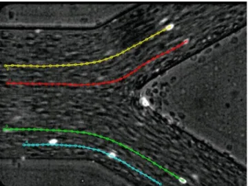
Fig. 5 (a) Paths displacement of fluorescent particles flowing in pure water; (b) Numerical trajec-tories using pure water
Fig. 6 Paths displacement of labeled RBCs (bright spots) flowing in physiological fluid with 14%
Hct.32/
particles and individual RBCs. Qualitative comparison between the experimental data from pure water (see Fig.5a) and the numerical simulation (see Fig.5b), shows that in both cases the trajectories do not exhibit any appreciable deviations in the transversal (yyaxis) direction.
Microscale Flow Dynamics of Red Blood Cells in Microchannels 305
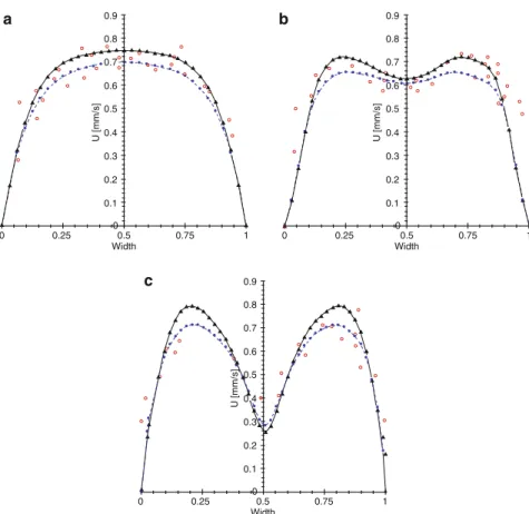
Fig. 7 Regions where the velocity profiles of the numerical and experimental results were compared
to suffer small deviations from the streamlines of the plasma flow probably due to flow perturbations caused by cell interactions in the neighbourhood of the apex of bifurcation.
Moreover numerical simulations of non-Newtonian models were performed around the bifurcation at the regions 1–5 (see Fig.7).
Figures8and9 show the velocity profiles for both computational and experi-mental results before and after the bifurcation, respectively.
Figures8and9suggest that the RBCs velocities close to the microchannel wall are higher than that obtained with the numerical models since slip at the walls of the channel was assumed to be non-existent in the latter case. It is well know, in microcirculation and other areas [4,5,13,20,28,32], that RCBs, macromolecules, colloids, etc., are excluded from the region of the channel with low velocity and this may explain the observed differences.
306 R. Lima et al. 0.5 0 0.1 0.2 0.3 0.4 0.5 0.6 0.7 0.8 0.9 0 0.1 0.2 0.3 0.4 0.5 0.6 0.7 0.8 0.9 0 0.1 0.2 0.3 0.4 0.5 0.6 0.7 0.8 0.9 Width a b c
U [mm/s] U [mm/s]
U [mm/s]
0.25
0 0.75 1 0.5
Width
0.25 0.75 1
0
0.5 Width
0.25 0.75 1
0
Fig. 8 Velocity profiles for both computational and experimental results before the bifurcation:
(a) region 1; (b) region 2; (c) region 3. (—) Newtonian; (––) Power Law;.N/Carreau Model; (ı)
Confocal micro-PTV RBC velocities
0.5 Width 0.25
0 0.75 1 0.5
Width 0.25
0 0.75 1
0.1 0 0.2 0.3 0.4 0.5 0.6 0.7 0.8 0.9 U [mm/s] 0.1 0 0.2 0.3 0.4 0.5 0.6 0.7 0.8 0.9 U [mm/s] a b
Fig. 9 Velocity profiles for both computational and experimental results after the bifurcation: (a)
region 4; (b) region 5. (—) Newtonian; (––) Power Law;.N/Carreau Model; (ı) Confocal
Microscale Flow Dynamics of Red Blood Cells in Microchannels 307
4 Limitations and Future Directions
The primary goal of the present work is to provide new insights on the rheo-logical properties of blood in microvascular network models. To accomplish it experimental flow studies was performed with a confocal micro-PTV system com-plemented with the most recent advances in microfabricated technologies. However, the soft-litography quality is strongly dependent not only on the photolitography methodology but also on the photomask resolution and its fidelity to the original design. In the present work we used a plastic photomask and according to our preliminary results we found small discrepancies between the tested in vitro model and the original design. Although these discrepancies were not taken into account in the present study we are aware about the possible influence of them on the re-sults shown. Hence, in the future we are planning to carry on the current research by taking into account such discrepancies.
In vitro experimentation has the potential to provide a more realistic information on the flow properties of blood when compared with numerical simulations. How-ever, once validated the numerical models can be a valuable tool to obtain more detailed insights about the blood rhelogical properties in microvascular networks. Generally, there are two major approaches to model of the non-Newtonian nature of blood [33]. One is based on the conventional continuum approach, in which a blood constitutive equation is assumed such as the casson model and the power-law model [36]. A more complex and realistic approach is based on a multiphase ap-proach, in which the blood is considered as a multiphase suspension of deformable particles and where levels of submodeling for the behaviour of blood components are introduced. Some examples for this type of approach are the boundary ele-ment method [23,26,34], the immersed boundary method [2,7,24,31], the lattice Boltzmann method [6,29] and the moving particle semi-implicit (MPS) method [19,30]). Recent reviews on these numerical methods can be found in Liu et al. [18], Yamaguchi et al. [33] and Lima et al. [14]. Although the multiphase flow ap-proach is a very promising method it requires massive computational power. Hence, only recently this latter approach is being actively pursued due to the advances of the computational techniques and the computing power.
In the current study we have only performed simulations by using a continuum computational approach to model of the non-Newtonian nature of blood. However for the size of the microcahnnel used in presented work we can not neglect the effect of the suspension of deformable cells on its flow behaviour. Hence, we expect in a near future to compare the obtained experimental results with multi-phase numerical models.
5 Conclusions
308 R. Lima et al.
Qualitative experimental observations suggested that RBC paths around the bi-furcation apex seems to suffer small deviations from the streamlines of the plasma flow probably due to cell interactions enhanced by the high local Hct originated at this region.
The simulations performed with a finite-element computational fluid dynamics (CFD) program POLYFLOW emphasized the need of developing a multiphase approach.
Acknowledgements This study was supported in part by the following grants: Grant-in-Aid for Science and Technology (BII/UNI/0532/EME/2008, BEB/108728/2008, PTDC/SAU-BEB/105650/2008 and PTDC/EME-MFE/099109/2008) from the Science and Technology Foun-dation (FCT) and COMPETE, Portugal and Grant-in-Aid for Scientific Research (S) from the Japan Society for the Promotion of Science (JSPS; No.19100008). We also acknowledge the sup-port from the 2007 Global COE Program “Global Nano-Biomedical Engineering Education and Research Network”. The authors would like also to thank Dr. C. Balsa for his valuable assistance and support for the MATLAB numerical calculations and Ms. B. Oliveira, Ms. D. Cidre and Mr. M. Lagoela for their valuable technical assistance in this research work.
References
1.Abramoff, M., Magelhaes, P., Ram, S.: Image processing with image J. Biophotonics Int.11,
36–42 (2004)
2.Bagchi, P.: Mesoscale simulation of blood flow in small vessels. Biophys. J.92, 1858–1877
(2007)
3.Chien, S., Usami, S., Skalak, R.: Blood flow in small tubes In: Renkins, M., Michel, C.C. (eds.)
Handbook of Physiology–The Cardiovascular System IV, pp. 217–249. American Physiologi-cal Society, Bethesda (1984)
4.Dias, R.P.: Size fractionation by slalom chromatography and hydrodynamic chromatography.
Recent Patents Eng.2, 95–103 (2008)
5.Dias, R.P., Fernandes, C.S., et al.: Starch analysis using hydrodynamic chromatography with a
mixed-bed particle column, Carbohydr. Polym.74, 852–857 (2008)
6.Dupin, M.M., Halliday, I., et al.: Modeling the flow of dense suspensions of deformable
parti-cles in three dimensions. Phys. Rev. E.75, 066707 (2007)
7.Eggleton, C.D., Popel, A.S.: Large deformation of red blood cell ghosts in a simple shear flow.
Phys. Fluids.10, 1834–1845 (1998)
8.Fernandes, C.S., Dias, R.P., et al.: Simulation of stirred yoghurt processing in plate heat
ex-changers. J. Food Eng.69, 281–290 (2005)
9.Fernandes, C.S., Dias, R.P., et al.: Laminar flow in chevron-type plate heat exchangers: CFD
analysis of tortuosity, shape factor and friction factor. Chem. Eng. Process.: Process Intensif.
46, 825–833 (2007)
10.Fernandes, C.S., Dias, R.P., et al.: Friction factors of power-law fluids in plate heat exchangers.
J. Food Eng.89, 441–447 (2008)
11.Goldsmith, H., Turitto, V.: Rheological aspects of thrombosis and haemostasis: Basic principles
and applications. ICTH-Report-Subcommittee on Rheology of the International Committee on
Thrombosis and Haemostasis. Thromb. Haemost.55, 415–435 (1986)
12.Johnston, B.M., Johnston, P.R., et al.: Non-Newtonian blood flow in human right coronary
arteries: Steady state simulations. J. Biomech.37, 709–720 (2004)
13.Lima, R., Ishikawa, T., et al.: Measurement of individual red blood cell motions under high
hematocrit conditions using a confocal micro-PTV system. Ann. Biomed. Eng.37, 1546–1559
Microscale Flow Dynamics of Red Blood Cells in Microchannels 309
14.Lima, R., Ishikawa, T., et al.: Blood flow behavior in microchannels: Advances and future
trends. In: Single and Two-Phase Flows on Chemical and Biomedical Engineering. Bentham (in press) (2011)
15.Lima, R., Wada, S., et al.: Confocal micro-PIV measurements of three dimensional profiles of
cell suspension flow in a square microchannel. Meas. Sci. Tech.17, 797–808 (2006)
16.Lima, R., Wada, S., et al.: In vitro confocal micro-PIV measurements of blood flow in a square
microchannel: The effect of the haematocrit on instantaneous velocity profiles. J. Biomech.40,
2752–2757 (2007)
17.Lima, R., Wada, S., et al.: In vitro blood flow in a rectangular PDMS microchannel:
Experimental observations using a confocal micro-PIV system. Biomed. Microdevices2(10),
153–167 (2008)
18.Liu, W.K., Liu, Y., et al.: Immersed finite element method and its applications to biological
systems. Comput. Methods Appl. Eng.195, 1722–1749 (2006)
19.Kondo, H., Imai, Y., et al.: Hemodynamic analysis of microcirculation in malaria infection.
Ann. Biomed. Eng.37, 702–709 (2009)
20.Maeda, N.: Erythrocyte rheology in microcirculation. Jpn. J. Physiol.46, 1–14 (1996)
21.Meijering, E., Smal, I., Danuser, G.: Tracking in molecular bioimaging. IEEE Signal Process.
Mag.3(23), 46–53 (2006)
22.Oliveira, B., Lagoela, M., et al.: Analyses of the blood flow in a microchannel with a
bifurca-tion. In: Proceedings of 3ı
Congresso Nacional de Biomecˆanica, Braganc¸a, Portugal (2009)
23.Omori, T., Ishikawa, T. et al.: Behavior of a red blood cell in a simple shear flow simulated by
a boundary element method, In: Proceedings of Bioengineering 08, London, UK (2008)
24.Peskin, C.S.: Numerical analysis of blood flow in the heart. J. Comput. Phys.25, 220–233
(1977)
25.Popel, A., Johnson, P.: Microcirculation and hemorheology. Annu. Rev. Fluid Mech. 37,
43–69 (2005)
26.Pozrikidis, C.: Numerical simulation of the flow-induced deformation of red blood cells. Ann.
Biomed. Eng.31, 1194–1205 (2003)
27.Pries, A., Secomb, T., et al.: Resistance to blood flow in microvessels in vivo. Circ. Res.75,
904–915 (1994)
28.Small, H.: Hydrodynamic chromatography a technique for size analysis of colloidal particles.
J. Colloid. Interface Sci.48, 147–161 (1974)
29.Succi, S.: The Lattice Boltzmann Equation for Fluid Mechanics and Beyond, Clarendon Press,
Oxford (2001)
30.Tsubota, K., et al.: Particle method for computer simulation of red blood cell motion in blood
flow. Comp. Methods Programs Biomed.83, 139–146 (2006)
31.Univerdi, S.O., Tryggvason, G.: A front-tracking method for viscous, incompressible
multi-fluid flows. J. Comput. Phys.100, 25–37 (1992)
32.Venema, E., Kraak, J.C., et al.: Packed-column hydrodynamic chromatography using 1-m
non-porous silica particles. J. Chromatogr. A740, 159–167 (1996)
33.Yamaguchi, T., Ishikawa, T., et al.: Computational blood flow analysis – new trends and
meth-ods. J. Biomech. Sci. Eng.1, 29–50 (2006)
34.Youngren, G.K., Acrivos, A.: Stokes flow past a particle of arbitrary shape: A numerical
method of solution. J. Fluid Mech.69, 377–403 (1975)
35.Wilkinson, W.L.: Non-Newtonian fluids: Fluid mechanics, mixing and heat transfer, pp. 61–63.
Pergamon Press, London (1960)
36.Zhang, J.B., Kuang, Z.B.: Study on blood constitutive parameters in different blood constitutive