Antipro life rative e ffe cts o f
1 ,2 5 -dihydro xyvitam in D
3
o n bre ast ce lls - A m ini re vie w
Disciplina de O ncologia, Departamento de Radiologia, Faculdade de Medicina, Universidade de São Paulo, São Paulo, SP, Brasil
P. Bortman, M.A.A.K. Folgueira, M.L.H. Katayama, I.M.L. Snitcovsky and M.M. Brentani
Abstract
The hormone 1,25-dihydroxyvitamin D3 (1,25-(OH)2D3), the active form of vitamin D3, is an important regulator of calcium homeostasis, exerts antiproliferative effects on various cell systems and can induce differentiation in some kinds of hematopoietic cells. These effects are triggered by its receptor, vitamin D receptor (VDR), a phosphoprotein member of the nuclear receptor superfamily, which functions as a transcriptional factor. VDR binds as a heterodimer with retinoid X receptor (R X R) to hexameric repeats, characterized as vitamin D-responsive elements present in the regulatory region of target genes such as osteocalcin, osteopontin, calbindin-D28K, calbindin-D9K, p21WAF1/CIP1, TGF-ß2 and vitamin D 24-hydroxylase. Many factors such as glucocorticoids, estrogens, retinoids, proliferation rate and cell transformation can modulate VDR levels. VDR is expressed in mammary tissue and breast cancer cells, which are potential targets to hormone action. Besides having antiproliferative properties, vitamin D might also reduce the invasiveness of cancer cells and act as an anti-angiogenesis agent. All of these antitumoral features suggest that the properties of vitamin D could be explored for chemopreventive and therapeutic purposes in cancer. However, hypercalcemia is an unde-sirable side effect associated with pharmacological doses of 1,25-(OH)2D3. Some promising 1,25-(OH)2D3 analogs have been devel-oped, which are less hypercalcemic in spite of being potent antiprolif-erative agents. They represent a new field of investigation.
Co rre spo nde nce
M.M. Brentani
Departamento de Radiologia Faculdade de Medicina, USP Av. Dr. Arnaldo, 455 01246-903 São Paulo, SP Brasil
Fax: + 55-11-3082-6580 Research supported by FAPESP (Nos. 98/16066-9 and 99/00900-2). P. Bortman received the “Young Scientist Luis Renato Caldas-2000” award from the Brazilian Society of Biophysics for this paper.
Received February 20, 2001 Accepted O ctober 18, 2001
Ke y wo rds ·Calcitriol
·Calcitriol receptor
·Calcitriol analog
·Breast tumor
·Cell proliferation
Me chanism o f actio n o f vitamin D
Vitamin D is in fact a secosteroid hor-mone which in its active form, 1,25-dihy-droxyvitamin D3 (1,25-(OH)2D3), is an
im-portant regulator of bone development and metabolism and calcium homeostasis. Be-sides these well-known functions on classi-cal target tissues (bone, kidneys, intestine, parathyroids), 1,25-(OH)2D3 plays an
impor-tant role in the regulation of cell growth and differentiation in cells other than its classical targets.
being subsequently hydroxylated in the liver and kidneys to 25-(OH)D3 and 1,25-(OH)2D3,
the active form (1).
The hormone exerts its effects via ge-nomic and non-gege-nomic mechanisms. In the first case, the response is triggered by its nuclear receptor - the vitamin D receptor or VDR, which is a trans-acting transcriptional factor and a member of the nuclear hormone receptor superfamily. The N-terminal do-main of VDR is configured into two zinc-coordinated fingers responsible for DNA recognition and binding, whereas the C-ter-minal domain binds the 1,25-(OH)2D3. VDR
binds selectively to DNA primarily as a het-erodimer with retinoid X receptor (R X R). The binding of the hormone and the receptor causes a conformational alteration in the linkage domain of the latter with consequent dissociation of co-repressors, facilitating the interactions between VDR and co-activator proteins such as the members of the p160, SRC-1, 2 and 3 families, and the protein complex named DRIP. These co-activators modulate the chromatin structure and the contact with the basal transcriptional factors (2). The receptor/1,25-(OH)2D3 complex
regulates gene transcription both positively and negatively through binding motifs in the promoter regions of target genes, designated vitamin D response elements, or VDREs. Several VDREs have been characterized and they generally consist of two direct repeats of six nucleotides (AGGTCA) separated by three aleatory nucleotides; however, neither the sequence of half-sites nor the spacing between them is well conserved. VDREs have been identified in genes such as calbindin-D28K (3) and calbindin-D9K, which
are calcium-binding proteins mainly present in mammalian kidney and intestine, respec-tively; osteocalcin (4) and osteopontin (5), which are bone matrix proteins produced by osteoblasts; vitamin D 24-hydroxylase (6), an enzyme that inactivates 1,25-(OH)2D3;
p21WAF1/CIP1
(7), a cyclin-dependent kinase inhibitor; c-fos, an early response gene, and
transforming growth factor ß2 (TGF-ß2) (8). VDR has been detected in numerous clas-sical and nonclasclas-sical target tissues of 1,25-(OH)2D3, in tumors of various origins and
cell lines such as NIH-3T3 mouse fibro-blasts and MCF-7 human breast cancer cells (9,10). The responsiveness of target cells to the hormone depends on the amount of VDR and many factors can modulate VDR levels. Previous studies by our group and others have shown, for example, that glucocorti-coids, prolactin, estrogens, retinoids and growth factors might influence VDR expres-sion in mammary and leukemic cells (9,11-14). In HL-60 myeloblastic cells, VDR con-tent correlated indirectly with the prolifera-tion rate expressed by the fracprolifera-tion of cells in the G0/G1 phase (13). However, in other leukemic cell lines such as U937 and K562, phorbol ester treatment caused growth ar-rest, which was not accompanied by VDR down-regulation (14). Our data for leukemic cells suggest that VDR expression is not consistently changed upon inhibition of cell proliferation.
In addition to this genomic pathway, vi-tamin D has also been reported to induce nontranscriptional responses involving acti-vation of transmembrane signal transduc-tion pathways. Moreover, 1,25-(OH)2D3
modulates voltage-dependent Ca2+
channel-mediated Ca2+
influx in cultured chick muscle cells by a non-genomic pathway involving G protein-dependent stimulation of both the adenylyl cyclase/cyclic AMP/PKA messen-ger system and a phosphoinositide-specific phospholipase C (PLC). The rapid activa-tion of PLC, in turn, generates diacylgly-cerol and inositol-1,4,5-triphosphate, pro-moting the activation of protein kinase C and rapid release of Ca2+
from endogenous stores (15). These effects could be mediated by a putative but as yet unidentified membrane receptor (16). Other findings support evi-dence that some actions of 1,25-(OH)2D3
3-kinase, a lipid 3-kinase, which forms a com-plex with VDR, suggesting that non-genom-ic and genomnon-genom-ic mechanisms could take place in concert (17).
Vitamin D and bre ast cance r
Epidemiological studies have shown that death rate and incidence of breast cancer tend to increase with increasing latitude, suggesting that solar radiation might play a protective role in breast cancer development. These studies also provide provocative data indicating an inverse relationship between decreased sunlight exposure and diminished vitamin D production on the skin and higher breast cancer incidence and mortality (18,19). According to this hypothesis, it was reported that white women affected by breast cancer show lower 1,25-(OH)2D3 blood levels than
unaffected ones (20). In addition, Mawer et al. (21), in a study on breast cancer patients, found the highest serum 1,25-(OH)2D3
lev-els in the early stage as compared to more advanced bone metastatic disease.
Another possible link between the vitamin D pathway and breast cancer was recently reported as an amplification of CYP24, lo-cated in a region of recurrent aberration at 20q13.2 in breast cancer. This gene encodes vitamin D 24-hydroxylase, an enzyme respon-sible for 1,25-(OH)2D3 degradation, and its
overexpression could lead to abrogation of growth control mediated by vitamin D (22). On the other hand, 25-(OH)D3-1a
-hydroxy-lase, responsible for 25-(OH)D3 activation,
was detected in normal human breast as well as in breast carcinoma samples, indicating that both normal and cancerous tissues could be capable of 25-(OH)D3-1a-hydroxylation (23)
and local synthesis of 1,25-(OH)2D3.
Receptors for 1,25-(OH)2D3 in MCF-7, a
cultured breast cancer cell line, were first shown by Eisman et al. (10) in 1979 and later VDR expression was reported in carcino-gen-induced rat mammary tumors as well (24). VDR was also detected in some normal
breast tissues such as the mammary gland of pregnant and lactating rabbits but not in virgin rabbits (25).
The antiproliferative effect of 1,25-(OH)2D3 was demonstrated in vitro in
MCF-7 cells and other estrogen receptor-positive as well as -negative breast cancer cell lines (11,26). The in vivo antitumor effect of 1,25-(OH)2D3 or its analogs on rat mammary
car-cinogen (7,12-dimethylbenzanthracene, or N-methyl-N-nitrosourea)-induced tumors was observed as reduced total tumor burden or extended tumor latency and lessened tu-mor incidence (27,28).
The presence of VDR was also demon-strated in normal human breast tissue (29) and in a large proportion, ranging from 75 to 93%, of breast tumor biopsy specimens, as assessed by specific 1,25-(OH)2D3 binding
in tumor extracts (25,30) or positive immu-nostaining using antibodies to VDR (29,31). In some small series of patients, correlations of breast cancer VDR status with prognosis were conflicting. Freake et al. (30) examined breast cancer samples from 56 patients using a hormone-binding assay, and VDR content (less or more than 8 fmol/mg protein) could not predict a difference in probability of survival. In contrast, Colston et al. (31) found a longer disease-free survival among pa-tients with VDR-positive breast tumors as evaluated by immunocytochemistry.
disease outcome as evaluated by axillary node status (data not shown).
Our next step was to test the action of vitamin D on normal and transformed mam-mary cells. HC11 is a spontaneously immor-talized lineage derived from the mammary gland of midpregnant BALB/c mice. These cells retain characteristics of normal cells such as growth inhibition by cell contact and are able to differentiate in vitro and synthe-size milk proteins (ß-casein) following stim-ulation with lactogenic hormones. HC11 cells
do not express estrogen receptors and lack wild-type p53 (33). On the other hand, HC11 cells transformed with the oncogene Ha-ras (HC11ras) are no longer growth inhibited upon cell contact, do not respond to lactoge-nic hormones, and are tumorigelactoge-nic when injected into nude mice (34). Our studies have demonstrated that only HC11 parental cells are growth inhibited upon 1,25-(OH)2D3
treatment, whereas HC11ras cells respond modestly to the hormone. This differential sensitivity seems to reflect the decreased VDR mRNA content of transformed cells as compared to parental cells (35). Our recent data indicate that even though both cell lines present a similar VDR mRNA transcription rate as evaluated by run off assays, VDR mRNA seems to be less stable in HC11ras than in parental cells (36).
We have also determined if less hyper-calcemic 1,25-(OH)2D3 analogs, EB1089
(seocalcitol), which presents a double bond in the C-17 side chain, and KH1060, a C-20 epimeric compound (both donated by Dr. Lise Binderup, Leo Pharmaceutical Prod-ucts, Ballerup, Denmark), exerted antipro-liferative effects on HC11 and HC11ras cells. VDR
GAPDH
N T N T N T N T N T N
H
B
4
A
M
D
A
-M
B
2
3
1
Figure 1. Vitamin D receptor (VDR) mRNA expression in breast cancer or normal adjacent mammary tissue from Brazilian patients as evaluated by Northern blot assays. Total mRNA w as separated electrophoretically and hybridization w as performed w ith 32P-labeled specif-ic probes. VDR mRNA w as detected in normal (N) and tumoral (T) samples as w ell as in M DA-M B231 (breast cancer) and HB4A (normal breast) human cell lines. GAPDH (glyceral-dehyde-3-phosphate dehydrogenase) w as used as mRNA load control.
Figure 2. Vitamin D receptor (VDR) expression in breast can-cer tissue as determined by im-m unohist ocheim-m ist ry assay. Strong VDR staining can be ob-served in the nuclei of epithelial cells. M agnification 600X.
HC11 cells were growth inhibited by both analogs, in contrast to HC11ras cells, as evidenced by the growth curves presented in Figure 3. A lower concentration of KH1060 (1 nM) as compared to 1,25-(OH)2D3 (10
nM) was able to double the duplication time of HC11 cells in a similar way to the parent compound (37).
The inhibitory effects of 1,25-(OH)2D3 on
breast cancer cell growth could be mediated by inducing the expression of cyclin-depend-ent kinase inhibitors such as p21WAF1/CIP1
(38) and p27KIP1
(39). A functional vitamin
D response element was described in the promoter region of the p21WAF1/CIP1
gene (7). On the other hand, 1,25-(OH)2D3 positive
regulation of the p27KIP1
gene does not di-rectly involve VDR but is mediated by the transcription factors Sp1 and NF-Y (40). Other potential molecular effectors of the antiproliferative actions of 1,25-(OH)2D3
could be TGF-ß1 (41), which exerts antipro-liferative actions on epithelial cells and its receptor type II (Tß-RII) (42). In addition, it was shown that the c-myc protooncogene could be down-regulated by 1,25-(OH)2D3
Figure 3. Grow th curves of HC11 parental and Ha-ras transformed cells (HC11ras). Cells (2 x 10⁴) w ere seeded onto 8.8-cm2 plates and maintained in the absence (control) or presence of 1, 10 or 100 nM EB1089 or 10 pm, 0.1 nM or 1 nM KH1060 and harvested at 24-h intervals. Tw o independent assays w ere performed in triplicate and mean cell number w as plotted on monolog graph paper.
1000
100
10
1
1000
100
10
1
1000
100
10
1
1000
100
10
1
0 24 48 72 96 120 144 0 24 48 72 96 120 144
0 24 48 72 96 120 144
0 24 48 72 96 120 144
HC11 HC11
HC11ras HC11ras
KH1060 EB1089
KH1060 EB1089
Time (h) 10 pM
1 nM Control
0.1 nM
1 nM
100 nM Control
10 nM
1 nM
100 nM Control
10 nM 10 pM
1 nM Control
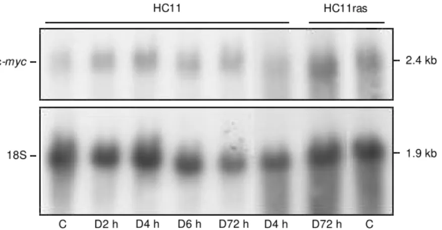
in breast cancer cells (43). The hormone enhances HOXB4 (a homeobox gene prod-uct) that binds to MIE1 sites located at intron 1 of the c-myc gene and as a result a tran-scriptional elongation block takes place (44). It was also demonstrated that 1,25-(OH)2D3
could inhibit the mitogenic activity of insu-lin and insuinsu-lin growth factor I-stimulated growth of MCF-7 cells (45) which may be related to insulin growth factor binding pro-tein (IGFBP)-5 (46) and IGFBP-3 (47) up-regulation. Furthermore, the antiprolifera-tive effect of 1,25-(OH)2D3 could be
modu-lated by induction of BRCA1 gene expres-sion, as recently reported (48). On the other hand, 1,25-(OH)2D3-induced growth
inhibi-tion may involve activainhibi-tion of apoptosis in vitro with up-regulation of genes associated with mammary gland apoptosis such as TRPM-2/clusterin and cathepsin B as well as with down-regulation of antiapoptotic genes such as bcl-2 (38). Vitamin D-stimu-lated apoptotic regression in mice bearing MCF-7 xenografts was also described (49). The underlying mechanism of the anti-proliferative effects of 1,25-(OH)2D3 on
HC11 cells has yet to be clarified. Our data suggest that it does not involve c-myc down-regulation (Figure 4) (50) or TGF-ß1 up-regulation (51). Cell cycle regulators such as cyclins D1 and D3 and p27KIP1
determined by Western blot were also not involved. We
have observed a slight increase of CDKI p21WAF1/CIP1 and cyclin E expression (data
not shown).
Vitamin D analogs have already been employed in some clinical studies and topi-cal treatment of patients with lotopi-cally ad-vanced or cutaneous metastatic breast can-cer with calcipotriol resulted in very few responses (52), whereas a phase I study in which patients with advanced breast and colorectal cancer received EB1089 showed a few cases of disease stabilization (53).
In addition to having effects on cell pro-liferation 1,25-(OH)2D3 has been shown to
inhibit the invasive potential of breast can-cer cells in vitro (54) and its analog EB1089 can prevent skeletal metastasis development in vivo (55) and angiogenesis in vitro and in vivo (56) in human breast carcinoma cells transplanted into nude mice. Furthermore, an interaction between 1,25-(OH)2D3 or
ana-logs with chemotherapeutic drugs active on breast cancer, such as doxorubicin (57) and paclitaxel (58), was also demonstrated, re-sulting in potentiation of cytotoxicity in MCF-7 cell cultures. In addition, topical 1,25-(OH)2D3 increased the antitumor effect of
cyclophosphamide in female mice inocu-lated with murine mammary tumor (59) and additive effects were observed when a vita-min D analog, CB1093, was advita-ministered together with paclitaxel to MCF-7 growing in immunodeficient mice (60).
Taken together, these studies indicate that vitamin D and its analogs have important antitumoral properties, which might be ex-plored in chemopreventive as well as in thera-peutic cancer approaches.
Co ncluding re marks
The involvement of the vitamin D3
path-way in breast carcinogenesis and cancer pro-gression has not been fully clarified. Besides the tumor growth-suppressive activity of vi-tamin D3 compounds in vitro and in vivo,
additional aspects may be involved in the 2.4 kb
1.9 kb
D4 h D6 h D72 h D4 h D72 h C
c-myc
18S
HC11 HC11ras
Figure 4. Expression of c-myc mRNA in HC11 and HC11ras cells exposed or not (C, control) to 100 nM 1,25-(OH)2D3 (D) for short (2, 4, 6 h) or long (72 h) periods of time, evaluated in Northern blot assays.
Re fe re nce s
1. Horst RL & Reinhardt TA (1997). Vitamin D metabolism. In: Feldman D, Glorieux FH & Pike JW (Editors), Vitamin D. Aca-demic Press, San Diego, CA, USA. 2. Rachez C, Gamble M , Chang CP, Atkins
GB, Lazar M A & Freedman LP (2000). The DRIP complex and SRC-1/p160 coactiva-tors share similar nuclear receptor bind-ing determinants but constitute function-ally distinct complexes. M olecular and Cellular Biology,20: 2718-2726. 3. Gill RK & Christakos S (1993).
Identifica-tion of sequence elements in mouse calbindin-D28K gene that confer 1,25-dihy-droxyvitamin D3 and butyrate inducible re-sponses. Proceedings of the National Academy of Sciences, USA, 90: 2984-2988.
4. Kerner SA, Scott RA & Pike JW (1989). Sequence elements in the human osteo-calcin gene confer basal activation and inducible response to hormonal vitamin D3. Proceedings of the National Academy
of Sciences, USA, 86: 4455-4459. 5. Noda M , Vogel RL, Craig AM , Prahl J,
DeLuca HF & Denhardt DT (1990). Identi-fication of a DNA sequence responsible for binding of the 1,25-dihydroxyvitamin D3 receptor and 1,25-dihydroxyvitamin D3 enhancement of mouse secreted phos-phoprotein 1 (Spp-1 or osteopontin) gene expression. Proceedings of the National Academy of Sciences, USA, 87: 9995-9999.
6. Ohyama Y, Noshiro M , Eggertsen G, Gotoh O, Kato Y, Bjorkhem I & Okuda K (1993). Structural characterization of the gene enconding rat 25-hydroxyvitamin D3 24-hydroxylase. Biochemistry, 32: 76-82. 7. Liu X, Lee M T, Cohen M , Bormakanti M & Freedman LP (1996). Transcriptional acti-vation of the CDK inhibitor p21 by vitamin
D3 leads to the induced differentiation of the myelomonocytic cell line U937. Genes and Development, 10: 142-153. 8. Wu Y, Craig TA, Lutz WH & Kumar R
(1999). Identification of 1a ,25-dihydroxy-vitamin D3 response elements in the hu-man transforming grow th factor ß2 gene.
Biochemistry, 38: 2654-2660.
9. Krishnan AV & Feldman D (1991). Stimu-lation of 1,25-dihydroxyvitamin D3 recep-tor gene expression in cultured cells by serum and grow th factors. Journal of Bone and M ineral Research, 6: 1099-1107.
10. Eisman JA, M artin TJ, M acIntyre I & M oseley JM (1979). 1,25-Dihydroxyvita-min-D-receptor in breast cancer cells. Lan-cet, 2: 1335-1336.
11. Escaleira M TF, Sonohara S & Brentani M M (1993). Sex steroids induced up-regu-lation of 1,25-(OH)2 vitamin D3 receptors in T47D breast cancer cells. Journal of Steroid Biochemistry and M olecular Biol-ogy, 45: 257-263.
12. Feldman J, Federico M HH, Sonohara S, Katayama M LH, Koike M AA, Da Silva M RP & Brentani M M (1993). Vitamin D3 binding activity during leukemic cell dif-ferentiation. Leukemia Research, 17: 97-101.
13. Folgueira M AAK, Federico M HH, Kata-yama M LH, Silva M RP & Brentani M M (1998). Expression of vitamin D receptor (VDR) in HL-60 cells is differentially regu-lated during the process of differentiation induced by phorbol ester, retinoic acid or interferon-g. Journal of Steroid Biochem-istry and M olecular Biology, 66: 193-201. 14. Folgueira M AAK, Federico M HH, Roela RA, M aistro S, Katayama M LH & Brentani M M (2000). Differential regulation of vita-min D receptor expression in distinct
leu-kemic cell lines upon phorbol ester-in-duced grow th arrest. Brazilian Journal of M edical and Biological Research, 33: 559-568.
15. Capiati DA, Vazquez G, Iñon M TT & Boland RL (2000). Role of protein kinase C in 1,25(OH)2-vitamin D3 modulation of intracellular calcium during development of skeletal muscle cells in culture. Journal of Cellular Biochemistry, 77: 200-212. 16. Nem ere I, Dorm anen M C, Ham m ond
M W, Okamura WH & Norman AW (1994). Identification of a specific binding protein for 1a,25-dihydroxyvitamin D3 in basal-lat-eral membranes of chick intestinal epithe-lium and relationship to transcaltachia.
Journal of Biological Chem istry, 269: 23750-23756.
17. Hmama Z, Nandan D, Knutson KL, Herrera-Velit P & Reiner NE (1999). 1a ,25-Dihy-droxyvitamin D3-induced myeloid cell dif-ferentiation is regulated by a vitamin D receptor-phosphatidylinositol 3-kinase sig-naling complex. Journal of Experimental M edicine, 190: 1583-1594.
18. Garland FC, Garland CF, Gorham ED & Young JF (1990). Geographic variation in breast cancer mortality in the United States: a hypothesis involving exposure to solar radiation. Preventive M edicine, 19: 614-622.
19. John EM , Schw artz GG, Dreon DM & Koo J (1999). Vitamin D and breast cancer risk: the NHANES epidem iologic follow -up study, 1971-1975 to 1992. National Health and Nutrition Examination Survey. Cancer Epidemiology, Biomarkers and Preven-tion, 8: 399-406.
20. Janow sky EC, Lester GE, Weinberg CR, M illikan RC, Schildkraut JM , Garrett PA & Hulka BS (1999). Association betw een low levels of 1,25-dihydroxyvitamin D and antitumoral properties of this vitamin.
Vita-min D3 seems also to induce apoptosis and to
inhibit the processes of angiogenesis, inva-sion and metastasis. Interactions between vitamin D3 and chemotherapeutic drugs have
been reported and represent another field to be explored. On the other hand, the pharma-cological doses of vitamin D3 necessary to
induce antiproliferative effects are associated with hypercalcemia in vivo. New vitamin D3
analogs, which are less hypercalcemic but are potent growth inhibitory agents, might be an option to fight cancer development.
Ackno wle dgm e nts
breast cancer risk. Public Health Nutrition, 2: 283-291.
21. M aw er EB, Walls J, How ell A, Davies M , Ratcliffe WA & Bundred NJ (1997). Se-rum 1,25-dihydroxyvitamin D may be re-lated inversely to disease activity in breast cancer patients w ith bone metastases.
Journal of Clinical Endocrinology and M e-tabolism, 82: 118-122.
22. Albertson DG, Ylstra B, Segraves R, Collins C, Dairkee SH, Kow bel D, Kuo WL, Gray JW & Pinkel D (2000). Quantitative mapping of amplicon structure by array CGH identifies CYP24 as a candidate on-cogene. Nature Genetics,25: 144-146. 23. Friedrich M , Reichrath J, Chen TC,
Tan-pricha V, Gherson I, Tilgen W, Schmidt W & Holick M F (2000). Expression of 25-hydroxyvit am in D3-1a-hydroxylase in breast tissue. In: Norman AW, Bouillon R & Thomasset M (Editors), Vitamin D En-docrine System Structural, Biological, Ge-netic and Clinical Aspects. Proceedings of the Eleventh Workshop on Vitamin D. Printing and Reprographics University of California, Riverside, CA, USA, 189-191. 24. Colston K, Wilkinson JR & Coombes RC
(1986). 1,25-Dihydroxyvitamin D3 binding in estrogen responsive rat breast tumor.
Endocrinology, 119: 397-403.
25. Eism an JA, M acInt yre I, M art in TJ, Frampton RJ & King RJ (1980). Normal and malignant breast tissue is a target organ for 1,25-(OH)2 vitamin D3. Clinical
Endocrinology, 13: 267-272.
26. Chouvet C, Vicard E, Devonec M & Saez S (1986). 1,25-Dihydroxyvitamin D3 inhibito-ry effect on the grow th of tw o human breast cancer cell lines (M CF-7, BT-20).
Journal of Steroid Biochemistry and M o-lecular Biology, 24: 373-376.
27. Iino Y, Hyoshida M , Sugamata N, M ae-mura M , Ohw ada S, Yokoe T, Ishikita T, Horiuchi R & M orishita Y (1992). 1Alpha-hydroxyvitamin D3, hypercalcemia, and grow th suppression of 7,12-dim ethyl-benz(a)anthracene-induced rat mammary tum ors. Breast Cancer Research and Treatment, 22: 133-140.
28. M ehta R, Haw thorne M , Uselding L, Albinescu D, M oriarty R, Christov K & M ehta R (2000). Prevention of N-methyl-N-nitrosourea-induced mammary carcino-genesis in rats by 1 alpha-hydroxyvitamin D(5). Journal of the National Cancer
Insti-tute, 92: 1836-1840.
29. Friedrich M , Rafi L, Tilgen W, Schmidt W & Reichrath JV (1998). Expression of 1,25-dihydroxyvitamin D3 receptor in breast carcinoma. Journal of Histochemistry and Cytochemistry, 46: 1335-1337.
30. Freake HC, Abeyasekera G, Iw asaki J, M arocci C, M acIntyre I, M cClelland RA, Skilton RA, Easton DF & Coombes RC (1984). M easurement of 1,25-dihydroxy-vitamin D3 receptors in breast cancer and relationship to biochemical and clinical in-dices. Cancer Research, 44: 1677-1681. 31. Colston KW, Berger U & Coombes RC
(1989). Possible role for vitamin D in con-trolling breast cancer cell proliferation.
Lancet, 1: 185-191.
32. Katayama M LH, Barboza EM & Brentani M M (2000). Expression of vitamin D re-ceptor and 24-hydroxylase genes in breast cancer. Cancer Detection and Prevention, 24: S126.
33. M erlo GR, Venesio T, Taverna D, M arte BM , Callahan R & Hynes NE (1994). Grow th suppression of normal mammary epithelial cells by w ild type p53. Onco-gene, 9: 443-453.
34. Hynes NE, Taverna D, Harw et h IM , Clardiello F, Salomon DS, Yamamoto T & Groner B (1990). Epidermal grow th factor receptor, but not c-erb-2, activation pre-vents lactogenic hormone induction of ß-casein gene in mouse mammary epitheli-al cells. M olecular and Cellular Biology, 10: 4027-4043.
35. Escaleira M TF & Brentani M M (1999). Norm al x Ha-ras-t ransf orm ed HC11 m ouse m am m ary cells: dif f erent re-sponses to the antiproliferative activity of vitamin D3 and modulation of its receptor.
Breast Cancer Research and Treatment, 54: 123-134.
36. Bortman P, Folgueira M AAK, Katayama M LH, Snitcovsky IM L & Brentani M M (2000). Regulação da expressão de RNAm de VDR e caracterização do envolvimento da via genôm ica e não genôm ica da vitamina D em células HC11 parentais ou transformadas com H-ras. Anais of the XV Annual M eeting of the Federação de So-ciedades de Biologia Experimental, Ca-xambu, M G, Brazil, August 23-26, 2000, 98-99.
37. Katayama M LH, Folgueira M AAK, Snitcov-sky IM L, Garcia EF, Apolinário D, Bortman P, Roela RA & Brentani M M (2000). HC11 mouse mammary cells are grow th inhib-ited by EB1089 and KH1060 w ith a late induction of TGF-ß expression w hile HC11 Ha-ras transformed cells are not affected by these compounds. In: Norman AW, Bouillon R & Thomasset M (Editors), Vita-min D Endocrine System Structural, Bio-logical, Genetic and Clinical Aspects. Pro-ceedings of the Eleventh Workshop on Vitamin D. Printing and Reprographics University of California, Riverside, CA,
USA, 407-410.
38. James SY, M ackay AG & Colston KW (1996). Effects of 1,25 dihydroxyvitamin D3 and its analogues on induction of ap-optosis in breast cancer cells. Journal of Steroid Biochemistry and M olecular Biol-ogy, 58: 395-401.
39. Wu G, Fan RS, Li W, Ko TC & Brattain M G (1997). M odulation of cell cycle control by vitamin D3 and its analogue, EB 1089, in human breast cancer cells. Oncogene, 15: 1555-1563.
40. Inoue T, Kamiyama J & Sakai T (1999). Sp1 and NF-Y synergistically mediate the effect of vitamin D3 in the p27Kip1 gene promoter that lacks vitamin D response elements. Journal of Biological Chemis-try, 274: 32309-32317.
41. Koli K & Keski-Oja J (1995). 1,25-Dihy-droxyvitamin D3 enhances the expression of transforming grow th factor beta 1 and its latent form binding protein in cultured breast carcinoma cells. Cancer Research, 55: 1540-1546.
42. Wu G, Fan RS, Li W, Srinivas V & Brattain M G (1998). Regulation of transforming grow th factor-beta type II receptor expres-sion in human breast cancer M CF-7 cells by vitamin D3 and its analogs. Journal of
Biological Chemistry, 273: 7749-7756. 43. Saunders DE, Christensen C, Wappler NL,
Schultz JF, Law rence WD, M alviya VK, M alone JM & Deppe G (1993). Inhibition of c-myc in breast and ovarian carcinoma cells by 1,25-dihydroxyvitamin D3, retinoic acid and dexam et hasone. Ant icancer Drugs, 4: 201-208.
44. Pan Q & Simpson RU (2001). Antisense knockout of HOXB4 blocks 1,25-dihy-droxyvitamin D3 inhibition of c-myc ex-pression. Journal of Endocrinology, 169: 153-159.
45. Vink-van Wijngaarden T, Pols HA, Buur-man CJ, Birkenhager JC & van Leeuw en JP (1996). Inhibition of and insulin-like grow th factor-I-stimulated grow th of human breast cancer cells by 1,25-dihy-droxyvitamin D3 and the vitamin D3 ana-logue EB1089. European Journal of Can-cer, 32A: 842-848.
46. Rozen F, Yang XF, Huynh H & Pollak M (1997). Antiproliferative action of vitamin D-related com pounds and insulin-like grow th-factor-binding protein 5 accumu-lation. Journal of the National Cancer In-stitute, 89: 652-656.
grow th factor binding protein-3. Journal of M olecular Endocrinology, 20: 157-162. 48. Campbell M J, Gombart AF, Kw ok SH, Park S & Koeffler HP (2000). The anti-proliferative effects of 1alpha,25(OH)2D3 on breast and prostate cancer cells are associated w ith induction of BRCA gene expression. Oncogene, 19: 5091-5097. 49. VanWeelden K, Flanagan L, Binderup L,
Tennisw ood M & Welsh JW (1998). Apop-totic regression of M CF-7 xenografts in nude mice treated w ith the vitamin D3 analog, EB1089. Endocrinology, 139: 2102-2110.
50. Folgueira M AAK, Katayama M LH, Snitcov-sky IM L & Brentani M M (1998). c-myc
and c-max mRNA expression in HC11 m ouse m am m ary cells exposed t o 1,25(OH)2D3 are events unrelated to grow th arrest. In: M oraes M , Brentani R & Bevilacqua R (Editors), 17th Interna-t ional Cancer Congress. M onduzzi Editore, Bologna, Italy.
51. Folgueira M AAK, Katayama M LH, Snitcov-sky IM L & Brentani M M (2000). The anti-proliferative effect of 1,25(OH)2D3 on HC11 mammary cells is not associated to induction of TGFß and p21WAF1/CIP1 or inhibition of c-myc expression. Breast Cancer Research and Treatment, 64: 115
(Abstract).
52. Bow er M , Colston KW, Stein RC, Hedley A, Gazet JC, Ford HT & Coombes RC (1991). Topical calcipotriol treatment in advanced breast cancer. Lancet, 337: 701-702.
53. Gulliford T, English J, Colston KW, M endy P, M oller S & Coombes RC (1998). A phase I study of the vitamin D analogue EB 1089 in patients w ith advanced breast and colorectal cancer. British Journal of Cancer, 78: 6-13.
54. Hansen CM , Frandsen TL, Brünner N & Binderup L (1994). 1,25-Dihydroxyvitamin D3 inhibits the invasive potential of hu-man breast cancer cells in vitro. Clinical and Experimental M etastasis, 12: 195-202.
55. El Abdaimi K, Dion N, Papavasiliou V, Car-dinal PE, Binderup L, Goltzman D, Ste-M arie LG & Kremer R (2000). The vitamin D analogue EB 1089 prevents skeletal metastasis and prolongs survival time in nude m ice t ransplant ed w it h hum an breast cancer cells. Cancer Research, 60: 4412-4418.
56. M antell DJ, Ow ens PE, Bundred NJ, M aw er EB & Canfield AE (2000). 1Alpha, 25-dihydroxyvitamin D3 inhibits angiogen-esis in vitro and in vivo. Circulation
Re-search, 87: 214-220.
57. Chaudhry M , Sundaram S, Gennings C, Carter H & Gew irtz DA (2001). The vita-min D3 analog, ILX-23-7553, enhances the response to adriamycin and irradiation in M CF-7 breast tumor cells. Cancer Che-motherapy and Pharmacology, 47: 429-436.
58. Wang Q, Yang W, Uytingco M S, Christa-kos S & Wieder R (2000). 1,25-Dihydroxy-vitamin D3 and all-trans-retinoic acid sen-sit ize breast cancer cells t o chem o-therapy-induced cell death. Cancer Re-search, 60: 2040-2048.
59. Chen G, Baechle A, Nevins TD, Oh S, Harmon C & Stacey DW (1998). Protec-tion against cyclophosphamide-induced alopecia and inhibition of tumor grow th by topical 1,25-dihydroxyvitamin D3 in mice. International Journal of Cancer, 2: 303-309.