Associated with the Degree of Valvular Aortic Stenosis
Thomas Wurster1*., Roland Tegtmeyer1., Oliver Borst1
, Dominik Rath1, Tobias Geisler1, Meinrad Gawaz1, Boris Bigalke1,2
1Medizinische Klinik III, Kardiologie und Kreislauferkrankungen, Eberhard-Karls-Universita¨t Tu¨bingen, Germany,2King’s College London, Division of Imaging Sciences and Biomedical Engineering, London, United Kingdom
Abstract
Background and Purpose:Platelet surface expression of stromal-cell-derived factor-1 (SDF-1) is increased during platelet activation and constitutes an important factor in hematopoetic progenitor cell trafficking at sites of vascular injury and ischemia. Enhanced platelet SDF-1 expression has been reported previously in patients suffering from acute coronary syndrome (ACS). We hypothesized that expression of platelet associated SDF-1 may also be influenced by calcified valvular aortic stenosis (AS).
Methods:We consecutively evaluated 941 patients, who were admitted to the emergency department with dyspnea and chest pain. Platelet surface expression of SDF-1 was determined by flow cytometry, AS was assessed using echocardiography and hemodynamic assessment by heart catheterization. A 1:1 propensity score matching was implemented to match 218 cases with 109 pairs adjusting for age, sex, cardiovascular risk factors, and medication including ACE inhibitors, angiotensin receptor blockers, beta blockers, statins, aspirin, clopidogrel, GPIIb/IIIa antagonists, and vitamin K antagonists.
Results:Patients with valvular AS showed enhanced platelet SDF-1 expression compared to patients without AS (non-valvular disease, NV) independent of ACS and stable coronary artery disease (SAP) [mean fluorescence intensity (MFI) for ACS (AS vs. NV): 75640.4 vs. 39.5623.3; P = 0.002; for SAP (AS vs. NV): 54.9644.6 vs. 24.3611.2; P = 0.008]. Moreover, the degree of AS significantly correlated with SDF-1 platelet surface expression (r = 0.462; P = 0.002).
Conclusions: Valvular AS is associated with enhanced platelet-SDF-1 expression; moreover the degree of valvular AS correlates with SDF-1 platelet surface expression. These findings may have clinical implications in the future.
Citation:Wurster T, Tegtmeyer R, Borst O, Rath D, Geisler T, et al. (2014) Platelet Expression of Stromal Cell-Derived Factor-1 Is Associated with the Degree of Valvular Aortic Stenosis. PLoS ONE 9(5): e97405. doi:10.1371/journal.pone.0097405
Editor:Ingo Ahrens, University Hospital Medical Centre, Germany
ReceivedJanuary 30, 2014;AcceptedApril 19, 2014;PublishedMay 16, 2014
Copyright:ß2014 Wurster et al. This is an open-access article distributed under the terms of the Creative Commons Attribution License, which permits unrestricted use, distribution, and reproduction in any medium, provided the original author and source are credited.
Funding:This study has been supported by the Deutsche Forschungsgemeinschaft -Klinische Forschergruppe KFO274 "Platelets – Molecular Mechanisms and Translational Implications" to M.G. and the German Cardiac Society (DGK) ‘‘molecular imaging of atherosclerotic plaques’’ to B.B. The funders had no role in study design, data collection and analysis, decision to publish, or preparation of the manuscript.
Competing Interests:The authors have declared that no competing interests exist. * E-mail: [email protected]
.These authors contributed equally to this work.
Introduction
Degenerative calcified valvular heart disease concerns a noteworthy group of patients in the Western world and increases with age. The more frequent appearance of aortic stenosis (AS) in an increasingly elderly population poses a growing challenge to clinicians and public healthcare [1]. Risk factors for the development of AS are similar to those associated with athero-sclerosis, and approximately half of the patients with severe AS feature significant coronary artery disease (CAD) [2]. Nevertheless, patients with aortic sclerosis are also likely to suffer from cardiovascular events [3]. To date, biomarkers play a subordinate role in the diagnosis and staging of AS. The chemokine stromal cell-derived factor-1 (SDF-1) captures an important role in the regeneration of ischemic tissue [4] and stem cell trafficking [5]. Both, in patients with AS [6] and acute coronary syndrome (ACS) [7] platelets show increased reactivity. However, platelets exhibit
an enhanced SDF-1 surface expression upon activation [8,9]. In a previous study, in a large cohort comprising 1,000 patients suffering from acute chest pain, our group demonstrated an enhanced SDF-1 expression on activated platelets in patients with ACS [10]. Hemodynamic alterations caused by AS are likely to cause platelet activation, therefore platelet-SDF-1 surface expres-sion might be associated with AS. The aim of the present study was to evaluate platelet SDF-1 surface expression in patients presenting symptomatic CAD and concomitant AS in the emergency care unit.
Methods
Study population and enrolment criteria
angiography and complete hemodynamic assessment by heart catheterization and echocardiographic analysis. After implemen-tation of a 1:1 propensity score matching adjusting for age, sex, cardiovascular risk factors and medication including ACE inhibitors, angiotensin receptor blockers, beta blockers, statins, aspirin, clopidogrel, GPIIb/IIIa antagonists, and vitamin K antagonists, 218 cases with 109 pairs were matched. All patients underwent ECG and serum examination for troponin-I, creatine kinase, C-reactive peptide and creatinine, measurement of blood pressure, clinical examination and echocardiography, as well as left heart catheterization. Exclusion and inclusion criteria are given inTable 1. All participant patients provided written informed consent. The study was conducted according to the principles of the Declaration of Helsinki and approved by the local ethics committee at the University Hospital Tu¨bingen, Germany (Ethics approval number: ‘‘292/2005V platelet activation in patients with coronary artery disease‘‘).
Definitions
ACS: ACS definition according to AHA/ACC guidelines [11]: ACS comprises ST-Elevation Myocardial Infarction (STEMI), non–ST-segment elevation myocardial infarction (NSTEMI) as well as unstable angina pectoris. The predetermined diagnostic threshold for troponin-I was$0.04mg/L [12].
SAP: Patients suffering from stable angina pectoris manifesting through chest pain with stable intensity and stress inducible
myocardial ischemia [13]. Furthermore patients showing CAD and presenting with non-elevated CK or troponin-I and without dynamic ECG changes.
AS: AS definition according to AHA/ACC guidelines: Mild AS (AS Iu) exhibits an aortic valve area (AVA) of more than 1.5 cm2 with a mean gradient of less than 25 mmHg, or a jet velocity less than 3.0 m per second. Moderate AS (AS IIu) exhibits an AVA of 1.0 to 1.5 cm2with a mean gradient of 25 to 40 mmHg, or a jet velocity of 3.0 to 4.0 m per second. Severe AS (AS IIIu) exhibits an AVA of less than 1.0 cm2 with a mean gradient greater than 40 mmHg, or a jet velocity greater than 4.0 m per second [14].
Sample collection, FACS analysis and AVA determination
On admission to the emergency care unit venous blood samples were drawn from patients with chest pain and dyspnea while standard routine laboratory assessment and filled into 5 mL citrate phosphate dextrose adenine vials (CPDA). Blood samples have been processed within 3 hours maximum after blood was drawn from the participant according to a predefined assay as described before [15,16]. In brief, CPDA-Blood was diluted with phosphate buffered saline (PBS) in relation 1:50. From this suspension, 35ml were put into test tubes and further 5ml PBS were added. Then 5ml solute containing either FITC labeled SDF-1 or
anti-CD62P antibodies following 5ml of PE-labeled anti-CD42b
antibodies were adjusted. The probes were incubated for 25 minutes at 21uCelcius in complete darkness. Afterwards 300ml of
Table 1.Inclusion and Exclusion Criteria.
Inclusion criteria Patients with emergency care unit admittance due to chest pain and/or dyspnea and further assessment including coronary angiography, complete hemodynamic assessment by heart catheterization and echocardiographic analysis
Exclusion criteria Additional hemodynamically significant valve lesions (moderate or severe) or patients with (paradoxical) low flow/low gradient aortic stenosis, age below 18 years, cardiac shock, stroke, malignancy, connective tissue disease, psychiatric disease, anorexia, pregnancy or hormone replacement therapy in women, lack or inability of informed consent
doi:10.1371/journal.pone.0097405.t001
0.5% paraformaldehyde were added. The probes were stored at 4uCelcius in complete darkness not later than 48 hours until two-color whole blood flow cytometry analysis was performed (FACS-Calibur Flow Cytometer, Becton-Dickinson, Heidelberg, Ger-many). Platelet surface expression of SDF-1, CD62P and GPIb (CD42) was analyzed using conjugated antibodies [SDF-1, R&D Systems, Minneapolis, MN, USA; clone 79014; fluorescein isothiocyanate (FITC); CD62P, Immunotec, Marseille, France; clone CLB-Thromb/6; FITC and CD42b, Immunotec, Marseille, France; clone SZ2; phycoerythrin (PE)]. CD42b-PE was used for identification of the platelet population as second color control antibody. The mean fluorescence intensity (MFI) served as quantity index of biomarker surface expression. AVA was estimated using echocardiography (iE33, Philips Medical Systems) according to the continuity equation including the velocity-time integral of the aortic and left ventricular outflow tract flows.
Statistics
We considered a probability value of less than 0.05 as statistically significant. Propensity score was applied for bias reduction. Therefore, we implemented a 1:1 propensity score
matching to balance 109 cases with 109 controls regarding age, sex, cardiovascular risk factors and medication including ACE inhibitors, angiotensin receptor blockers, beta blockers, statins, aspirin, clopidogrel, GPIIb/IIIa antagonists and vitamin K antagonists. Raynald’s SPSS tool for propensity score matching was used. We analyzed Categorical data by chi-square test. For comparison of differences between groups Kruskal–Wallis test was used for non-normally distributed data. For evaluation of associations concerning events and variables a log-rank test (Mantel-Cox) was implemented. Correlation between AS and SDF-1 or CD62P expression was analyzed using Pearson correlation. All statistical analyses were performed using SPSS version 19.
Results
A consecutive cohort of 941 patients with underlying coronary artery disease [ACS,n = 498; SAP,n = 443] suffering from chest pain and dyspnea was evaluated on hospital admission. All patients were evaluated by heart catheterization and transthoracic echocardiography. Platelet surface expression of SDF-1 was determined by flow cytometry. Finally, a 1:1 propensity score
Table 2.Baseline Patients’ Characteristics and Medical Treatment on Admission.
Characteristics Before Matching After Matching
Total (n = 941) Non-valvular Disease (n = 109) Aortic Stenosis (n = 109)
Age – years 65.5 66.7 62.7
Gender – no. (%)
Female 636 (67.6%) 79 (72.5%) 76 (69.7%)
Male 305 (32.4%) 30 (27.5%) 33 (30.3%)
ACS 613 (65.1%) 87 (79.8%) 55 (50.5%)
SAP 328 (34.9%) 22 (20.2%) 54 (49.5%)
Cardiovascular Risk Factors – no. (%)
Arterial hypertension 714 (75.9%) 81 (74.3%) 78 (71.6%)
Hyperlipidaemia 506 (53.8%) 59 (54.1%) 62 (56.9%)
Diabetes 253 (26.9%) 22 (20.2%) 36 (33%)
Family history of CVD 158 (16.8%) 11 (10.1%) 19 (17.4%)
Smoking 321 (34.1%) 47 (43.1%) 43 (39.4%)
Atrial fibrillation 147 (15.6%) 9 (8.2%) 22 (20.2%)
Medication – no. (%)
ACE inhibitors 88 (9.4%) 22 (20.2%) 10 (9.2%)
Angiotensin receptor blockers 64 (6.8%) 7 (6.4%) 6 (5.5%)
Beta blockers 67 (7.1%) 7 (6.4%) 6 (5.5%)
Statins 67 (7.1%) 7 (6.4%) 6 (5.5%)
Aspirin 70 (7.4%) 7 (6.4%) 8 (7.3%)
Clopidogrel 66 (7%) 6 (5.5%) 7 (6.4%)
Vitamin K antagonists 67 (7.1%) 4 (3.7%) 5 (4.6%)
Medication on hospital discharge– no. (%)
ACE inhibitors 682 (72.5%) 78 (71.6%) 80 (73.3%)
Angiotensin receptor blockers 172 (18.3%) 7 (6.4%) 8 (7.3%)
Beta blockers 777 (82.6%) 84 (77.1%) 79 (72.5%)
Statins 695 (73.9%) 86 (78.9%) 75 (68.8%)
Aspirin 762 (81%) 85 (78%) 77 (70.6%)
Clopidogrel 680 (72.3%) 82 (75.2%) 72 (66.1%)
Vitamin K antagonists 185 (19.7%) 3 (2.8%) 18 (16.5%)
matching was implemented to match 218 cases with 109 pairs comprising age, gender, cardiovascular risk factors, as well as medication including ACE inhibitors, angiotensin receptor block-ers, beta blockblock-ers, statins, aspirin, clopidogrel, GPIIb/IIIa antagonists and vitamin K antagonists (Fig. 1). The demographic details are given inTable 2.
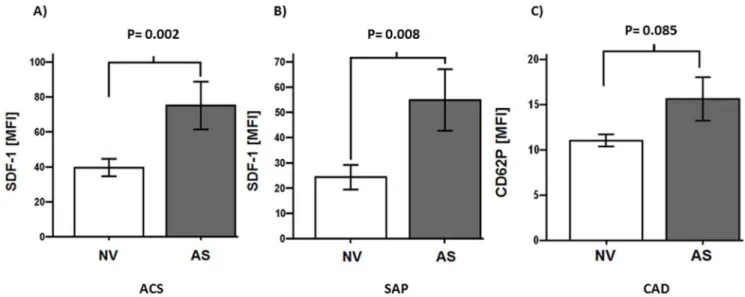
Patients with valvular AS showed enhanced platelet SDF-1 expression compared to patients with non-valvular disease (NV) both with ACS and SAP [mean fluorescence intensity (MFI) for ACS (AS vs. NV): 75640.4 vs. 39.5623.3; P = 0.002; for SAP (AS
vs. NV): 54.9644.6 vs. 24.3611.2; P = 0.008] (Fig. 2A,B). CD62P mean fluorescence intensity showed a trend for increased platelet activation in CAD patients with AS compared to non-valvular disease (NV) [AS vs. NV: 15.6612.6 vs. 1163.6; P = 0.085] (Fig. 2C). Among the 109 patients featuring AS, 75 were classified as AS Iu(69%), 21 were classified as AS IIu (19%) and 13 were classified as AS IIIu(12%) (Fig. 3A). The degree of AS correlated with SDF-1 platelet surface expression (r = 0.462; P = 0.002) (Fig. 3B).
Figure 2. Platelet surface expression of SDF-1 in acute coronary syndrome (ACS) (A), stable angina pectoris (SAP) (B) subgroups and platelet CD62P expression in patients with symptomatic coronary artery disease (CAD) (C).(A, B) SDF-1 mean fluorescence intensity (MFI) in patients with non-valvular disease (NV) compared to patients with aortic stenosis (AS) [ACS (AS vs. NV): 75640.4 vs. 39.5623.3; P = 0.002; SAP (AS vs. NV): 54.9644.6 vs. 24.3611.2; P = 0.008]. (C) CD62P mean fluorescence intensity showed a trend for increased platelet activation in CAD patients with aortic stenosis (AS) compared to patients with non-valvular disease (NV) [AS vs. NV: 15.6612.6 vs. 1163.6; P = 0.085].
doi:10.1371/journal.pone.0097405.g002
Figure 3. Platelet SDF-1 surface expression according to the degree of aortic stenosis (AS) (A) and association between platelet SDF-1 expression and the degree of AS (B).(A) Platelet SDF-1 surface expression depicted by mean fluorescence intensity (MFI6standard deviation) in patients with non-valvular disease (NV; n = 109; 36.3622.1) and patients with mild (AS Iu; n = 75; 54.2642.2), moderate (AS IIu; n = 21; 81.8647.9) and severe (AS IIIu; n = 13; 100.7655.9) aortic stenosis. (B) Association of platelet SDF-1 surface expression in patients with non-valvular disease (NV) and patients with mild AS (Iu), moderate AS (IIu) and severe AS (IIIu) (r = 0.462; P = 0.002).
Discussion
The major findings of the present study are: 1) Patients with valvular AS show enhanced platelet SDF-1 expression compared to patients with non-valvular disease both in patients with ACS and SAP; 2) The degree of AS correlates with surface expression of platelet SDF-1.
SDF-1, also known as CXCL12 is a prominent CXC-chemokine that binds to its receptors CXCR4 and CXCR7 and regulates chemotaxis, migration and differentiation of inflamma-tory cells including monocytes and hematopoetic progenitor cells [9]. Among other cells, platelets have been shown to store and release substantial amounts of SDF-1 upon activation [9,17,18]. In resting platelets, SDF-1 is stored in cytosolic granules. Upon platelet activation and peripheralization of granules, a markedly enhanced platelet surface expression of SDF-1 can be observed [8]. Previous studies demonstrated a positive correlation between plasma levels of SDF-1 and platelet SDF-1 surface expression [19,20]. However, experimental data suggest an imprecise relation between SDF-1 detachment and SDF-1 expression [8]. Therefore, the study focused on platelet associated SDF-1 expression.
In a large study including patients suffering from chest pain, our group demonstrated an enhanced SDF-1 expression on the platelet surface in patients with ACS [10]. Moreover, our group observed an association between enhanced platelet SDF-1 expression with reduced left ventricular function and the number of circulating CD34+ progenitor cells, predominantly in patients suffering from acute myocardial infarction (MI) [19,21].
Circulating platelets are critically influenced by altered hemo-dynamics and increased shear stress in individuals with AS [6,22– 25], moreover high shear forcesin vivoand in vitroare associated with platelet activation [26]. Experimental data suggests the release of antithrombotic agents, such as nitric oxide (NO) and prostacyclin from normal aortic valves [27,28], whereas increased platelet reactivity as well as thrombus formation have been observed on severely calcified and stenotic aortic valves [29]. In previous studies the expression of several biomarkers in patients suffering from AS has been observed. Dimitrow et al. showed enhanced concentrations of thrombin, thrombin–antithrombin complexes (TAT), prothrombin fragment 1+2 (F1+2), soluble CD40 ligand (sCD40L) and beta-thromboglobulin (beta-TG) in patients with AS [30]. Furthermore, Luszczak et al. observed detectable plasma tissue factor (TF) and factor XIa activity associated with thrombin generation in patients with especially severe AS [31]. Increased plasma thrombin formation and platelet activation in patients with moderate to severe AS has also been reported by Natorska et al. in patients additionally deficient for high molecular weight multimers of von Willebrand factor (HMWM vWF) [24]. In fact, platelet activation via thrombin receptor PAR-1, as well as adenosine diphosphate (ADP) receptors P2Y1/P2Y12 and glycoprotein VI (GPVI)-dependent pathways result in increased platelet surface expression and release of SDF-1 [8]. Compared to our preceding study in patients with ACS, subgroup analysis in the present study reveals an even more increased platelet SDF-1 expression in patients with ACS featuring
AS compared to patients with ACS and non-valvular disease. Coherently, platelet SDF-1 expression is enhanced in patients with SAP and AS compared to non-valvular SAP. Therefore, AS resembles an independent co-variate associated with systemic platelet activation. The present study revealed moreover a significant correlation between increased SDF-1 platelet surface expression and the degree of AS. Thus, high shear stress associated with AS may be a determining factor regarding platelet activation. Previous studies revealed an association between high shear stress featuring AS and platelet aggregation due to increased binding of plasma HMWM of vWF to the platelet membrane [23,32]. Comparatively elevated shear stress due to obstruction of the left ventricular outflow tract in hypertrophic obstructive cardiomyop-athy is also associated with platelet activation and augmented thrombin generation [33].
In the present study, in contrast to SDF-1, platelet expression of CD62P in CAD patients showed merely a trend for increased platelet surface expression in patients with AS compared to NV (P = 0.085). Correspondingly, previous studies also revealed only weak associations between CD62P and AS severity or elevated gradients of the left ventricular outflow tract [24,33]. Despite evaluation of several biomarkers in AS, currently no biomarker for AS detection, progression or prognosis is used in clinical routine so far. However, there are several promising biomarkers which might provide future diagnostic and therapeutic implications [34]. To our knowledge, this is the first study demonstrating a significant association between platelet SDF-1 expression and the severity of AS in symptomatic patients. Thus, biomarker panels including platelet associated SDF-1 may offer new diagnostic options in diagnosis and staging of patients with AS in the future and might be especially interesting in the emergency setting. Further studies as well as simplified assays are needed to assess the impact of platelet SDF-1 expression in patients with AS especially in the preclinical setting.
Conclusions
To date, clinical examination, echocardiography and heart catheterization represent common standard in AS detection and staging. Our study shows increased platelet SDF-1 expression in patients with AS compared to patients with non-valvular disease both in ACS and SAP in a large cohort of patients after admittance to the emergency care unit. Therefore, detection of enhanced platelet SDF-1 expression in patients with chest pain or dyspnea on hospital admission might raise the suspicion of AS as well as concomitant ACS. In conclusion, platelet SDF-1 might represent a novel biomarker in AS to indicate risk and therefore should be evaluated for its prognostic value in aortic valve disease.
Author Contributions
Conceived and designed the experiments: TW MG BB. Performed the experiments: TW RT DR OB. Analyzed the data: BB TG. Contributed reagents/materials/analysis tools: BB TG. Wrote the paper: TW.
References
1. Nkomo VT, Gardin JM, Skelton TN, Gottdiener JS, Scott CG, et al. (2006) Burden of valvular heart diseases: a population-based study. Lancet 368: 1005– 1011.
2. Stewart BF, Siscovick D, Lind BK, Gardin JM, Gottdiener JS, et al. (1997) Clinical factors associated with calcific aortic valve disease. Cardiovascular Health Study. J Am Coll Cardiol 29: 630–634.
3. Aronow WS, Ahn C, Shirani J, Kronzon I (1999) Comparison of frequency of new coronary events in older subjects with and without valvular aortic sclerosis. Am J Cardiol 83: 599–600, A598.
4. Yamaguchi J, Kusano KF, Masuo O, Kawamoto A, Silver M, et al. (2003) Stromal cell-derived factor-1 effects on ex vivo expanded endothelial progenitor cell recruitment for ischemic neovascularization. Circulation 107: 1322–1328. 5. Kucia M, Reca R, Miekus K, Wanzeck J, Wojakowski W, et al. (2005)
6. Chirkov YY, Holmes AS, Willoughby SR, Stewart S, Horowitz JD (2002) Association of aortic stenosis with platelet hyperaggregability and impaired responsiveness to nitric oxide. Am J Cardiol 90: 551–554.
7. Theroux P, Fuster V (1998) Acute coronary syndromes: unstable angina and non-Q-wave myocardial infarction. Circulation 97: 1195–1206.
8. Chatterjee M, Huang Z, Zhang W, Jiang L, Hultenby K, et al. (2011) Distinct platelet packaging, release, and surface expression of proangiogenic and antiangiogenic factors on different platelet stimuli. Blood 117: 3907–3911. 9. Chatterjee M, Gawaz M (2013) Platelet-Derived CXCL12 (SDF-1alpha): Basic
Mechanisms and Clinical Implications. J Thromb Haemost 11: 1954–67. 10. Wurster T, Stellos K, Haap M, Seizer P, Geisler T, et al. (2013) Platelet
expression of stromal-cell-derived factor-1 (SDF-1): an indicator for ACS? Int J Cardiol 164: 111–115.
11. Anderson JL, Adams CD, Antman EM, Bridges CR, Califf RM, et al. (2007) ACC/AHA 2007 guidelines for the management of patients with unstable angina/non ST-elevation myocardial infarction: a report of the American College of Cardiology/American Heart Association Task Force on Practice Guidelines (Writing Committee to Revise the 2002 Guidelines for the Management of Patients With Unstable Angina/Non ST-Elevation Myocardial Infarction): developed in collaboration with the American College of Emergency Physicians, the Society for Cardiovascular Angiography and Interventions, and the Society of Thoracic Surgeons: endorsed by the American Association of Cardiovascular and Pulmonary Rehabilitation and the Society for Academic Emergency Medicine. Circulation 116: e148–304.
12. Waxman DA, Hecht S, Schappert J, Husk G (2006) A model for troponin I as a quantitative predictor of in-hospital mortality. J Am Coll Cardiol 48: 1755– 1762.
13. Gibbons RJ, Balady GJ, Bricker JT, Chaitman BR, Fletcher GF, et al. (2002) ACC/AHA 2002 guideline update for exercise testing: summary article: a report of the American College of Cardiology/American Heart Association Task Force on Practice Guidelines (Committee to Update the 1997 Exercise Testing Guidelines). Circulation 106: 1883–1892.
14. Bonow RO, Carabello BA, Chatterjee K, de Leon AC Jr, Faxon DP, et al. (2008) 2008 Focused update incorporated into the ACC/AHA 2006 guidelines for the management of patients with valvular heart disease: a report of the American College of Cardiology/American Heart Association Task Force on Practice Guidelines (Writing Committee to Revise the 1998 Guidelines for the Management of Patients With Valvular Heart Disease): endorsed by the Society of Cardiovascular Anesthesiologists, Society for Cardiovascular Angiography and Interventions, and Society of Thoracic Surgeons. Circulation 118: e523– 661.
15. Gawaz M, Neumann FJ, Schomig A (1999) Evaluation of platelet membrane glycoproteins in coronary artery disease: consequences for diagnosis and therapy. Circulation 99: E1–E11.
16. Gawaz M, Reininger A, Neumann FJ (1996) Platelet function and platelet-leukocyte adhesion in symptomatic coronary heart disease. Effects of intravenous magnesium. Thromb Res 83: 341–349.
17. Massberg S, Konrad I, Schurzinger K, Lorenz M, Schneider S, et al. (2006) Platelets secrete stromal cell-derived factor 1alpha and recruit bone marrow-derived progenitor cells to arterial thrombi in vivo. J Exp Med 203: 1221–1233. 18. Stellos K, Langer H, Daub K, Schoenberger T, Gauss A, et al. (2008) Platelet-derived stromal cell-Platelet-derived factor-1 regulates adhesion and promotes
differentiation of human CD34+cells to endothelial progenitor cells. Circulation 117: 206–215.
19. Stellos K, Bigalke B, Langer H, Geisler T, Schad A, et al. (2009) Expression of stromal-cell-derived factor-1 on circulating platelets is increased in patients with acute coronary syndrome and correlates with the number of CD34+progenitor cells. Eur Heart J 30: 584–593.
20. Stellos K, Rahmann A, Kilias A, Ruf M, Sopova K, et al. (2012) Expression of platelet-bound stromal cell-derived factor-1 in patients with non-valvular atrial fibrillation and ischemic heart disease. J Thromb Haemost 10: 49–55. 21. Geisler T, Fekecs L, Wurster T, Chiribiri A, Schuster A, et al. (2012) Association
of platelet-SDF-1 with hemodynamic function and infarct size using cardiac MR in patients with AMI. Eur J Radiol 81 (4): e486–490.
22. Blackshear JL, Wysokinska EM, Safford RE, Thomas CS, Stark ME, et al. (2013) Indexes of von Willebrand factor as biomarkers of aortic stenosis severity (from the Biomarkers of Aortic Stenosis Severity [BASS] study). Am J Cardiol 111: 374–381
23. Goto S, Ikeda Y, Saldivar E, Ruggeri ZM (1998) Distinct mechanisms of platelet aggregation as a consequence of different shearing flow conditions. J Clin Invest 101: 479–486.
24. Natorska J, Bykowska K, Hlawaty M, Marek G, Sadowski J, et al. (2011) Increased thrombin generation and platelet activation are associated with deficiency in high molecular weight multimers of von Willebrand factor in patients with moderate-to-severe aortic stenosis. Heart 97: 2023–2028. 25. Peterson DM, Stathopoulos NA, Giorgio TD, Hellums JD, Moake JL (1987)
Shear-induced platelet aggregation requires von Willebrand factor and platelet membrane glycoproteins Ib and IIb-IIIa. Blood 69: 625–628.
26. O’Brien JR, Etherington MD, Brant J, Watkins J (1995) Decreased platelet function in aortic valve stenosis: high shear platelet activation then inactivation. Br Heart J 74: 641–644.
27. Biberthaler P, Mendler N, Ettner U, Meisner H (1998) Endothelial prostacyclin (PGI-2) production of human and porcine valve allografts related to ischemic history. Eur J Cardiothorac Surg 14: 503–507.
28. De Meyer E, Van Hove CE, Feng XJ, Rampart M, Herman AG (1995) Thrombin triggers the de novo expression of an inducible NO synthase in porcine aortic valve endothelial cells. Eur J Pharmacol 291: 67–72.
29. Chirkov YY, Mishra K, Chandy S, Holmes AS, Kanna R, et al. (2006) Loss of anti-aggregatory effects of aortic valve tissue in patients with aortic stenosis. J Heart Valve Dis 15: 28–33.
30. Dimitrow PP, Hlawaty M, Undas A, Sniezek-Maciejewska M, Sobien´ B, et al. (2009) Effect of aortic valve stenosis on haemostasis is independent from vascular atherosclerotic burden. Atherosclerosis 204: e103–108.
31. Luszczak J, Undas A, Gissel M, Olszowska M, Butenas S (2011) Activated factor XI and tissue factor in aortic stenosis: links with thrombin generation. Blood Coagul Fibrinolysis 22: 473–479.
32. Moake JL, Turner NA, Stathopoulos NA, Nolasco LH, Hellums JD (1986) Involvement of large plasma von Willebrand factor (vWF) multimers and unusually large vWF forms derived from endothelial cells in shear stress-induced platelet aggregation. J Clin Invest 78: 1456–1461.
33. Dimitrow PP, Undas A, Bober M, Tracz W, Dubiel JS (2008) Obstructive hypertrophic cardiomyopathy is associated with enhanced thrombin generation and platelet activation. Heart 94: e21.