Dynamic Three-Dimensional Shoulder Mri
during Active Motion for Investigation of
Rotator Cuff Diseases
Christine Tempelaere1,2, Jérome Pierrart1,2, Marie-Martine Lefèvre-Colau3,
Valérie Vuillemin4, Charles-André Cuénod4, Ulrich Hansen5, Olivier Mir6, Wafa Skalli1,
Thomas Gregory2,5,6*
1Laboratory of Biomechanics, Arts et métiers ParisTech, Paris, France,2Upper Limb Surgery, European Hospital Georges Pompidou, APHP, Université Paris Descartes, Sorbonne Paris Cité, Faculté de Médecine, Paris, France,3Physical Medicine and Rehabilitation Unit, Cochin Hospital, APHP, Université Paris Descartes, Sorbonne Paris Cité, Faculté de Médecine, Paris, France,4Radiology Unit, European Hospital Georges Pompidou, APHP, Université Paris Descartes, Sorbonne Paris Cité, Faculté de Médecine, Paris, France,5Department of Mechanical Engineering, Imperial College London, London, United Kingdom, 6Institut MOVEO, Université Paris Descartes, Sorbonne Paris Cité, Faculté de Médecine, Paris, France
Abstract
Background
MRI is the standard methodology in diagnosis of rotator cuff diseases. However, many patients continue to have pain despite treatment, and MRI of a static unloaded shoulder seems insufficient for best diagnosis and treatment. This study evaluated if Dynamic MRI provides novel kinematic data that can be used to improve the understanding, diagnosis and best treatment of rotator cuff diseases.
Methods
Dynamic MRI provided real-time 3D image series and was used to measure changes in the width of subacromial space, superior-inferior translation and anterior-posterior translation of the humeral head relative to the glenoid during active abduction. These measures were investigated for consistency with the rotator cuff diseases classifications from standard MRI.
Results
The study included: 4 shoulders with massive rotator cuff tears, 5 shoulders with an isolated full-thicknesssupraspinatustear, 5 shoulders with tendinopathy and 6 normal shoulders. A change in the width of subacromial space greater than 4mm differentiated between rotator cuff diseases with tendon tears (massive cuff tears andsupraspinatustear) and without tears (tendinopathy) (p = 0.012). The range of the superior-inferior translation was higher in the massive cuff tears group (6.4mm) than in normals (3.4mm) (p = 0.02). The range of the anterior-posterior translation was higher in the massive cuff tears (9.2 mm) and
a11111
OPEN ACCESS
Citation:Tempelaere C, Pierrart J, Lefèvre-Colau M-M, Vuillemin V, Cuénod C-A, Hansen U, et al. (2016) Dynamic Three-Dimensional Shoulder Mri during Active Motion for Investigation of Rotator Cuff Diseases. PLoS ONE 11(7): e0158563. doi:10.1371/ journal.pone.0158563
Editor:Vince Grolmusz, Mathematical Institute, HUNGARY
Received:February 7, 2016
Accepted:June 19, 2016
Published:July 19, 2016
Copyright:© 2016 Tempelaere et al. This is an open access article distributed under the terms of the
Creative Commons Attribution License, which permits unrestricted use, distribution, and reproduction in any medium, provided the original author and source are credited.
Data Availability Statement:All relevant data are within the paper.
Funding:The authors received no specific funding for this work.
supraspinatustear (9.3 mm) shoulders compared to normals (3.5mm) and tendinopathy (4.8mm) shoulders (p = 0.05).
Conclusion
The Dynamic MRI enabled a novel measure;‘Looseness’, i.e. the translation of the humeral head on the glenoid during an abduction cycle. Looseness was better able at differentiating different forms of rotator cuff disease than a simple static measure of relative glenohumeral position.
Introduction
Musculoskeletal diseases of the shoulder are frequent, rotator cuff diseases alone [1] affecting up to 30% of the population. Currently, MRI with its ability to evaluate the soft tissues of the rotator cuff is the standard imaging technique to aid the detection and best treatment of rotator cuff diseases. Still, half of all rotator cuff diseases patients have persistent pain despite 12 to 18 months of treatment [2] and it seems rotator cuff diseases are still poorly understood.
Shoulder motion is the result of the synergy and combined movement of the scapula-humeral and the scapula-thoracic joints. The rotator cuff muscles are key activators for the control of this motion and rotator cuff diseases are associated with alteration of this motion [3]. The suggestion of this work is that a technique which could assess the changed kinematics due to rotator cuff diseases, as well as a simultaneous assessment of the loaded soft tissues of the rotator cuff would provide an improved ability to detect rotator cuff diseases as well as an aid to decide on the best treatment.
Cadaver studies of shoulder kinematics inherently provide limited information about the complex muscle activation pattern of the shoulder during active arm elevation [4].In vivo
methods based on external markers are questionable because of the movement between the skin and the bone structures during shoulder motion and the difficulty of interpreting EMG signals from deep muscles [5,6]. Conventional X-rays [7–9], low dose Stereography System™ -and EOS [10] as well as bi-planar fluoroscopy [11–13] have all been used to analyze the humeral head translation relative to the glenoid. However, these methods are all limited by the involved radiation exposure as well as the inability to assess the status of the soft tissues of the rotator cuff.
Ultrasound scanning [14] has the ability to assess both the kinematics of joint movement and the status of the soft tissues but has the disadvantage of being operator dependent. In-vivo three-dimensional (3D) MRI techniques [3,15–22] have investigated shoulder kinematics by simulating the physiological movement as a series of static positions and performing an MRI scan at each position. The shoulder muscles were loaded in an attempt to produce physiological kinematics but the loading was isometric due to the static position. The authors recognized that isotonic muscle loading and continuous dynamic movement of the arm may produce more realistic results. The scan time at each position was 4 min and it may also have been diffi-cult for the patient to maintain a consistent level of muscle activity for this length of time.In vivotwo-dimensional (2D) MRI techniques have allowed investigations during active and con-tinuous arm abduction [22]. However, these techniques rely on a predefined abduction plane and can only analyze movement within this 2D plane. The difficulty for the patient to adhere to the predefined abduction plane is an additional limitation of this method.
active arm elevation. The 3D nature of the Dynamic-MRI avoided the problems associated the 2D MRI techniques mentioned above. The acquisition process of the Dynamic-MRI technique was fast enough to carry out multiple scans while the patient abducted the shoulder in a contin-uous motion. The resulting kinematics was that of an isotonically and naturally loaded shoul-der joint [23]. While the fast acquisition resulted in a quality of the images insufficient for assessment of the loaded soft tissues it seems likely that further development will lead to better quality images.
Pierrart et al. evaluated the Dynamic-MRI technique only in normal shoulders. The objec-tive of this paper was to evaluate if Dynamic MRI provides novel kinematic data that can be used to improve the understanding, diagnosis and best treatment of rotator cuff diseases.
Methods
Patient population and selection
Patients presenting with pre-existing MRI scans showing cuff tear disease were selected for the study on a consecutive basis, from June to December 2014. In all cases but one (patient 7), the physiopathology of rotator cuff diseases was degenerative. In patient 7, the onset was traumatic. Patients with a past history of surgery or algodystrophia were excluded. Each participant was given an informed consent form to read and sign and the local ethics committee (CPP Paris-Ile-De-France 2) approved all parts of this study.
Global function was classified according to the Constant score [24] and the active forward flexion and abduction were measured.
The quality of the Dynamic-MRI scans were not adequate for assessing the rotator cuff but appropriate for tracking the bone. Therefore, prior to Dynamic-MRI, the status of the rotator cuff was assessed from standard shoulder MRI scans by a radiologist with expertise in musculo-skeletal diseases (VV) and the shoulders divided into 4 grades of cuff disease: normals (group N), tendinopathy in thesupraspinatustendon but not involving any full-thickness tears (group tendinopathy), isolated full-thicknesssupraspinatustear (group SST) and massive rotator cuff tear involving at least two tendons. In cases of full thickness tears, the tendon retraction on the frontal plane was classified according to Patte et al. [25]. For all groups and all rotator cuff ten-dons except for theteres minor, two additional parameters were assessed: muscle atrophy, scored using the three stage classification of Thomazeau et al. [26,27] and the muscle fatty degeneration, scored according to the five stages classifications of Goutallier et al. [28].
3D Dynamic-MRI technique
depending on patient-specific factors such as weight and size but was on average 38 (20–56, 8)°, 52 (42–62,3)°, 49 (40,2–67)° and 43(30–60)°, for the massive cuff tearssupraspinatustear, tendinopathy and control groups, respectively. The exception was patient 6 whom had limited abduction (20°) also outside the MRI.
Through the MRI scanner window, the following parameters of the patient motion were monitored and readjusted if needed: direction (abduction in the plane of the scapula), pace (maximal abduction in 28 seconds) and uninterrupted motion. The sequence of 28 seconds was repeated two or three times to obtain an optimal fiesta sequence. Subsequently, a standard shoulder MRI including 4 sequences (T1 coronal, T1 sagittal, T1 axial, and T2 sagittal FAT-SAT) was performed at rest. For each patient, the overall examination time, including the time to explain, read and sign the consent form and time to get in and out the MRI, lasted 20 to 25 minutes.
3D reconstruction and registration
The following steps were performed blinded to the rotator cuff diseases groups. Commercial medical imaging software (AVIZO, Visualisation Science Group,VSG; Burlington,MA) was used to reconstruct 3D shoulder models from coronal T1 sequences as previously described [23]. Using the Fiesta sequence, 8 shoulders reconstructions, corresponding to 8 successive positions during abduction, were obtained. Using best-fit alignment another software package (Geomagic, Morrisville, NC) semi-automatically registered these models to the reference model (0° abduction). From the reconstructed models, the position of anatomical areas (the humeral head, shaft and greater tuberosity of the humerus, acromion process, and the glenoid of the scapula) were determined.
MathLab (MathWorksVR, software) was used to compute, animate, and analyze the kine-matics of the gleno-humeral joint as follows: A mean least squares ellipse was fitted onto the contour of the glenoid region and subsequently the glenoid coordinate system was character-ized: minor axis as the anterior-posterior axis (X), major axis as the superior-inferior axis (Y) and orthogonal to the X-axis, and the Z-axis as the cross product of the X- and Y-axes. The center of the ellipse was used as the center of the glenoid coordinate system and X- and Y- axes defined the glenoid plane. For determining the central point of the humeral head, a sphere was fitted to the humeral surface of the humeral head. This central point was projected perpendicu-larly onto the glenoid plane and its location was defined in the glenoid coordinate system. Gle-nohumeral abduction was defined as the angle formed by the longitudinal axis of the humerus and the glenoid plane.
The width of the subacromial space was defined as the shortest distance between the supe-rior aspect of the proximal humerus contour (humeral head or greater tuberosity) and the infe-rior aspect of the acromion. The translation of the humeral head on the glenoid was defined as the movements in the X- and Y-directions (roughly anterior-posterior and superior-inferior directions, respectively) on the glenoid plane of the projected of center of the humeral head. The intra-observer reproducibility was tested for one intermediate position of one normal shoulder. The 3D reconstruction of the same intermediate position was repeated 6 times, each reconstruction followed by the registration step. The difference between the largest values was calculated for three parameters: coordinates on the X and Y-axes, width of the subacromial space and measure of the gleno-humeral abduction.
Statistical analysis
order to compare quantitative variables of two groups (independent variables), a Student parametric test was used when the variables had a normal distribution, and a Mann-Withney test was used if the distribution was not normal. To compare quantitative variables of more than two groups, a Kruskal-Wallis test was used. The post-hoc analysis, which compares a group with another, was performed with the Dunn Test.
Results
This prospective study involved 14 shoulders from 11 patients with rotator cuff diseases (mean age 67, range: 53–79) years, 10 females, 4 right shoulders) and a control group of 6 normal shoulders from 4 volunteers (mean age 34.2 (30–45) years, 3 females, 4 right shoulders).
Table 1depicts the clinical evaluation of patients. No patient had a past history of shoulder instability. Patients Body mass index remained between 20 and 25 kg/m2(Table 1).
Muscle atrophy, scored using the three stage classification of Thomazeau et al. [26,27] and the muscle fatty degeneration, scored according to the five stages classifications of Goutallier et al. [28]. The result of this assessment for the massive cuff tears,supraspinatustear and tendi-nopathy groups is shown inTable 2. Theteres minorin all specimens of the massive cuff tears group was hypertrophic, but showed normal trophicity in thesupraspinatustear and tendino-pathy groups. In the control group of normal shoulders, no tears or muscle atrophy or degener-ation was observed.
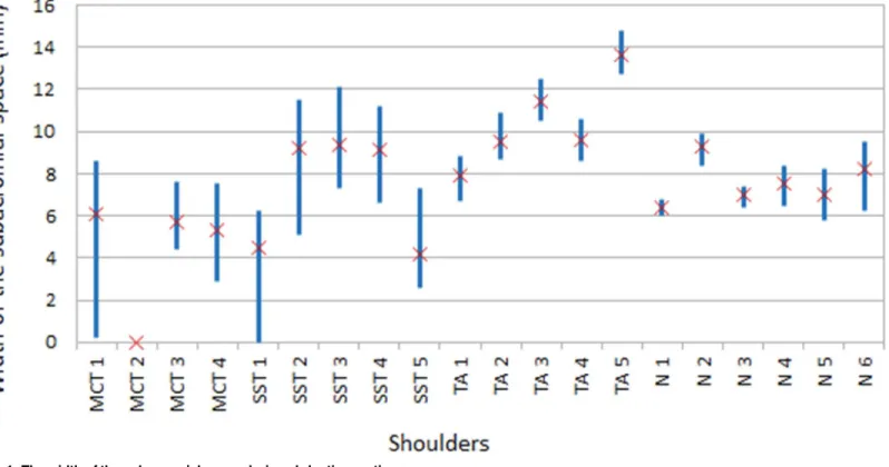
The width of the subacromial space was recorded at 4 sec intervals throughout the 28 sec abduction cycle, thus producing 7 measurements for each shoulder. The average, WSSavg, and range, WSSrange, of these 7 measurements for each specimen are shown inFig 1. The mean of
Table 1. Description of clinical evaluation of patients and healthy volunteers.
Patient Shoulder specimen
Sex Age (years) Shoulder Height (cm) Weight (kg) flexion/abduction/external rotation (°)
Constant score (/100)
1* N1 Male 30 right 175 60 180/120/80 92
1* N2 Male 30 left 175 60 180/130/80 95
2* N3 Female 35 right 160 45 180/120/70 97
2* N4 Female 45 right 162 52 180/130/80 93
3* N5 Female 33 right 161 54 180/110/80 92
4* N6 Female 33 left 161 54 180/120/80 95
5 MCT1 Male 81 right 170 81 180/90/90 65
5 MCT2 Male 81 left 170 81 100/60/40 63
6 MCT3 Female 59 right 170 88 150/70/70 65
7 MCT4 Female 76 right 168 62 180/80/90 64
8 SST1 Female 63 right 157 64 180/80/90 75
9 SST2 Female 65 left 153 55 160/90/80 73
10 SST3 Female 68 right 150 65 180/85/70 76
7 SST4 Female 76 left 168 67 180/75/80 76
11 SST5 Female 71 right 155 60 180/50/70 77
12 TA1 Female 73 right 160 70 180/90/90 82
13 TA2 Female 53 right 158 68 160/90/80 74
14 TA3 Female 66 right 158 54 140/90/70 83
6 TA4 Female 59 left 170 88 100/40/30 75
15 TA5 Female 65 right 165 70 180/90/90 84
*: healthy subjects
Abbreviations: N, normal; MCT, massive rotator cuff tear; SST, supraspinatus tear; TA, tendinopathy.
WSSavgand WSSrangeof each of the rotator cuff diseases groups are presented inTable 3. WSSavgwas lower in the massive cuff tears group than in any other group (p = 0.012) while a WSSrangeof more than 4 mm differentiated control and tendinopathy shoulders from supraspi-natustear and massive cuff tears shoulder, i.e. between rotator cuff disease with and without tendon tears (p = 0.012).
Corresponding to the above description, the average and range of translation of the humeral head in the approximately superior-inferior direction (Y-direction) and anterior-posterior direction (X-direction) for each shoulder were termed Yavgand Yrange, and Xavgand Xrange, respectively. The Yavgand Yrangefor each specimen are shown inFig 2while Xavgand Xrangeare shown inFig 3. Essentially, Yavgand Xavgdescribes where the humeral head is located for most of the time during the abduction movement while Yrangeand and Xrangedescribes how much the humeral head‘wobbles’around during the abduction movement. The mean of Yavg, Yrange, Xavg, Xrangeof each of the specimen groups are presented inTable 3.
Yrangewas higher in massive cuff tears shoulders compared to the control group (6.4 vs. 3.4 mm, p = 0.02), but not when compared to the tendinopathy (p = 0.11) andsupraspinatustear groups (p = 0.28). Finally, Xrangewas higher in massive cuff tears andsupraspinatustear shoul-ders when compared to the control and tendinopathy groups (9.2 and 9.3 vs. 4.8 and 3.5 mm, p = 0.05) (Table 3).
With respect intra-observer reproducibility test, the difference between extreme values was 2 mm in the X-direction, 1.9 mm in Y-direction, 1.3 mm for the width of the subacromial space, and 1.3° for the measure of the gleno-humeral abduction.
Fig 4summarize the monitoring of the humeral head center projection on to the glenoid for each of the 14 shoulders during abduction, for respectively massive rotator cuff tear (Fig 4A),
supraspinatustear (Fig 4B), tendinopathy alone (Fig 4C) and normal shoulders (Fig 4D). Table 2. Assessment of rotator cuff.
Shoulder specimen
Extent of tendon retraction(Patte24) Level of muscle atrophy(Thomazeau26, 27) Level of fatty degeneration(Goutallier
et al28)
Subscapularis Supra-spinatus
Infra-spinatus
Subscapularis Supra-spinatus
Infra-spinatus
Subscapularis Supra-spinatus
Infra-spinatus
MCT1 I III III II III III II III IV
MCT2 I III III III III III IV III IV
MCT3 - III III - III III - IV IV
MCT4 I III III III III III IV IV IV
SST1 - I PT - - -
-SST2 - I - - -
-SST3 - I PT - - -
-SST4 - I PT - II II - II II
SST5 - I - - II II - II II
TA1 - - -
-TA2 - PT - - -
-TA3 - PT - - -
-TA4 - - -
-TA5 - - -
-Abbreviations : N, normal; MCT, massive rotator cuff tear; SST, supraspinatus tear; TA, tendinopathy; PT: partial tear;‘-‘, normal.
Discussion
The objective of this paper was to evaluate if Dynamic MRI provides novel kinematic data that can be used to improve the understanding, diagnosis and best treatment of rotator cuff dis-eases. The long-term aim of this work is to develop the Dynamic-MRI technique as a tool that can provide kinematic data and simultaneous MRI assessment of the loaded soft tissues of the rotator cuff during arm movement.
The most important finding of this work was that the Dynamic-MRI technique enabled a novel measure;‘Looseness’, i.e. the translation of the humeral head on the glenoid during an abduction cycle (Xrangeand Yrange). Looseness was better able at differentiating rotator cuff dis-ease than a simple static measure of relative gleno-humeral position.
This study found that the subacromial space narrowed with rotator cuff disease (Fig 1and
Table 3) which is consistent with other studies [17,29]. The reported width of the subacromial space varies between studies (5 to 9 mm for healthy shoulders [11,17,29]) and are not incon-sistent with the values found here (Table 3).
Fig 1. The width of the subacromial space during abduction motion.
doi:10.1371/journal.pone.0158563.g001
Table 3. Mean of kinematic measures for each rotator cuff disease group
Group WSSavg(mm) WSSrange(mm) Yavg(mm) Yrange(mm) Xavg(mm) Xrange(mm)
MCT 4.3 4.0 3.8 6.4 -0.8 9.2
SST 7.3 5.3 2.0 5.0 0.9 9.3
TA 10.4 2.1 0.3 4.4 -1.6 4.8
N 7.7 1.9 1.0 3.4 1.1 3.5
Abbreviations : N, normal; MCT, massive rotator cuff tear; SST, supraspinatus tear; TA, tendinopathy.
The finding that superior-inferior excursion in torn shoulders is larger than in normals (Yrange,Table 3) is related to the narrowing of the subacromial space and seems sensible consider-ing the superiorly directed deltoid force and the reduced ability of the rotator cuff to compress and stabilise the joint. While this finding was not surprising it does provide some confidence in the Dynamic-MRI kinematic measurements. In contrast, the notable anterior-posterior excur-sion (Fig 3andTable 3) was not expected. Most studies investigating the effect of rotator cuff dis-ease have not considered the anterior-posterior gleno-humeral translation. However Bey et al. [11] did report that the anterior-posterior excursion was not restored (was larger than normal) by rotator cuff repair. To our knowledge, this study is the first to report that the range of anterior posterior motion (Xrange) increases significantly with rotator cuff diseases.
In fact, the superior-inferior motion (Yrange) was less affected than anterior-posterior trans-lation (Table 3) which may seem surprising. However, Bey et al. [11] showed that the superior-inferior excursion was restored by rotator cuff repair whereas anterio-posterior translation was not, which also indicate that anterior-posterior motion may be more affected by rotator cuff diseases. Previous studies [30,31] have demonstrated that the glenoid is“flatter”in the rior-posterior direction than in the superior-inferior direction and joint excursion in the ante-rior-posterior direction may depend more on the stability provided by the rotator cuff and, consequently, may be more affected by rotator cuff disease. Therefore analysis and improved understanding of excursion in the anterior-posterior direction may be critical to diagnosis and treatment of different conditions of rotator cuff diseases.
The dynamic MRI technique also allowed us to measure the ranges of the variables (WSSrange, Yrange, Xrange) during the motion; effectively how loose or unstable the joint was. Fig 2. Translation of the humeral head along the Y-axis (superior-inferior direction) of glenoid coordinate system
This looseness was shown to be a better measure for identifying and specifying cuff tear disease than the average measures used in static techniques.
Although an analysis of the subacromial space combined with an analysis of the translations of the humeral head allowed identification of cuff tears and the degree of cuff tear pathology, statistical significance was not consistently found between tendinopathy shoulders and healthy shoulders. This may be due to the relatively few specimens in the study, which may have pre-vented statistically significant differences to be found for both analyses.
The abduction cycle was prescribed to take 28 seconds, which clearly does not represent physiological arm motion. However, image noise is inversely proportional to the acquisition speed and an acquisition phase lasting for 4 seconds, resulting in 7 acquisitions during the abduction motion, was chosen as a balance between image quality and realistic abduction times. Consequently, the MRI dynamic technique enabled kinematics analysis of the bone structures but not simultaneous visualization of the tendons. Visualization of the soft structures is a priority for future work.
Another limitation was the use of a closed-bore scanner. This scanner was chosen because of its common availability but resulted in the elbow abutting against the wall of the scanner during the motion, thus restricting humero-thoracic abduction to approximately 45°. However, the protocol could be easily transferred to an open-bore MRI scanner that would provide, in upright position, the full range of shoulder abduction.
Fig 3. Translation of the humeral head along the X-axis (anterior-posterior direction) of glenoid coordinate system
This study did assess the intra-observer reproducibility of the technique. Considering all cri-teria (data on X-axis and Y-axis of the humeral head projection and width of the subacromial space), the difference between the extreme values were less than 2 mm, i.e., showing a good consistency. The inter-observer reproducibility was not investigated because the segmentation of the images, in step two of the protocol, is very time-consuming. Consequently, this protocol needs to be enhanced including improved computer-based segmentation of the images.
Thus, before being used in routine practice, this protocol needs to be transferred to an open-bore MRI scanner and needs to be improved by shortening the acquisition times, by visu-alizing the tendons and by developing a the computer-based segmentation of the images.
Conclusion
The Dynamic-MRI technique identified kinematic differences between groups of patients with various degrees of cuff tears.
Fig 4. Monitoring of the humeral head center projection on to the glenoid for each of the 14 shoulders during abduction.The size of the glenoids was standardized so that each fit with a circle of 200% diameter (-100% to +100%). The locations of the humeral head center projections on to the glenoid are expressed in percentile. Fig 4A, massive rotator cuff tear; Fig 4B,Supraspinatustear; Fig 4C, Tendinopathy alone; Fig 4D, normal shoulders.
Relative to standard MRI, the Dynamic-MRI has the advantage of providing kinematic data to improve the understanding, diagnosis and best treatment of rotator cuff diseases.
The technique enabled a novel measure;‘Looseness’, i.e. the translation of the humeral head on the glenoid during an abduction cycle (Xrangeand Yrange). Looseness was better able at dif-ferentiating rotator cuff disease than a simple static measure of relative gleno-humeral position.
The study showed that anterior-posterior gleno-humeral motion increases with rotator cuff disease and it is suggested that a better understanding of this relationship may help improve treatment for rotator cuff diseases.
Author Contributions
Conceived and designed the experiments: TG CT JP WS VV CAC MMLC. Performed the experiments: TG CT JP WS VV CAC MMLC. Analyzed the data: TG OM UH. Contributed reagents/materials/analysis tools: TG OM UH. Wrote the paper: TG CT JP WS VV CAC MMLC UH OM.
References
1. Bodin J, Ha C, Chastang JF, Descatha A, Leclerc A, Goldberg M, et al. Comparison of risk factors for shoulder pain and rotator cuff syndrome in the working population. Am J Ind Med 2012; 55: 605–15. doi:10.1002/ajim.22002PMID:22213435
2. Van der Windt DA, Koes BW, de Jong BA, Bouter LM. Shoulder disorders in general practice: inci-dence, patient characteristics, and management. Ann Rheum Dis 1995; 54: 959–64. PMID:8546527 3. Graichen H, Bonel H, Stammberger T, Haubner M, Rohrer H, Englmeier KH. Three-dimensional
analy-sis of the width of the subacromial space in healthy subjects and patients with impingement syndrome. AJR Am J Roentgenol 1999; 172: 1081–6. PMID:10587151
4. Billuart F, Devun L, Gagey O, Skalli W, Mitton D. 3D kinematics of the glenohumeral joint during abduc-tion moabduc-tion: an ex vivo study. Surg Radiol Anat 2007; 29: 291–5. PMID:17460813
5. Benoit DL, Ramsey DK, Lamontagne M, Xu L, Wretenberg P, Renström P. Effect of skin movement arti-fact on knee kinematics during gait and cutting motions measured in vivo. Gait Posture 2006; 24: 152– 64. PMID:16260140
6. Hill AM, Bull AM, Dallalana RJ, Wallace AL, Johnson GR. Glenohumeral motion: review of measure-ment techniques. Knee Surg Sports Traumatol Arthrosc 2007; 15: 1137–43. PMID:17431588 7. Deutsch A, Altchek DW, Schwartz E, Otis JC, Warren RF. Radiologic measurement of superior
dis-placement of the humeral head in the impingement syndrome. Journal of shoulder and elbow surgery / American Shoulder and Elbow Surgeons 1996; 5: 186–93.
8. Paletta GA Jr., Warner JJ, Warren RF, Deutsch A, Altchek DW. Shoulder kinematics with two-plane x-ray evaluation in patients with anterior instability or rotator cuff tearing. Journal of shoulder and elbow surgery / American Shoulder and Elbow Surgeons 1997; 6: 516–27.
9. Yamaguchi K, Sher JS, Andersen WK, Garretson R, Uribe JW, Hechtman K, et al. Glenohumeral motion in patients with rotator cuff tears: a comparison of asymptomatic and symptomatic shoulders. Journal of shoulder and elbow surgery / American Shoulder and Elbow Surgeons 2000; 9: 6–11. 10. Lagace PY, Billuart F, Ohl X, Skalli W, Tétreault P, de Guise J, et al. Analysis of humeral head
displace-ments from sequences of biplanar X-rays: repeatability study and preliminary results in healthy sub-jects. Comput Methods Biomech Biomed Engin 2011; 15: 221–9. doi:10.1080/10255842.2010. 522185PMID:21506033
11. Bey MJ, Peltz CD, Ciarelli K, Kline SK, Divine GW, van Holsbeeck M, et al. In vivo shoulder function after surgical repair of a torn rotator cuff: glenohumeral joint mechanics, shoulder strength, clinical out-comes, and their interaction. Am J Sports Med 2011; 39: 2117–29. doi:10.1177/0363546511412164 PMID:21737834
12. Giphart JE, van der Meijden OA, Millett PJ. The effects of arm elevation on the 3-dimensional acromio-humeral distance: a biplane fluoroscopy study with normative data. Journal of shoulder and elbow sur-gery / American Shoulder and Elbow Surgeons 2012; 21: 1593–600.
14. Illyes A, Kiss RM. Shoulder joint kinematics during elevation measured by ultrasound-based measuring system. J Electromyogr Kinesiol 2007; 17: 355–64. PMID:16624576
15. Graichen H, Bonel H, Stammberger T, Englmeier KH, Reiser M, Eckstein F. Subacromial space width changes during abduction and rotation—a 3-D MR imaging study. Surg Radiol Anat 1999; 21: 59–64. PMID:10370995
16. Graichen H, Bonel H, Stammberger T, Heuck A, Englmeier KH, Reiser M, et al. A technique for deter-mining the spatial relationship between the rotator cuff and the subacromial space in arm abduction using MRI and 3D image processing. Magn Reson Med 1998; 40: 640–3. PMID:9771582 17. Graichen H, Hinterwimmer S, von Eisenhart-Rothe R, Vogl T, Englmeier KH, Eckstein F. Effect of
abducting and adducting muscle activity on glenohumeral translation, scapular kinematics and suba-cromial space width in vivo. J Biomech 2005; 38: 755–60. PMID:15713296
18. Graichen H, Stammberger T, Bonel H, Haubner M, Englmeier KH, Reiser M et al. Magnetic resonance-based motion analysis of the shoulder during elevation. Clin Orthop Relat Res 2000; 370: 154–63. PMID:10660709
19. Graichen H, Stammberger T, Bonel H, Englmeier KH, Reiser M, Eckstein F. Glenohumeral translation during active and passive elevation of the shoulder—a 3D open-MRI study. J Biomech 2000; 33: 609– 13. PMID:10708782
20. Graichen H, Stammberger T, Bonel H, Wiedemann E, Englmeier KH, Reiser M et al. Three-dimensional analysis of shoulder girdle and supraspinatus motion patterns in patients with impingement syndrome. J Orthop Res 2001; 19: 1192–8. PMID:11781023
21. Sahara W, Sugamoto K, Murai M, Tanaka H, Yoshikawa H. The three-dimensional motions of gleno-humeral joint under semi-loaded condition during arm abduction using vertically open MRI. Clin Bio-mech (Bristol, Avon) 2007; 22: 304–12.
22. Sans N, Richardi G, Fourcade D, Assoun J, Chiavassa H, Giron J, et al. Cine-MRI of the shoulder. Nor-mal aspects. J Radiol 1996; 77: 117–23. PMID:8729339
23. Pierrart J, Lefevre-Colau MM, Skalli W, Vuillemin V, Masmejean EH, Cuénod CA, et al. New dynamic three-dimensional MRI technique for shoulder kinematic analysis. J Magn Reson Imaging 2014; 39: 729–34. doi:10.1002/jmri.24204PMID:23723138
24. Constant CR, Murley AH. A clinical method of functional assessment of the shoulder. Clin Orthop Relat Res 1987; 214: 160–4. PMID:3791738
25. Patte D. Classification of rotator cuff lesions. Clin Orthop Relat Res 1990; 254: 81–6. PMID:2323151 26. Thomazeau H, Rolland Y, Lucas C, Duval JM, Langlais F. Atrophy of the supraspinatus belly.
Assess-ment by MRI in 55 patients with rotator cuff pathology. Acta Orthop Scand 1996; 67: 264–8. PMID: 8686465
27. Zanetti M, Gerber C, Hodler J. Quantitative assessment of the muscles of the rotator cuff with magnetic resonance imaging. Invest Radiol 1998; 33: 163–70. PMID:9525755
28. Goutallier D, Postel JM, Bernageau J, Lavau L, Voisin MC. Fatty muscle degeneration in cuff ruptures. Pre- and postoperative evaluation by CT scan. Clin Orthop Relat Res 1994; 304:78–83. PMID: 8020238
29. Nove-Josserand L, Boulahia A, Levigne C, Noel E, Walch G. Coraco-humeral space and rotator cuff tears. Rev Chir Orthop Reparatrice Appar Mot 1999; 85: 677–83. PMID:10612131
30. McPherson EJ, Friedman RJ, An YH, Chokesi R, Dooley RL. Anthropometric study of normal gleno-humeral relationships. Journal of shoulder and elbow surgery / American Shoulder and Elbow Sur-geons 1997; 6: 105–12.