Agnollitto PM et al. / Glenohumeral interposition of rotator cuff stumps
Radiol Bras. 2016 Jan/Fev;49(1):53–55 53
0100-3984 © Colégio Brasileiro de Radiologia e Diagnóstico por Imagem
Case Report
Glenohumeral interposition of rotator cuff stumps: a rare
complication of traumatic rotator cuff tear
*
Interposição dos cotos do manguito rotador na articulação glenoumeral: uma complicação rara da lesão traumática do manguito rotador
Agnollitto PM, Chu MWK, Lorenzato MM, Zatiti SCA, Nogueira-Barbosa MH. Glenohumeral interposition of rotator cuff stumps: a rare complication of traumatic rotator cuff tear. Radiol Bras. 2016 Jan/Fev;49(1):53–55.
Abstract
R e s u m o
The present report describes a case where typical findings of traumatic glenohumeral interposition of rotator cuff stumps were surgically confirmed. This condition is a rare complication of shoulder trauma. Generally, it occurs in high-energy trauma, frequently in association with glenohumeral joint dislocation. Radiography demonstrated increased joint space, internal rotation of the humerus and coracoid process fracture. In addition to the mentioned findings, magnetic resonance imaging showed massive rotator cuff tear with interposition of the supraspinatus, infraspinatus and subscapularis stumps within the glenohumeral joint. Surgical treatment was performed confirming the injury and the rotator cuff stumps interposition. It is important that radiologists and orthopedic surgeons become familiar with this entity which, because of its rarity, might be neglected in cases of shoulder trauma.
Keywords: Interposition; Rotator cuff; Tear; Trauma; Shoulder; Magnetic resonance imaging.
O objetivo deste trabalho é relatar um caso com os achados de imagem característicos da lesão traumática do manguito rotador com interposição de múltiplos cotos de tendões do manguito rotador entre a glenoide e o úmero, confirmada cirurgicamente. Esta condição é uma rara complicação do trauma do ombro. Em geral ocorre no trauma de alta energia, frequentemente associada com luxação da articulação glenoumeral. No caso em questão não houve documentação de luxação franca da articulação glenoumeral. As radiografias mostraram aumento do espaço articular, rotação interna do úmero e fratura do coracoide. Nas imagens de ressonância magnética, além das alterações observadas nas radiografias, foi identificada ruptura massiva do manguito rotador com interposição dos cotos dos ten-dões do supraespinal, do infraespinal e do subescapular no interior da articulação glenoumeral. Foi realizado tratamento cirúrgico con-firmando a lesão e a interposição dos cotos do manguito. É importante que radiologistas e ortopedistas estejam familiarizados com esta entidade, que, pela sua raridade, pode ser negligenciada no atendimento do trauma do ombro.
Unitermos: Interposição; Manguito rotador; Ruptura; Trauma; Ombro; Ressonância magnética.
* Study developed at Hospital das Clínicas – Faculdade de Medicina de Ribeirão Preto da Universidade de São Paulo(HCFMRP-USP), Ribeirão Preto, SP, Brazil.
1. Physician Assistant at Division of Radiology, Hospital das Clínicas – Faculdade de Medicina de Ribeirão Preto da Universidade de São Paulo (HCFMRP-USP), Ribei-rão Preto, SP, Brazil.
2. Volunteer Physician, Fellow of Musculoskeletal Radiology, Division of Radiology at Hospital das Clínicas – Faculdade de Medicina de Ribeirão Preto da Universidade de São Paulo (HCFMRP-USP), Ribeirão Preto, SP, Brazil.
3. MD, Radiologist, Clínica Radiologia Especializada, Ribeirão Preto, SP, Brazil. 4. MD, Orthopedist, Hospital Especializado de Ribeirão Preto, Ribeirão Preto, SP, Brazil.
5. Associate Professor of Radiology, Centro de Ciências das Imagens e Física Médica (CCIFM) – Faculdade de Medicina de Ribeirão Preto da Universidade de São Paulo (FMRP-USP), Ribeirão Preto, SP, Brazil.
Mailing Address: Dr. Paulo Moraes Agnollitto. Divisão de Radiologia / CCIFM, FMRP-USP. Avenida Bandeirantes, 3900, Monte Alegre. Ribeirão Preto, SP, Brazil, 14048-900. E-mail: [email protected].
Received October 31, 2013. Accepted after revision April 10, 2014.
the case of rotator cuff stumps interposition in the gleno-humeral joint (GHJ), the most common clinical presenta-tion is articular blockage, many times with persistence of subluxation and/or irreducible luxation of the GHJ(4–8).
The objective of this report is to describe a surgically confirmed case with characteristic imaging findings of trau-matic rotator cuff injury with multiple stumps interposition between the glenoid and the humerus.
CASE REPORT
A female, 27-year-old patient was referred to the emer-gency unit, at another service, with a history of motorcycle accident occurred about two hours before her arrival at the unit. She presented with pain and limited range of motion in her right shoulder. At the initial approach, there was no report of GHJ dislocation, and the patient was discharged with analgesic medication. No supplementary tests or imag-ing studies were performed.
The patient evolved with right shoulder pain and block-age and, 15 days after the episode, she sought specialized as-sistance, undergoing plain radiography of her right shoulder. Paulo Moraes Agnollitto1, Marcio Wen King Chu2, Mario Muller Lorenzato3, Salomão Chade Assan Zatiti4,
Marcello Henrique Nogueira-Barbosa5
http://dx.doi.org/10.1590/0100-3984.2013.0011 INTRODUCTION
Agnollitto PM et al. / Glenohumeral interposition of rotator cuff stumps
Radiol Bras. 2016 Jan/Fev;49(1):53–55 54
The analysis of the images showed coracoid process fracture, GHJ space widening, and internal humeral rotation (Figure 1). No signs of glenohumeral dislocation or instability were found at the radiographic images.
The hypothesis of traumatic rotator cuff tear was raised on the basis of the plain radiography findings, and the pa-tient was submitted to magnetic resonance imaging (MRI) whose images demonstrated the presence of a traumatic in-jury with interposition of the supraspinatus, infraspinatus and subscapularis tendons stumps in the GHJ (Figures 2 and 3). Coracoid process fracture, diffuse periarticular edema, and edema of intermuscular fat planes were also identified.
Then, the patient was submitted to open surgical explo-ration, with diagnostic confirmation and rotator cuff rein-sertion (Figure 4).
The patient presented a good evolution over the imme-diate postoperative period and was discharged. Three months
after the surgery, she presented with external rotation restric-tion and was submitted to arthroscopy that revealed the pres-ence of adhesions, which were released. After this new peocedure, the patient evolved satisfactorily, without any new complication.
DISCUSSION
Traumatic rotator cuff stumps interposition in the GHJ is a very rare complication in shoulder trauma. In general, it occurs as a result from high-energy trauma, and frequently is associated with either anterior or posterior GHJ luxation. Relatively few articles on this subject are found in the litera-ture, and most of them are case reports.
Difficulty or incapacity to reduce GHJ luxation is not common(1–3). Irreducible GHJ luxation may be related to bone tissue or soft tissues interposition(1,2,4,6–10). Amongst the causes associated with soft tissues interposition, one can
Figure 1. Radiography of right shoulder. Anteroposte-rior view showing glenohumeral joint space widening (continuous arrows). The humerus presented with in-ternal rotation. The dashed arrow indicates fracture of the coracoid process.
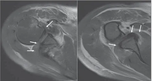
Figure 2. MRI. Axial sections images acquired with intermediate weighting demonstrates rotator cuff stumps interposition (arrows) between the glenoid and the humerus, explaining the blockage and the joint space widening.
Figure 3. MRI, coronal section, intermediate weight-ing. The arrows identify rotator cuff stumps interposi-tion between the glenoid and the humerus. The white arrow indicates the supraspinatus tendon and the black arrow shows the subscapularis tendon.
Figure 4.A: Intraoperative finding demonstrating uncovered humeral head (continuous arrow) and retracted and interposed rotator cuff stumps in the glenohumeral joint (dashed arrow). B: Intraoperative finding dem-onstrating rotator cuff (continuous arrows), repaired by surgical thread (dashed arrow) coursing up to the site of insertion of the greater humeral tuberosity.
Agnollitto PM et al. / Glenohumeral interposition of rotator cuff stumps
Radiol Bras. 2016 Jan/Fev;49(1):53–55 55
mention, for example, interposition of the long head of bi-ceps(9) and interposition of the musculocutaneous nerve(10); however, interposition of rotator cuff tendons, particularly the subscapularis tendon, is highlighted(2,4,5,7,8). Soft tissues interposition and bone tissue interposition may occur con-comitantly(2).
Most cases reported in the literature are associated with high-energy trauma, with episodes of traumatic GHJ lux-ation(1–5); however, like in the present case, the history of luxation is not always well established(3). As our patient was initially assisted in other service, such a possibility cannot be completely ruled out.
The clinical presentation of glenohumeral interposition of rotator cuff stumps also includes pain and varied degrees of functional limitation or joint blockage. The clinical diag-nosis is difficult to be made, and suspicious should be raised in cases where previous radiographic images and those ob-tained after articular reduction attempts demonstrate persis-tence of subluxation or articular space widening(2). The ra-diological signs are subtle, but should be taken into consid-eration in the clinical context.
In the present case, as well as in the literature review, the authors highlight the role played by MRI in the identification of post-trauma interposition of soft tissues in the GHJ(1,2,5). Such a role is not restricted to cases involving the shoulders, and MRI has been utilized, for example, to detect post-trauma periosteal interposition in growth cartilage fracture in chil-dren and adolescents(11,12). In case of irreducible GHJ lux-ation, computed tomography may be utilized to better iden-tify bone fragments blocking the reduction, but such a method is limited to evaluate soft tissues(2–5).
The management of traumatic rotator cuff injury with tendons entrapment in the GHJ should be surgical, and an early diagnosis can minimize the damages to the involved muscle bellies and tendons, improving the postoperative results(2,5).
Because of its rarity, such a condition may be easily neglected at emergency settings(1–3), and a previous
knowl-edge about this entity by radiologists and orthopedists is critical for a correct diagnosis and institution of an appro-priate treatment.
In the present case, the authors conclude that the evalu-ation by MRI was appropriate to identify the post-trauma rotator cuff tendons entrapment in the GHJ.
REFERENCES
1. Lin CL, Su WR, Jou IM, et al. Occult interpositional rotator cuff – an extremely rare case of traumatic rotator cuff tear. Korean J Radiol. 2012;13:98–101.
2. Walch G, Boulahia A, Robinson AH, et al. Posttraumatic sublux-ation of the glenohumeral joint caused by interposition of the rota-tor cuff. J Shoulder Elbow Surg. 2001;10:85–91.
3. Rickert M, Loew M. Glenohumeral interposition of a torn rotator cuff in a young motorcyclist. Arch Orthop Trauma Surg. 2006;126: 184–7.
4. Connolly S, Ritchie D, Sinopidis C, et al. Irreducible anterior dis-location of the shoulder due to soft tissue interposition of subscapu-laris tendon. Skeletal Radiol. 2008;37:63–5.
5. Ilaslan H, Bilenler A, Schils J, et al. Pseudoparalysis of shoulder caused by glenohumeral interposition of rotator cuff tendon stumps: a rare complication of posterior shoulder dislocation. Skeletal Radiol. 2013;42:135–9.
6. Davies MB, Rajasekhar C, Bhamra MS. Irreducible anterior shoul-der dislocation: the greater tuberosity Hill-Sachs lesion. Injury. 2000;31:470–1.
7. Bridle SH, Ferris BD. Irreducible acute anterior dislocation of the shoulder: interposed subscapularis. J Bone Joint Surg Br. 1990;72: 1078–9.
8. Tietjen R. Occult glenohumeral interposition of a torn rotator cuff. A case report. J Bone Joint Surg Am. 1982;64:458–9.
9. Mihata T, Doi M, Abe M. Irreducible acute anterior dislocation of the shoulder caused by interposed fragment of the anterior glenoid rim. J Orthop Sci. 2000;5:404–6.
10. Gudena R, Iyengar KP, Nadkarni JB, et al. Irreducible shoulder dislocation – a word of caution. Orthop Traumatol Surg Res. 2011; 97:451–3.
11. Moura MVT. Trapped periosteum in a distal femoral physeal in-jury: magnetic resonance imaging evaluation. Radiol Bras. 2012;45: 184–6.