355
Pulmonary paracoccidioidomycosis:a two-case report
Radiol Bras 2007;40(5):355–357
Cases Report
PULMONARY PARACOCCIDIOIDOMYCOSIS AND REVERSED HALO
SIGN: A TWO-CASE REPORT*
Matias de Freitas Filho1
, Fabrício Guimarães Gonçalves2
, Marcello Antônio Rezende Basílio2 , Alexandre Dias Mançano3
, Bruno Cherulli4
, Márcia Rocha Carneiro Barreiros4
The present study reports two histologically proven cases of pulmonary paracoccidioidomycosis where a reversed halo sign was found. The reversed halo, initially described as a pathognomonic sign of cryptogenic organizing pneumonia, has later been found and described in paracoccidioidomycosis.
Keywords: Reversed halo sign; Paracoccidioidomycosis; Cryptogenic organizing pneumonia.
Paracoccidioidomicose pulmonar: relato de dois casos enfatizando o sinal do halo invertido.
Este trabalho relata dois casos de paracoccidioidomicose pulmonar comprovados histologicamente, nos quais foi encontrado o sinal radiológico do halo invertido. Este sinal, descrito inicialmente como patognomônico da pneumonia criptogênica em organização, foi posteriormente encontrado e descrito na paracoccidioido-micose.
Unitermos: Halo invertido; Paracoccidioidomicose; Pneumonia criptogênica em organização. Abstract
Resumo
* Study developed at Hospital Regional de Taguatinga – Se-cretaria de Estado da Saúde, Brasília, DF, Brazil.
1. MD, Trainee at Service of Imaging Diagnosis, Hospital Re-gional de Taguatinga – Secretaria de Estado da Saúde, Brasília, DF, Brazil.
2. MDs, Residents at Service of Imaging Diagnosis, Hospital Regional de Taguatinga – Secretaria de Estado da Saúde, Brasília, DF, Brazil.
3. Coordinator for Medical Residency in Radiology and Imaging Diagnosis at Hospital Regional de Taguatinga – Secretaria de
INTRODUCTION
Paracoccidioidomycosis is the most prevalent systemic fungal infection in Latin America, particularly in Brazil(1). This
dis-ease is caused by the single dimorphic spe-cies Paracoccidioides brasiliensis and is
the most significant type of mycosis in our environment. As a deep mycosis, it is not limited to the epithelial surface of the or-ganism, invading conjunctive tissue and viscera(2). Pulmonary, cutaneous, mucosal
are the predominant clinical presentations of this disease. Lungs are affected in ap-proximately 75% of cases(2), and in 10% of
these cases the reversed halo sign may be found on high-resolution computed tomog-raphy (HRCT)(3).
CASES REPORT Case 1
A male, 37-year-old peasant living in Mato Grosso do Sul and presenting with progressive dyspnea for one week, has been referred to the service to be submit-ted to chest compusubmit-ted tomography (CT).
Chest radiography had demonstrated bilat-eral reticulonodular opacity, predominantly in the upper thirds, with consolidation in the middle third of the left pulmonary field. The patient presented in a good general condition, eupneic, ruddy, and afebrile, complaining of odynophagia and pain in the mesogastrium. He denied thoracic pain and cough. At physical examination, pain-ful, bilateral submandibular lymph nodes enlargement (measuring about 1.0 cm) was found attached to the deep planes, with absent trophic alterations of the adjacent skin. The soft palate presented a granulo-matous hyperemic lesion. Bilateral, ulcer-ative lesions were found on the jugal mu-cosa. Unaltered cardiopulmonary symp-tomatology, and painful abdomen under hypogastric palpation.
Chest HRCT showed diffuse nodular opacities, cavitated images, consolidation, ground-glass attenuation and subpleural reversed halo sign. The patient has also been submitted to abdominal ultrasonogra-phy and spirometry that have demonstrated no alteration.
Incisional biopsy of granulomatous
le-sion of the mouth revealed squamous epi-thelial hyperplasia, intraepiepi-thelial micro-abscesses and granulomatous reaction with epithelial histiocytes, multinucleated giant cells, and central accumulation of polymor-phonuclear neutrophils. Elongated and round fungi with thick and refractive cap-sules were observed inside the giant cells. The histopathological diagnosis was paracoccidioidomycosis.
The patient has been medicated with itraconazol (200 mg/ day), and was dis-charged with no complaint, after improve-ment of the pulmonary condition. He has been under clinical follow-up for two months, and using prophylactic suphame-toxazole + trimethoprim.
Case 2
A male, thirty-nine-year old peasant, born in Guairá, SP, Brazil, has been re-ferred to the service to be submitted to chest radiography because of a productive cough, hemoptysis and progressive dysp-nea that had initiated for one month and a half.
The patient presented in a good general condition, with a history of irregular fever, lesions in the oral cavity and 10 kg weight loss. At clinical examination, a granuloma-tous lesion was observed on the right lip commissure involving jugal mucosa and gingiva, besides a limited opening of the mouth. At pulmonary auscultation, a ve-sicular murmur was observed, with bron-Estado da Saúde, MD, Radiologist at Radiologia Anchieta,
Brasília, DF, Brazil.
4. Preceptors for Medical Rasidency in Radiology and Imaging Diagnosis at Hospital Regional de Taguatinga – Secretaria de Estado da Saúde, Brasília, DF, Brazil.
Mailing address: Dr. Alexandre Dias Mançano. SQSW 100, Bloco G, ap. 401. Brasília, DF, Brazil, 70670-017. E-mail: [email protected]
356
Freitas Filho M et al.
Radiol Bras 2007;40(5):355–357
chophony over the middle third of the left lung, and hypersonority under auscultatory percussion. Cardiac and abdominal symp-tomatology with no clinical evidence. Ab-sence of palpable enlarged lymph nodes.
Plain chest radiograph has demon-strated diffuse and bilateral gross, reticulo-nodular opacities with sparse foci of con-solidation on the lower and middle pulmo-nary fields. HRCT has demonstrated sig-nificant architectural distortion, extensive foci of subpleural consolidation, nodular cavitation, interlobular septal thickening, parenchymal bands, sparse foci of ground-glass attenuation, air-space nodular
opaci-ties and some areas with reversed halo sign in lower lobes.
Bronchoscopy has demonstrated the presence of hyaline secretion in the whole bronchial tree, a pale mucosa with signs of bronchitis, and increased diameter of bron-chial segments. Endobronbron-chial washing, brushing and biopsy were performed. Bronchial brushing has demonstrated the presence of columnar cells, large amount of neutrophils, eosinophils and numerous histiocytes, with presence of multinuclear, giant cells, including round structures with birefractive membrane compatible with P. brasiliensis. The typical resemblance of the
fungus to a ship’s steering wheel was found. Malignancy has not been found. Negative test for alcohol-acid resistant bacilli in the bronchial washing and negative tuberculo-sis test. Biopsy of the granulomatous lesion on the oral mucosa has diagnosed paracoc-cidioidomycosis.
The patient has been medicated with itraconazol and sulphametoxazole + trime-thoprim, with an excellent clinical progress.
DISCUSSION
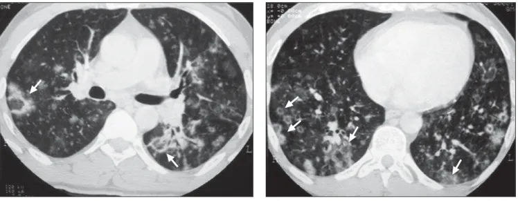
Paracoccidioidomycosis predominantly affects young men and peasants living in Figure 1. Case 1. HRCT showing reversed halo sign (arrows). Besides small, sparse cavitated nodules.
A B
A B
Figure 2. Case 2. HRCT showing reversed halo sign (arrows), besides interlobular septal thickening and thick-walled cavitated nodules.
357
Pulmonary paracoccidioidomycosis:a two-case report
Radiol Bras 2007;40(5):355–357
rural zones. The disease is acquired by in-halation of infectious particles of the fun-gus P. brasiliensis involving lungs, upper
respiratory and digestive mucosas, central nervous system, suprarenal glands and lymph nodes(4).
Main clinical presentations include fe-ver, cough, weight loss, hemoptysis, ody-nophagia, lymph node enlargement, ulcer-ative or granulomatous lesions on the up-per digestive and respiratory mucosas.
Radiological pulmonary findings in-clude interlobular septal thickening, nodu-lar opacities, thickening of the peribron-chovascular interstice, intralobular lines, ground-glass attenuation, cavitations, air-space consolidation, traction bronchiecta-sis, irregular increase of the air-space(2,5)
and reversed halo sign(3).
The reversed halo sign is defined as a focal round area of ground-glass attenua-tion and surrounding air-space consolida-tion of crescent (forming more than three quarters of a circle) or ring (a complete circle) shapes(3,6). Histologically, central
ground-glass attenuation corresponds to an inflammatory infiltrate in the alveolar sep-tum with macrophages, lymphocytes, plas-matic cells and some giant cells with a rela-tive preservation of alveolar spaces. Periph-eral consolidation consists of a dense and homogeneous, intra-alveolar cellular infil-trate. Evidence of organizing pneumonia is not found. Presence of P. brasiliensis is
observed inside alveolar septa and air-spaces. These findings indicate that the reversed halo sign may be found in
indi-viduals affected by active infection by P. brasiliensis(3).
Central ground-glass attenuation sur-rounded by dense consolidation of crescent or ring shapes as a finding of HRCT was reported in 1996 by Voloudaki et al.(7) in
patients affected by cryptogenic organizing pneumonia. Histological studies have dem-onstrated that ground-glass attenuation corresponded to alveolar septal inflamma-tion and cellular débris, and peripheral opacity of crescent or ring shapes, to con-solidation and areas of organizing pneumo-nia inside alveolar ducts(3).
In 2003, Kim et al.(6) reviewed patients
affected by cryptogenic organizing pneu-monias with HRCT findings of focal ground-glass attenuation surrounded by consolidation of crescent and ring shapes with the same histological characteristics described by Voloudaki et al.(7), and called
this finding “reversed halo sign”(6).
Recently, Gasparetto et al.(3) described
the association between reversed halo sign and paracoccidioidomycosis. In a study of 148 patients diagnosed with paracocci-dioidomycosis, 15 presented with this find-ing. In two cases, the reversed halo sign was the only finding on HRCT. Three pa-tients presented only one image of reversed halo sign, one had two lesions, and the oth-ers presented with multiple lesions. The prevalent sites of presentation were middle and lower pulmonary fields. Also, the re-versed halo sign was predominantly found in peripheral zones, with diameters ranging between 10 and 50 mm (mean 20 mm)(3).
CONCLUSION
Recent studies suggest that active paracoccidioidomycosis may course with reversed halo sign. About 10% of patients with active infection by P. brasiliensis may present with this finding that is not specific of cryptogenic organizing pneumonia.
REFERENCES
1. Gonzáles FM, Faucz RA, Paes Jr. AJO, Caval-cante AJW, Souza RP. Aspecto da tomografia computadorizada de alta resolução do tórax na pa-racoccidioidomicose em paciente com SIDA: re-lato de caso e revisão da literatura. Rev Imagem 2004;26:241–245.
2. Muniz MAS, Marchiori E, Magnago M, Moreira LBM, Almeida Jr JG. Paracoccidioidomicose pul-monar: aspectos na tomografia computadorizada de alta resolução. Radiol Bras 2002;35:147–154. 3. Gasparetto EL, Escuissato DL, Davaus T, et al. Reversed halo sign in pulmonary paracoccidioi-domycosis. AJR Am J Roentgenol 2005;184: 1932–1934.
4. Capone D, Jansen JM, Tessarollo B, Lopes AJ, Mogami R, Marchiori E. Micoses pulmonares. In: Santos AASMD, Nacif MS. Radiologia e diag-nóstico por imagem: aparelho respiratório. 1ª ed. Rio de Janeiro, RJ: Livraria e Editora Rubio, 2005;163–169.
5. Funari M, Kavakama J, Shikanai-Yasuda MA, et al. Chronic pulmonary paracoccidioidomycosis (South American blastomycosis): high-resolution CT findings in 41 patients. AJR Am J Roentgenol 1999;173:59–64.
6. Kim SJ, Lee KS, RyuYH, et al. Reversed halo sign on high-resolution CT of cryptogenic organizing pneumonia: diagnostic implications. AJR Am J Roentgenol 2003;180:1251–1254.