184
Carvalho BV et al. Persistence of stapedial artery: a case report
Radiol Bras. 2013 Mai/Jun;46(3):184–186
Persistence of stapedial artery: a case report
*
Persistência da artéria estapedial artery: relato de caso
Bruna Vilaça de Carvalho1, Juliana Oggioni Gaiotti1, Renata Lopes Furletti Caldeira Diniz2, Marcelo Almeida Ribeiro2, Emília Guerra Pinto Coelho Motta2, Wanderval Moreira2
Persistent stapedial artery is a rare congenital anomaly that occurs by a failure in the involution of such artery. Most patients with persistent stapedial artery are asymptomatic. The imaging diagnosis is made principally by means of multidetector computed tomography. In the present case, persistent stapedial artery was an incidental computed tomography finding. The authors discuss the embryogenesis, computed tomography findings and the importance of an early diagnosis of such anomaly.
Keywords: Persistent stapedial artery; Congenital; Tomography.
A artéria estapedial persistente é uma rara anomalia congênita que ocorre por uma falha na sua involução. A maioria dos pacientes com artéria estapedial persistente é assintomática. O diagnóstico de imagem é feito principalmente pela tomografia computadorizada multidetectores. Neste estudo a artéria estapedial persistente foi um achado ocasional no exame tomográfico. São discutidos a embriologia, os achados tomográficos e a importância do seu diagnóstico precoce. Unitermos: Artéria estapedial persistente; Congênita; Tomografia.
Abstract
Resumo
* Study developed in the Unit of Radiology and Imaging Diagnosis – Hospital Mater Dei, Belo Horizonte, MG, Brazil.
1. MDs, Trainees of Radiology and Imaging Diagnosis – Hospital Mater Dei, Belo Horizonte, MG, Brazil.
2. MDs, Radiologists, Hospital Mater Dei, Belo Horizonte, MG, Brazil.
Mailing Address: Dra. Bruna Vilaça de Carvalho. Rua Ala-goas, 904, ap. 1204, Savassi. Belo Horizonte, MG, Brazil, 30130-160. E-mail: [email protected].
Received September 27, 2012. Accepted after revision January 11, 2013.
Carvalho BV, Gaiotti JO, Diniz RLFC, Ribeiro MA, Motta EGPC, Moreira W. Persistence of stapedial artery: a case report. Radiol Bras. 2013 Mai/Jun;46(3):184–186.
0100-3984 © Colégio Brasileiro de Radiologia e Diagnóstico por Imagem
CASE REPORT
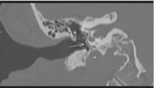
the presence of a subtle, apparently vascu-lar, tube-shaped structure originating from the vertical segment of the internal carotid artery at right, with an ascending course along the cochlear promontory, crossing the oval window (Figure 1), passing adjacent to the stapes crura (Figure 2) towards the middle cranial fossa, with enlargement of the anterior tympanic segment of the facial nerve (Figure 3). Associated absence of the CASE REPORT
Female, 50-year-old patient complain-ing of left hypoacusis for three months. The patient denied sonitus and dizziness. There was no positive family history for ear dis-eases or trauma/surgery involving such site. Audiometry study evidenced left-sided hearing loss, without air-bone gap. Multi-slice computed tomography demonstrated INTRODUCTION
Persistent stapedial artery (PSA) is a rare congenital anomaly, with prevalence calculated to be of 0.02%–0.05%(1) in sur-gical series and of 0.48% in temporal bone studies(2). The stapedial artery is a vascu-lar structure that is transiently present dur-ing the developmental stage of a fetus where it makes anastomosis between the external and internal branches of the carotid arteries. During the normal embryogenesis, the stapedial artery atrophies at the third gestational month.
The imaging diagnosis may be made by means of multidetector computed tomog-raphy or by angiogtomog-raphy. Magnetic nance imaging as well as magnetic reso-nance angiography play a limited role in the diagnosis of PSA, because of the reduced dimensions of the stapedial artery (diameter between 1.5 and 2.0 mm)(3).
Figure 1. Multislice computed tomography image with coronal reconstruction. Persistent stapedial artery (arrow) originating from the internal carotid artery (asterisk), with an ascending course adjacent to the cochlear promontory, passing through the oval window.
185
Carvalho BV et al. Persistence of stapedial artery: a case report
Radiol Bras. 2013 Mai/Jun;46(3):184–186 ipsilateral spinous foramen was observed, which is compatible with PSA. No evi-dence of association with aberrant carotid artery was observed. No significant alter-ation was observed in the side ipsilateral to the reported symptoms.
DISCUSSION
The embryonic stapedial artery persists because of a failure in its involution. Such artery is an integral part of the second bra-chial arch and originates from the hyoid artery that is a branch of the internal carotid artery. Then, it extends cranially and passes through the mesenchymal primordium of the stapes, forming the obturator foramen
of the stapes(3), becoming the middle meningeal artery, in the extradural region of the middle cranial fossa(4). When the stape-dial artery fails to involute, it originates the middle meningeal artery, and the spinous foramen that normally includes de middle meningeal artery will be absent (Figure 4). In most of times, patients with PSA are asymptomatic, but reports about cases of conductive hypoacusis, sonitus, retro-tympanic pulsatile mass and, rarely, neuro-sensory hearing loss are found in the litera-ture(5). Also, such condition has been asso-ciated with other diseases such as trisomy 13, 15 and 21, Paget’s disease, otosclero-sis, anencephaly, congenital immunodefi-ciency and neurofibromatosis(4,5).
For an accurate detection of such vas-cular anomaly, it is important to know the persistent stapedial artery trajectory. Its course is typical, originating from the pe-trous segment of the internal carotid artery, entering anteromedially into the hypotym-panum, coursing adjacent to the promon-tory and then cranially, through the obtu-rator foramen of the stapes, entering into the facial nerve canal through a dehiscence right behind the cochleariform process(3,4). The stapedial artery courses anteriorly to the facial nerve canal and leaves the canal immediately before the geniculate ganglion and continuing through the extradural space in the middle cranial fossa. In the lit-erature, there are reports of cases where the
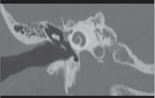
Figure 4. Normal embryological development of the stapedial artery (A,B) and anatomical variation with persistent stapedial artery (C). Figure 2. Axial multislice computed tomography.
Persistent stapedial artery (arrow) adjacent to the anterior crura and to the stapes footplate.
186
Carvalho BV et al. Persistence of stapedial artery: a case report
Radiol Bras. 2013 Mai/Jun;46(3):184–186 stapedial artery may either course through
a canal parallel to the facial nerve or extend into such canal, through a short segment(5). The tomographic findings suggestive of this anomaly are the following: 1) small canaliculus originating from the carotid canal; 2) linear structure passing through the middle ear space, adjacent to the prom-ontory; 3) enlargement of the tympanic seg-ment of the facial nerve canal or a separate canal paralleling the facial nerve; 4) ab-sence of the spinous foramen(4,5). Such con-dition may be associated with other abnor-malities involving the stapes, the facial nerve and the internal carotid artery. The association with aberrant internal carotid artery is the most common association(5–7). Intraoperative or post-mortem diagno-sis of such abnormality has been a rule(5). However, with the progress in imaging
methods, the rate of early detection of such abnormality has increased. The radiologist plays a fundamental role in the diagnosis of this vascular anomaly, aiding in the dif-ferential diagnosis of facial nerve tumors and in the surgical planning.
The previous knowledge of the anomaly by the otorhinolaryngologist allows the coagulation of this malformation with la-ser, facilitating the surgery and avoiding complications(8).
Persistent stapedial artery does not re-quire treatment, except in cases of intense sonitus, where endovascular ligation can be performed.
REFERENCES
1. Stott CC, Royer MF, Olmedo RO, et al. Resulta-dos auditivos de estapeResulta-dostomías en platinas com-plicadas. Rev Otorrinolaringol Cir Cabeza Cuello. 2006;66:89–94.
2. Jain R, Gandhi D, Gujar S, et al. Case 67: Persis-tent stapedial artery. Radiology. 2004;230:413–6. 3. Silbergleit R, Quint DJ, Mehta BA, et al. The per-sistent stapedial artery. AJNR Am J Neuroradiol. 2000;21:572–7.
4. Thiers FA, Sakai O, Poe DS, et al. Persistent sta-pedial artery: CT findings. AJNR Am J Neuro-radiol. 2000;21:1551–4.
5. Dimmick SJ, Faulder KC. Normal variants of the cerebral circulation at multidetector CT angiogra-phy. Radiographics. 2009;29:1027–43. 6. Moreno BA, García AU, Sampériz LC, et al.
Ca-rótida interna aberrante como causa de tumoración pulsátil en el oído médio. ORL ARAGON. 2010; 13:14–6.
7. Shimizu S, Sasahara G, Iida Y, et al. Aberrant in-ternal carotid artery in the middle ear with a defi-ciency in the origin of the anterior cerebral artery: a case report. Auris Nasus Larynx. 2009;36:359– 62.