J Appl Oral Sci.
240
ABSTRACT
www.scielo.br/jaos
Visibility of the mandibular canal on CBCT
cross-sectional images
Christiano OLIVEIRA-SANTOS1, Ana Lúcia Álvares CAPELOZZA1, Mariela Siqueira Gião DEZZOTI1,
Cássia Maria FISCHER2, Marcelo Lupion POLETI3, Izabel Regina Fischer RUBIRA-BULLEN1
1- DDS, MSc, PhD, Department of Stomatology, Bauru School of Dentistry, University of São Paulo, Bauru, SP, Brazil. 2- DDS, MSc, PhD, Dental School, Federal University of Goiás, Goiânia, GO, Brazil.
3- DDS, MSc, Department of Stomatology, Bauru School of Dentistry, University of São Paulo, Bauru, SP, Brazil.
Corresponding address: Christiano Oliveira-Santos - Al. Dr. Octávio Pinheiro Brisolla, 9-75 - Vila Universitária - 17012-901 - Bauru, SP - Brasil - Phone: +55-14-3235-8254 - e-mail: [email protected]
Received: August 24, 2009 - Modiication: March 26, 2010 - Accepted: May 25, 2010
T
he identiication of the mandibular canal (MC) is an important prerequisite for surgicalprocedures involving the posterior mandible. Cone beam computed tomography (CBCT) represents an advance in imaging technology, but distinguishing the MC from surrounding
structures may remain a delicate task. Objectives: The aim of this study was to assess
the visibility of the MC in different regions on CBCT cross-sectional images. Material and methods: CBCT cross-sectional images of 58 patients (116 hemi-mandibles) were analyzed, and the visibility of the MC in different regions was assessed. Results: The MC
was clearly visible in 53% of the hemi-mandibles. Dificult and very dificult visualizations
were registered in 25% and 22% of the hemi-mandibles, respectively. The visibility of the MC on distal regions was superior when compared to regions closer to the mental foramen. No differences were found between edentulous and tooth-bearing areas. Conclusions: The MC presents an overall satisfactory visibility on CBCT cross-sectional images in most cases. However, the discrimination of the canal from its surrounds becomes less obvious towards the mental foramen region when cross-sectional images are individually analyzed.
Key words: Mandible. Mandibular nerve. Cone-beam computed tomography.
INTRODUCTION
The identiication of the mandibular canal (MC)
is of fundamental importance for preoperative planning of surgical procedures involving the
posterior mandible2,7,10.Depiction of the MC
on imaging examinations is a requirement for endosseous implants surgeries, since the available height of the edentulous site is determined by the distance between the alveolar ridge and the MC2.
Several imaging modalities have been used to assess the course of the MC, including panoramic radiography2,9,14,17, conventional tomography9,
computed tomography (CT)11,14, and the most recently
introduced cone-beam computed tomography
(CBCT)6,10,17. Compared to conventional
two-dimensional techniques, CBCT imaging presents as main advantages the elimination of superimposition of neighboring structures, and absence of image
magniication. Furthermore, CBCT presents short
scanning time, and radiation dose up to 15 times lower than multislice CT (MSCT)16. The technology
is becoming increasingly more available in dental and radiological practices2,6-8,11-13,16-17,19.
The identiication of the MC is a delicate task.
The radiographic appearance usually involves a radiolucent zone lined by superior and inferior borders. The cortication of the canal is variable, which may explain why in some cases the MC is not well-visualized1-5,10,13-14,18. CBCT has been shown to
be superior to conventional imaging modalities for the depiction of the MC, however the visibility of
this structure may vary signiicantly, even within
the same individual1-5,7,18. Therefore, the aim of this
study was to determine the visibility of the MC on CBCT cross-sectional images in different regions of the mandible.
J Appl Oral Sci.
241
MATERIAL AND METhODS
Fifty-eight CBCT exams from patients referred to the Oral Imaging Clinic at Bauru School of Dentistry were randomly selected. The sample was composed of 33 females and 25 males with mean age of 47 years. Patients presenting edentulous regions in the posterior mandible with history of recent tooth extraction (within 5 years) were not included.
CBCT imaging was performed (i-CAT, Imaging
Sciences International, Hatield, Pennsylvania, USA)
with voxel size 0.3 mm, exposure cycle of 20 s. Cross-sectional images perpendicular to the occlusal
plane were reformatted (0.3 mm thickness) using the
software i-CAT Vision. A total of 116 hemi-mandibles were examined by one experienced calibrated Oral and Maxillofacial radiologist on a 20” monitor (eizo
Flexscan, Eizo Nanao Corporation, Ishikawa, Japan).
The visibility of the MC on the cross-sectional images was assessed in six mandibular regions: distal to third molar (D3M), third molar (3M), second molar
(2M), irst molar (1M), second premolar (2PM), and
just distal to the mental foramen (MF). Consecutive cross-sectional views were examined for each region. The observations were repeated twice, with at least one month time interval between them.
The visibility of the MC was registered as either positive or negative (possible or not possible, respectively, to undoubtedly differentiate the canal from surroundings, e.g. marrow spaces, bony lesions) (Figure 1). The scores of the regions were then clustered together so that each hemi-mandible received an overall visibility score: e (easy
identiication of the MC - 5 or 6 positive scores), D (dificult identiication – 3 or 4 positive scores), and VD (very dificult identiication – 0-2 positive
scores).
Information about missing teeth was also recorded. Regions 3M, 2M, 1M and, 2PM were additionally grouped as either “dentate” or “edentulous”, and the visibility of the MC for those groups was then registered. Descriptive statistical analysis was applied to the data. Mann-Whitney test was applied to test differences between dentate and edentulous groups, as well as eventual difference between right and left sides. The intra-observer agreement was calculated by Kappa index. This study has been approved by the Human Research ethics Committee at the University of São Paulo – Bauru School of Dentistry.
RESULTS
The visibility of the MC was registered as e (easy
identiication – i.e. positive visibility scores for 5-6
regions) in 53% of the hemi-mandibles (62/116). D and VD scores were recorded in 25% (29/116) and 22% (25/116) of the hemi-mandibles, respectively. The percentages of hemi-mandibles that showed a positive visibility for the MC, according to the mandibular region, are shown in Table 1. There was
not a statistically signiicant difference between right
and left sides (Mann-Whitney, p>0.05).
Among regions 3M, 2M, 1M and 2PM 202 regions were classified as “dentate”, whilst 262 were “edentulous”. Positive visibility of the MC was found for 65% of the dentate regions (131/202). Similarly, 68% of the edentulous regions (179/262) showed positive visibility. Kappa index for intra-observer agreement was 0.88 (almost perfect).
Region Positive Visibility
D3M 87.9% (102/116)
3M 74.1% (86/116)
2M 67.2% (78/116)
1M 66.4% (77/116)
2PM 62.9% (73/116)
MF 64.7% (75/116)
Table 1- Percentage of hemi-mandibles showing positive visibility scores for the mandibular canal (MC), according to the mandibular region
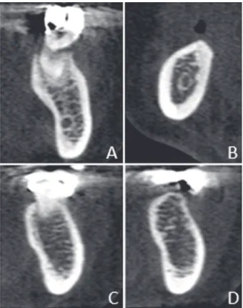
Figure 1- Cross-sectional cone beam computed
tomography (CBCT) images showing examples of: positive visibility of the mandibular canal (MC) in a dentate region (A); positive visibility of the MC in an edentulous region (B); negative visibility of the MC in a dentate region (C); negative visibility of the MC in an edentulous region (D)
Visibility of the mandibular canal on CBCT cross-sectional images
J Appl Oral Sci.
242
DISCUSSION
The introduction of new imaging technologies has allowed the visualization of anatomical structures in different plans without image superimposition. CBCT is a promising technique for the detailed evaluation of important bony structures, providing diagnostic images with good resolution while demanding relatively low radiation dose 2,6-8,11-13,16-17,19.
The quality of the image and the contrast between adjacent structures are important factors
in the reliability of the identiication of different landmarks11. The accuracy of multi-slice CT in
the analysis of important anatomical landmarks,
such as the mandibular canal pathway, has been shown15. CBCT has been attested as well suited for
imaging of the maxillofacial region, displaying high contrast, thus extremely useful for assessing bone. Cancellous bone is more sharply visualized in the cross-sectional images of CBCT than Spiral CT7.
In the present study, the identiication of the
MC on CBCT cross-sectional images was considered
a relatively easy task (i.e. the canal could be
visualized and discriminated from surroundings in nearly every region throughout its extension) in 53% of the hemi-mandibles. However, in 47%
of the hemi-mandibles the identiication of the MC
was not as readily feasible, which indicates that deciding which hypodense area corresponds to the actual MC on CBCT cross-sectional images is not always so evident.
Although the MC has been described as a radiolucent zone lined by radiopaque borders on radiographs, distinct bony-walled channels with definite borders do not seem to be a regular feature1,3-5,13-14,18.Carter & Keen3 (1970) studied
the intramandibular course of the inferior alveolar nerve and noted that in a number of cases, vessels and nerve branches may be spread out so that a distinct bone canal is not present. Neurovascular components may course through the mandible as a single entity or as a plexus, presenting a range of different-sized bundles, which do not necessarily travel within a bony canal from the mandibular foramen to the mental foramen1.
The MC walls are usually not formed by compact bone4-5,13-14,18. Instead, they are composed of a
coalescence of trabecular bone, ranging from dense to very delicate structures4-5,18. Additionally, the
trabeculation varies among individuals and also among different locations in the mandible1,3-4,9,18.
In the present study, the MC was readily visible on CBCT cross-sectional images of more posterior regions (3M and D3M). Visibility decreased
towards the mental foramen. These indings are in
accordance with those previous studies that found more unreliable radiographic visibility of the MC
near the mental foramen due to the lack of deinite
walls in the anterior portion of the canal4-5,9. The
posterior segment of the MC, i.e. closer to the mandibular foramen and extending apical to the
third molar region, is usually more identiiable due
to increased density of its walls4-5,18.
Similarly, Angelopoulus, et al.2 (2008) compared
digital and conventional panoramic radiographs and CBCT reformatted panoramic images in the delineation of the MC course in different areas of the mandible. CBCT was found superior to the other
modalities for such task, regardless of the location.
The posterior third of the MC was best visualized on all tested modalities, followed by the median third (rated second), and anterior third of the canal.
even though CBCT images may present more suitable images for the appreciation of the MC, the
identiication of this structure seems to be more linked to the bone density of its walls. The visibility
of the MC may be more dependable on anatomic features of the canal itself than on the technique used. Thus, it seems reasonable that if the MC is not well visualized in one technically satisfactory
exam, some degree of dificulty should be expected in the identiication of the canal on other imaging
modalities1,14,18.
Moreover, it has been suggested that the neurovascular bundle in the posterior regions
is usually in contact with and makes a discrete
depression in the lingual cortical plate, which may also account for a better depiction of that portion of the MC on radiographs5. On reformatted
cross-sectional CT images, grooving of the endosteal surface of the lingual cortical plate may be the only guide to the location of the MC when corticated walls
are not identiiable1,13.
Dissections have indicated that edentulous mandibular regions may present a reduction in size of the neurovascular bundle and the blood vessels
are more dificult to be identiied than in dentate
mandibles18. However, the presence or absence of
teeth did not seem to inluence the visibility of the
MC on CBCT cross-sectional images in this study, since the MC could be clearly visualized in around two thirds of both dentate and edentulous regions. Although some degree of bilateral asymmetry may be expected for mandibular structures, statistically significant differences on visibility of the MC between right and left sides were not found.
Lofthag-Hansen, et al.10 (2008) evaluated the
visibility of the MC and alveolar ridge on cross-sectional CBCT images. The visibility of the marginal crest of the alveolar ridge was considered superior compared to the MC. When assessing only one predetermined cross-sectional image, observers
marked the MC as “clearly visible” only in one third
of the cases. The visibility increased when raters had access to more images. This result points out OLIVEIRA-SANTOS C, CAPELOZZA ALÁ, DEZZOTI MSG, FISCHER CM, POLETI ML, RUBIRA-BULLEN IRF
J Appl Oral Sci.
243
for the importance of assessing every sequential image available in order to improve the localization of the MC.
CONCLUSIONS
The MC presented an overall satisfactory visibility on CBCT cross-sectional images in over half of the hemi-mandibles evaluated in this study. However, the discrimination of the canal from its surrounds became increasingly less clear towards the mental foramen region. Visibility of the MC clearly increased on cross-sectional images of more distal regions of the canal. Differences in visibility of the MC on CBCT cross-sectional images between edentulous and tooth-bearing regions were not found.
REFERENCES
1- Anderson LC, Kosinsk TF, Mentag PJ. A review of the
intraosseous course of the nerves of the mandible. J Oral Implantol. 1991;17:394-403.
2- Angelopoulous C, Thomas SL, Hechler S, Parissis N, Hlavacek
M. Comparison between digital panoramic radiography and
cone-beam computed tomography for the identiication of the
mandibular canal as part of presurgical dental implant assessment. J Oral Maxillofac Surg. 2008;66:2130-5.
3- Carter RB, Keen eM. The intramandibular course of the inferior alveolar nerve. J Anat. 1971;108:433-40.
4- Denio D, Torabinejad M, Bakland LK. Anatomical relationship
of the mandibular canal to its surrounding structures in mature mandibles. J endod. 1992;18:161-5.
5- Gowgiel JM. The position and course of the mandibular canal. J Oral Implantol. 1992;18:383-5.
6- Kamburoğlu K, Kiliç C, Özen T, Yüksel SP. Measurements
of mandibular canal region obtained by cone-beam computed tomography: a cadaveric study. Oral Surg Oral Med Oral Pathol Oral Radiol endod. 2009;107:e34-e42.
7- Kobayashi K, Shimoda S, Nakagawa Y, Yamamoto A. Accuracy
in measurement of distance using limited cone-beam computerized tomography. Int J Oral Maxillofac Implants. 2004;19:228-31.
8- Liang X, Jacobs R, Hassan B, Li L, Pauwels R, Corpas L, et al. A comparative evaluation of Cone Beam Computed Tomography (CBCT) and Multi-Slice CT (MSCT). Part I. On subjective image quality. eur J Radiol. 2010;75(2):265-9.
9- Lindh C, Petersson A. Radiologic examination for location of the mandibular canal: a comparison between panoramic radiography and conventional tomography. Int J Oral Maxillofac Implants. 1989;4:249-53.
10- Lofthag-Hansen S, Gröndahl K, Ekestubbe A. Cone-beam
CT for preoperative implant planning in the posterior mandible:
visibility of anatomic landmarks. Clin Implant Dent Relat Res.
2009;11:246-55.
11- Lou L, Lagravere MO, Compton S, Major PW, Flores-Mir C.
Accuracy of measurements and reliability of landmark identiication
with computed tomography techniques in the maxillofacial area: a systematic review. Oral Surg Oral Med Oral Pathol Oral Radiol endod. 2007;104:402-11
12- Loubele M, Jacobs R, Maes F, Denis K, White S, Coudyzer W, et al. Image quality vs radioation dose of four cone beam computed tomography scanners. Dentomaxillofac Radiol. 2008;37:309-19. 13- Monsour PA, Dudhia R. Implant radiography and radiology. Aust Dent J. 2008;53(Sp. issue 1):S11-25.
14- Naitoh M, Katsumata A, Kubota Y, Hayashi M, Ariji e. Relationship between cancellous bone density and mandibular canal depiction. Implant Dent. 2009;18:112-8.
15- Paes Ada SF, Moreira CR, Sales MAO, Cavalcanti MGP. Comparative study of single and multislice computed tomography for assessment of the mandibular canal. J Appl Oral Sci. 2007;15:220-4.
16- Scarfe WC, Farman AG, Sukovic P. Clinical applications of
cone-beam computed tomography in dental practice. J Can Dent Assoc. 2006;72:75-80.
17- Tantanapornkul W, Okouchi K, Fujiwara Y, Yamashiro M, Maruoka Y, Ohbayashi N, et al. A comparative study of cone-beam
computed tomography and conventional panoramic radiography in assessing the topographic relationship between the mandibular canal and impacted third molars. Oral Surg Oral Med Oral Pathol Oral Radiol endod. 2007;103:253-9.
18- Wadu SC, Penhall B, Townsend GC. Morphological variability of the human inferior alveolar nerve. Clin Anat. 1997;10:82-7. 19- Ziegler CM, Woertche R, Brief J, Hassfeld S. Clinical indications for digital volume tomography in oral and maxillofacial surgery. Dentomaxillofac Radiol. 2002;31:126-30.
Visibility of the mandibular canal on CBCT cross-sectional images