Elastographic Evaluation of Indeterminate
Breast Masses on Ultrasound
Avaliação pela elastogra
fi
a dos nódulos mamários
indeterminados na ultrassonogra
fi
a
Luciana Graziano
1Almir Galvão Vieira Bitencourt
1Marcela Pecora Cohen
1Camila Souza Guatelli
1Miriam Rosalina Brites Poli
1Juliana Alves Souza
1Elvira Ferreira Marques
11Department of Imaging, AC Camargo Cancer Center, São Paulo, Brazil
Rev Bras Ginecol Obstet 2017;39:72–79.
Address for correspondence Luciana Graziano, MD, Department of Imaging, AC Camargo Cancer Center, Rua Prof. Antônio Prudente, 211, São Paulo/SP, Brazil 01509-010 (e-mail: [email protected]).
Keywords
►
breast neoplasms
►
mammary
ultrasonography
►
elastography
►
lesions
Abstract
Objective
To evaluate the diagnostic accuracy of elastography for breast cancer
identi
fi
cation in patients with indeterminate lesions on ultrasound.
Methods
This prospective, descriptive study included patients with indeterminate
breast lesions in the ultrasound and with indication for percutaneous or surgical biopsy.
The elastography was evaluated by qualitative analysis and by two methods for the
semi quantitative analysis.
Results
We evaluated 125 female patients with 159 lesions, with a mean age of
47 years, and a range of 20
–
85 years. Ultrasound has shown to be a method with good
sensitivity (98.1%), but with a lower speci
fi
city (40.6%). On the elastography qualitative
analysis, the speci
fi
city and accuracy were of 80.2% and 81.8% respectively. The mean
size of the lesions showed no difference in classi
fi
cation by elastography. For the
semiquantitative elastography, the mean values of the malignant lesions were
statistically higher when compared with the subcutaneous tissue or the adjacent
fi
broglandular tissue. The analysis of the receiver operating characteristic (ROC) curves
for these two semiquantitative methods showed that both are considered satisfactory,
with an area under the curve above 0.75 and statistical signi
fi
cance (
p
<
0.0001). The
best results were obtained when using the
fi
ndings of combined conventional
ultrasound and qualitative elastography, with 100% sensitivity and 63.2% speci
fi
city.
Conclusions
Elastography can be a useful complementary method, increasing the
speci
fi
city and diagnostic accuracy of conventional ultrasound for the diagnosis of
breast cancer in patients with indeterminate breast lesions.
Resumo
Objetivo
Avaliar a acurácia diagnóstica da elastogra
fi
a para identi
fi
cação do câncer
de mama em pacientes com lesões indeterminadas por ultrassom.
Métodos
Estudo prospectivo, descritivo, com pacientes com lesões mamárias
inde-terminadas no ultrassom e indicação de biópsia percutânea ou cirúrgica. A elastogra
fi
a
foi avaliada por análise qualitativa e dois métodos de análise semiquantitativa.
received May 8, 2016 accepted November 8, 2016 published online December 27, 2016
DOI http://dx.doi.org/ 10.1055/s-0036-1597753. ISSN 0100-7203.
Copyright © 2017 by Thieme-Revinter Publicações Ltda, Rio de Janeiro, Brazil Original Article
Introduction
Imaging methods have a fundamental role in the manage-ment of patients with breast cancer, especially in the early diagnosis of non-palpable breast lesions. The conventional image methods (that is, mammography and ultrasound [US]) already present high sensitivity; however, there is still a large number of false positive results.1 The biopsy rate with positive cancer is only 10–30%, and this means that most breast biopsies performed result on benignfindings, causing unnecessary discomfort and anxiety to the patient, and increasing the costs for health care systems.2
Ultrasonography is often used to complement mam-mography, especially in young patients or those with dense breasts. However, conventional US is known to have a high rate of false positive results, and its specificity varies from 24 to 98.8%.3 Elastography is a new tool available in some US devices that measures the degree of elasticity or deformation of a tissue. Combined with the morphological criteria evaluated during the examination of the US, it can aid in the differential diagnosis between benign and malignant lesions.4–7This technique relies on the fact that the tissue of malignant lesions is more resistant to compression than the surrounding normal parenchyma and benign lesions.8There are two different techniques available for clinical use, compression or
“strain” elastography, and “shear-wave” elastography, and both have a good diagnostic performance in the evaluation of breast lesions.9
Although this technology is already being studied for the evaluation of breast lesions, it only recently became available for use in the clinical practice, and there are few studies on its performance and real benefit in the evaluation of patients with breast lesions. The objective of this study was to evaluate the diagnostic accuracy of elastography for breast cancer identification in patients with indeterminate lesions on conventional US.
Methods
This prospective, descriptive study included 125 patients with 159 indeterminate breast lesions in ultrasonography, and with an indication for percutaneous or surgical biopsy, in the Imaging Department of a cancer center, from June 2013 to May 2015. The study was approved by the institution’s Ethics Review Board, and all patients signed a written informed consent before enrollment. A standardized data sheet was completed for all patients, with clinical information, ultrasound findings and histological analysis. The imagingfindings of other methods, such as mammography and magnetic resonance imaging, were not analyzed, as they were not available for most patients, and to avoid influence on lesion characterization by US.
The Breast Imaging Reporting and Data System (BI-RADS) lexicon (5th edition) was used to describe the lesions’
characteristics, including shape, margins, orientation, echo pattern and posterior features. Lesions classified in BI-RADS categories 3, 4a, 4b, 4c and 5 were considered indeterminate and included in the study. Category 3 lesions included hypoechoic, isoechoic or heterogeneous echo pattern, oval shape, circumscribed margins and parallel orientation masses, or isolated grouped micro cysts. Category 4a lesions included round masses with circumscribed masses and any posterior features. Category 4b included non-mass lesions with architectural distortion, oval or rounded masses with indistinct margins, intraductal masses and complex cystic and solid masses. Category 4c included non-mass lesions with architectural distortion and micro calcifications, and round or irregular hypoechoic masses with angular or micro lobulated margins. Category 5 included irregular hypoechoic mass with spiculated margins and posterior shadowing.
Patients were submitted to an ultrasonographic examina-tion with elastography before the percutaneous procedure (needle biopsy or preoperative localization). Ultrasounds were performed in a specific device (Aplio 500; Toshiba America Medical Systems, Minato-ku, Tokyo 105–8001,
Resultados
Avaliamos 125 pacientes do sexo feminino com 159 lesões, com média de
idade de 47 anos, variando de 20 a 85 anos. O ultrassom mostrou ser um método com
boa sensibilidade (98,1%), mas com menor especi
fi
cidade (40,6%). Na elastogra
fi
a da
análise qualitativa, a especi
fi
cidade e acurácia foram de 80,2% e 81,8%,
respectiva-mente. A dimensão média das lesões não mostrou diferença na classi
fi
cação por
elastogra
fi
a. Para a elastogra
fi
a semiquantitativa, os valores médios das lesões
malignas foram estatisticamente maiores quando comparados ao tecido subcutâneo
ou
fi
broglandular adjacente. A análise das curvas ROC para estes dois métodos
semiquantitativos mostrou que ambos são considerados satisfatórios, com área abaixo
da curva acima de 0,75 e signi
fi
cância estatística (
p
<
0,0001). Os melhores resultados
foram obtidos com os achados de ultrassonogra
fi
a combinada convencional e
elasto-gra
fi
a qualitativa, com sensibilidade de 100% e especi
fi
cidade de 63,2%.
Conclusões
A elastogra
fi
a pode ser um método complementar útil, aumentando a
especi
fi
cidade e a precisão diagnósticas do ultrassom convencional para o diagnóstico
de câncer de mama em pacientes com lesões mamárias indeterminadas.
Palavras-chave
►
neoplasias da mama
►
ultrassonogra
fi
a
mamária
►
elastogra
fi
a
Japan), using the“strain”elastography technique, performed by a single radiologist with expertise in breast US. The exam was performed in real time, with the probe positioned per-pendicular to the skin over the region of interest (ROI), with normal respiratory movements of the patient, and associated with repetitive movements of slight pressure. The ROI area for the elastography evaluation was selected including subcuta-neous fat and the pectoralis muscle, and more than 5 mm from the side edges. The elastographyfindings were evaluated for the qualitative and semi quantitative analyses.
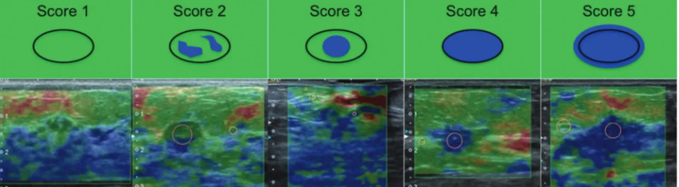
For the qualitative analysis of the elastography, a color scale was used, in which tissues with lower compressibility appeared as blue, more compressible tissues as red, and tissues with intermediate compressibility as green/yellow. According to the criteria proposed by Itoh et al,10the lesions were classified in 5 different scores, and considered as probably benign (scores 1, 2 and 3) or suggesting malignancy (scores 4 and 5)10,11(►Fig. 1):
• Score 1 - Uniformly compressible lesion, suggesting benignity.
• Score 2 - Highly compressible lesion with some areas of lesser compressibility, also suggesting benignity.
• Score 3 - Lesion with greater compressibility in the periphery, indicating the probability of a benign lesion.
• Score 4–Absence of compressibility all over the lesion; suspicion of malignancy.
• Score 5 - Absence of compressibility all over the lesion and also in the surrounding tissues, suggesting malignancy.
For the semi quantitative analysis, we used a ratio that compares the “strain” tension rate between two ROI areas selected manually. The compressibility within the lesion was compared with the compressibility in the subcutaneous tissue (lesion/subcutaneous tissue ratio), and also with the adjacent normalfibroglandular breast tissue (lesion/adjacentfi brogland-ular tissue ratio) (►Fig. 2). This“strain”rate reflects the relative
Fig. 1 Examples of lesions classified in each compressibility score for the qualitative elastography, according to the criteria proposed by Itoh et al.
lesion stiffness, which is directly proportional to the probability of malignancy.12–14
The Statistical analysis was performed using softwares STATA 11 SE (StataCorp LP, College Station, TX, USA), SPSS 16.0 (IBM, Armonk City, NY, USA) and MedCalc 15.6.1 (MedCalc Software bvba, Ostend, Belgium). In order to evaluate the diagnostic accuracy of the elastography, the histological result was considered as the gold standard. The Receiver Operating Characteristic (ROC) curve was used to determine the cut-off points on the semi quantitative analy-sis of the elastography, including the evaluation of the area under the curve (AUC), the standard error (SE), the 95% confidence interval (CI) and thepvalue. The normality of the variables was tested by the Shapiro-Wilk test, and the associations were tested by chi-square test or Fisher's exact test when necessary. Continuous variables were evaluated using the unpaired T-Student, ANOVA and non-parametric Mann-Whitney and Kruskal-Wallis tests, with a 5% signifi -cance level. Therefore, the results were considered statisti-cally significant when the value ofp< 0.05.
Results
Sample Description
The mean age of the 125 included patients was 47 years (standard deviation: 11 years), ranging from 20 to 85 years. Most of the patients were aged less than 40 years (70.4%). Twenty-three patients (18.4%) had breast cancer family history, and 10 (8.0%) had a previous history of breast cancer. Six patients (4.8%) had breast implants. Ninety-seven pa-tients (77.6%) had a single lesion, and 28 (22.4%) had more than one lesion in the breast.
We evaluated 159 indeterminate breast lesions at US: 46 (28.9%) were palpable, and 113 (71.1%) were non-palpable. The mean major size of the lesions was 15.6 mm (standard deviation: 11 mm), ranging between 3 mm and 68 mm. The morphological characteristics and BI-RADS category of the lesions are described in ►Table 1. ►Table 2describes all histological biopsies results, used as reference.
►Table 3shows the relationship of the BI-RADS categories
with the histopathologic results. Of the 106 benign lesions at histology, 43 (40.6%) were classified as probably benign (BI-RADS 3), and 63 (59.4%) were classified as suspect (BI-RADS 4 or 5) on US. Of the 53 malignant lesions at histology, 52 lesions (98.0%) were classified as suspect (BI-RADS 4 or 5) and 1 lesion was classified as probably benign (BI-RADS 3) on US. Thus, using the BI-RADS classifi -cation, US showed a sensitivity of 98.1%, a specificity of 40.6%, a positive predictive value of 45.2%, a negative predic-tive value of 97.0%, and a diagnostic accuracy of 59.7%.
Elastography
The qualitative classification of the elastography based on the criteria of Itoh et al10is described in►Table 4. It was observed that 91.4% of patients classified as probably benign confirmed this diagnosis, while 68.2% of patients classified as suspicious for malignancy had their results confirmed (p<0.01).
In the semi quantitative analysis, malignant lesions had a mean compression ratio higher than the benign lesions, when compared with the subcutaneous tissue and when compared with the adjacentfibroglandular tissue (►Table 5). The ROC curve analysis (►Fig. 3) showed no significant difference on sensitivity and specificity for the diagnosis of malignant lesions (p¼0.77) between these ratios: lesion/ subcutaneous tissue (AUC: 0.788; SE: 0.393;p<0.0001; 95% CI: 0.715 to 0.849); and lesion/adjacentfibroglandular tissue (AUC: 0.799; SE: 0.375;p<0.0001; 95% CI: 0.727 to 0.858).
Table 1 Characteristics of the lesions on conventional
ultrasonography, according to the BI-RADS lexicon (5th edition)
Characteristic Frequency (n) %
Lesion
Mass 132 83.0
Others 27 17.0
Echo pattern
Hypoechoic 125 78.6
Isoechoic 20 12.6
Hiperechoic 1 18.2
Heterogeneous 13 0.6
Shape
Oval 85 53.5
Round 30 18.9
Irregular 44 27.7
Margin
Circumscribed 64 40.2
Indistinct 43 27.0
Angular 11 6.9
Microlobulated 10 6.2
Spiculated 31 19.4
Orientation
Parallel 93 86.1
Not parallel 15 13.9
Posterior features
None 120 75.5
Enhancement 31 19.5
Shadowing 8 5.0
BI-RADS
3 44 27.7
4 a 32 20.1
4 b 36 22.6
4 c 13 8.2
5 34 21.4
Total 159 100
Using the data obtained by the ROC curve, the best cut-off points were 3.00 for the lesion/subcutaneous tissue ratio (sensitivity: 71.7%; specificity: 75.0%; accuracy: 73.9%) and 2.15 for the lesion/adjacentfibroglandular tissue ratio (sen-sitivity: 83.0%; specificity: 70.8%; accuracy: 72.3%).
Combination of Conventional Ultrasound and Elastography
For the combination of thefindings of the elastography and the conventional US, the following criteria were considered:
• Probably benign US (BI-RADS 3) and probably benign elastography: probably benign combination;
• Probably benign US (BI-RADS 3) and suspicious elastog-raphy: suspicious for malignancy combination;
• Low-suspicion US (BI-RADS 4a) and probably benign elastography: probably benign combination (►Fig. 4);
• Low-suspicion US (BI-RADS 4a) and elastography suspi-cion: suspicious for malignancy combination;
Table 2 Histological diagnosis of benign and malignant breast
lesions
Histological diagnosis Frequency (n) %
Benign lesions 106 66.7
Fibroadenoma 44 41.5
Stromalfibrosis 16 15.1
Papilloma 8 7.5
Fibrocystic changes 7 6.6
Malignant lesions 59 33.3
Invasive Carcinoma NST 35 66
Ductal carcinoma in situ 8 15.1
Invasive lobular carcinoma 7 13.2
Papillary Carcinoma 1 1.9
Tubular Carcinoma 2 3.7
Note:NST: no special type (ancient invasive ductal carcinoma).
Table 3 Correlation of histological results and BI-RADS
classification on conventional ultrasonography
BI-RADS Histological results
Benign (n) Malignant (n)
3 43 (97.7%) 1 (2.3%)
4a 32 (100%) 0 (0%)
4b 27 (75%) 9 (25.0%)
4c 3 (23.1%) 10 (76.9%)
5 1 (2.9%) 33 (97.1%)
Total 106 (66.7%) 53 (33.3%)
Abbreviation: BI-RADS, Breast Imaging Reporting and Data System.
Table 4 Correlation of histological results and qualitative
elastography analysis, according to Itoh et al criteria
Score Histological results
Total n (%)
Benign n (%) Malignant n (%)
1 21 (100%) 0 (0%) 21(13.2%)
2 55 (88.7%) 9 (11.3%) 62 (39.0%)
3 9 (90%) 1 (10%) 10 (6.3%)
4 21 (46.7%) 24 (53.3%) 45 (28.3%)
5 0 (0%) 21 (100%) 21 (13.2%)
Probably benign (1, 2 or 3)
85 (91.4%) 8 (8.6%) 93 (58.5%)
Suspicion of malignancy (4 or 5)
21 (31.8%) 45 (68.2%) 66 (41.5%)
Table 5 Correlation of histological results and semi
quantitative elastography ratios
Ratios Histological results p
Benign Mean (SD)
Malignant Mean (SD)
Lesion/Adipose
tissue 3.69 (4.4) 8.28 (7.5)
<0,001
Lesion/Adjacent fibroglandular tissue
2.15 (1.7) 7.18 (8.1) <0,001
Abbreviation: SD, standard deviation. Note:Data unknown for 2 patients (super
ficial lesions).
• Intermediate or high-suspicion US (BI-RADS 4b, 4c and 5), regardless of the elastography: suspicious for malignancy combination;
►Table 6 describes the sensitivity, specificity, positive
predictive value (PPV), negative predictive value (NPV) and accuracy for the conventional ultrasound, the qualitative and semiquantitative elastography analyses, and for their com-bination. Thus, we observed that the best results were obtained when using thefindings of the combined conven-tional US and the qualitative elastography, with 100% sensi-tivity and 63.2% specificity (versus 40.6% on the conventional US).
Discussion
In the literature, the sensitivity and specificity of the elas-tography ranged from 72 to 83.3%, and from 86.7 to 98.5% respectively.10,11,13,15–18In our study, the sensitivity (84.9%) was similar to the one found in the literature; however, the specificity (80.2%) was found to be slightly lower. Still, the
association of the US with the elastography showed an increase in specificity and diagnostic accuracy when com-pared with the isolated conventional US assessment. Similar findings observed in the literature showed that combined conventional US and elastography present a sensitivity of 89.1 to 96.9%, and a specificity of 50.5 to 95.7%.3
Studies that assessed semi quantitative elastography had different approaches, using subcutaneous adipose tissue and/ or adjacent fibroglandular tissue to assess the lesion com-pressibility ratio. The subcutaneous fat was considered the most suitable for the calculation of the deformity, because it is not influenced by other factors such as breast density, hor-monal status, lactation and cycle phase.3,14,19–21 In a study published by Zhou et al,22the lesion/adipose tissue ratio (with a cut-off point of 2.78) showed 82.9% sensitivity and 75.6% specificity, while the lesion/glandular tissue ratio (with a cut-off point of 1.54) showed a sensitivity of 77.1% and a specificity of 65.9%.22Similarly, in the present study, the lesion/subcuta-neous tissue ratio showed slightly superior results than the lesion/adjacentfibroglandular tissue ratio; however, this dif-ference was not statistically significant.
Fig. 4 Example of a suspiciousfinding at conventional ultrasound with probably benignfindings in both qualitative and semi quantitative elastographies. Conventional ultrasonography showed a hypoechoic round mass, considered suspicious for malignancy (A). The qualitative elastography showed a score 2 based on the criteria of Itoh et al, and the semi quantitative analysis showed a lesion/subcutaneous tissue ratio of 1.82 (B) and a lesion/adjacentfibroglandular tissue ratio of 1.07 (C), suggesting a probably benign lesion. The histological results were compatible withfibroadenoma.
Table 6 Sensitivity, specificity, positive predictive value (PPV), negative predictive value (NPV) and accuracy of conventional
ultrasound, qualitative and semi quantitative elastography analysis, and combination of these methods
Method Sensibility Specificity PPV NPV Accuracy
Conventional ultrasound 98.1% 40.6% 45.2% 97.7% 69.7%
Qualitative elastography 84.9% 80.2% 68.2% 91.4% 81.8%
Lesion/subcutaneous tissue ratio 71.7% 75.0% 59.4% 83.9% 73.9%
Lesion/adjacentfibroglandular tissue ratio 83.0% 70.8% 56.3% 85.2% 72.3%
Combined conventional ultrasound and qualitative elastography
100% 63.2% 57.6% 100% 75.5%
Combined conventional ultrasound and lesion/subcutaneous tissue ratio
98.1% 53.8% 51.5% 98.3% 68.6%
Combined conventional ultrasound and lesion/adjacentfibroglandular tissue ratio
100% 55.6% 53.0% 100% 70,4%
Our results showed that the qualitative analysis of the elastography showed better results than the semi quantita-tive assessment, regardless of the approach used. These data are consistent with thefindings published by Stachs et al.23 It is worth mentioning that elastography can also have false-negative and false-positive results. Not all cancers are more rigid than the healthy tissue, and the stiffness is different depending on the type of histological and clinical presentations, such as the association with necrosis, which can make them softer.24 Fur-thermore, elastography has some limitations, such as the size of the lesion: the higher the lesion, the less accurate is the elastog-raphy, with a higher performance on lesions smaller than 1 cm.25 Due to the high percentage of malignancy in lesions in categories 4b, 4c and 5 BI-RADS, biopsy should always be performed, regardless of thefinding of the elastography. How-ever, in lesions with low suspicion for malignancy (BI-RADS 3 and 4a), elastography can help define the best management, reducing the number of false-negative and false-positive results.
In our study, only one probably benign (BI-RADS 3) lesion on conventional US was diagnosed as malignant on biopsy. However, this lesion showed suspicious findings in the elastography, and that could be used to reclassify it as BI-RADS 4a, which would avoid a delay in diagnosis. Moreover, in our sample,84% of lesions classified as BI-RADS 4a on conventional US had probably benignfindings in the elas-tography, and could be reclassified as BI-RADS 3, reducing the number of unnecessary biopsies in this group. For Raza et al,26all BI-RADS 4a lesions classified as probably benign in the elastography have benign histological diagnoses.
This study has some limitations. Because we used only one observer, it was not possible to evaluate the variability of the interpretation of the elastography, which may be a challenge in the clinical practice, where there are sonographers with varying levels of experience. Moreover, we did not assess the influence of breast size, lesion depth or proximity to the papilla in the elastography results.
It is important to emphasize that elastography is a comple-mentary tool for US examination, and should not be used as a single method; thefinal diagnosis should always be done in combination with the morphological characteristics. In addition, in patients with lesions of intermediate suspicion in the conven-tional US, with a benign histological result after the percutaneous biopsy, the elastographyfindings could help in the radio-patho-logical correlation. Therefore, we believe that this method has the potential to effectively improve the management of breast lesions.
In conclusion, elastography can be a useful complemen-tary method, increasing the level of confidence in thefinal evaluation of breast lesions at US. The results presented in this study showed that elastography may increase the speci-ficity and diagnostic accuracy of conventional US for the diagnosis of breast cancer in patients with indeterminate breast lesions. The combination of conventional US and qualitative elastography showed higher specificity and accu-racy values, without reducing the sensitivity in our sample, and it could be used to decrease unnecessary biopsy rates.
References
1 Wojcinski S, Boehme E, Farrokh A, Soergel P, Degenhardt F, Hill-emanns P. Ultrasound real-time elastography can predict malig-nancy in BI-RADS®-US 3 lesions. BMC Cancer 2013;13(1):159 2 Zhi H, Xiao XY, Ou B, et al. Could ultrasonic elastography help the
diagnosis of small (2 cm) breast cancer with the usage of sono-graphic BI-RADS classification? Eur J Radiol 2012;81(11):3216–3221 3 Lee JH, Kim SH, Kang BJ, et al. Role and clinical usefulness of elastography in small breast masses. Acad Radiol 2011;18(1): 74–80
4 Balleyguier C, Ciolovan L, Ammari S, et al. Breast elastography: the technical process and its applications. Diagn Interv Imaging 2013; 94(5):503–513
5 Barr RG. Sonographic breast elastography: a primer. J Ultrasound Med 2012;31(5):773–783
6 Ricci P, Maggini E, Mancuso E, Lodise P, Cantisani V, Catalano C. Clinical application of breast elastography: state of the art. Eur J Radiol 2014;83(3):429–437
7 Yerli H, Yılmaz T, Ural B, Gülay H. The diagnostic importance of evaluation of solid breast masses by sonoelastography. Ulus Cerrahi Derg 2013;29(2):67–71
8 Yoon JH, Kim MH, Kim EK, Moon HJ, Kwak JY, Kim MJ. Interobserver variability of ultrasound elastography: how it affects the diagnosis of breast lesions. AJR Am J Roentgenol 2011;196(3):730–736 9 Chang JM, Won JK, Lee KB, Park IA, Yi A, Moon WK. Comparison of
shear-wave and strain ultrasound elastography in the differenti-ation of benign and malignant breast lesions. AJR Am J Roentgenol 2013;201(2):W347-56
10 Itoh A, Ueno E, Tohno E, et al. Breast disease: clinical application of US elastography for diagnosis. Radiology 2006;239(2):341–350 11 Kumm TR, Szabunio MM. Elastography for the characterization of
breast lesions: initial clinical experience. Cancer Contr 2010; 17(3):156–161
12 Goddi A, Bonardi M, Alessi S. Breast elastography: A literature review. J Ultrasound 2012;15(3):192–198
13 Carlsen JF, Ewertsen C, Lönn L, Nielsen MB. Strain elastography ultrasound: an overview with emphasis on breast cancer diagno-sis. Diagnostics (Basel) 2013;3(1):117–125
14 Thomas A, Degenhardt F, Farrokh A, Wojcinski S, Slowinski T, Fischer T. Significant differentiation of focal breast lesions: cal-culation of strain ratio in breast sonoelastography. Acad Radiol 2010;17(5):558–563
15 Schaefer FKW, Heer I, Schaefer PJ, et al. Breast ultrasound elastography–results of 193 breast lesions in a prospective study with histopathologic correlation. Eur J Radiol 2011;77(3): 450–456
16 Tan SM, Teh HS, Mancer JFK, Poh WT. Improving B mode ultra-sound evaluation of breast lesions with real-time ultraultra-sound elastography–a clinical approach. Breast 2008;17(3):252–257 17 Tardivon A, El Khoury C, Thibault F, Wyler A, Barreau B,
Neuensch-wander S. [Elastography of the breast: a prospective study of 122 lesions]. J Radiol 2007;88(5 Pt 1):657–662
18 Thomas A, Fischer T, Frey H, et al. Real-time elastography–an advanced method of ultrasound: First results in 108 patients with breast lesions. Ultrasound Obstet Gynecol 2006;28(3):335–340 19 Cho N, Moon WK, Park JS, Cha JH, Jang M, Seong MH. Nonpalpable
breast masses: evaluation by US elastography. Korean J Radiol 2008;9(2):111–118
20 Barr RG, Nakashima K, Amy D, et al. WFUMB guidelines and recommendations for clinical use of ultrasound elastography: Part 2: breast. Ultrasound Med Biol 2015;41(5):1148–1160 21 Farrokh A, Wojcinski S, Degenhardt F. [Diagnostic value of strain
ratio measurement in the differentiation of malignant and benign breast lesions]. Ultraschall Med 2011;32(4):400–405
23 Stachs A, Hartmann S, Stubert J, et al. Differentiating between malignant and benign breast masses: factors limiting sonoelasto-graphic strain ratio. Ultraschall Med 2013;34(2):131–136 24 Fleury EdeF, Assunção-Queiros MdoC, Roveda D Jr. Breast
carci-nomas: variations in sonoelastographic appearance. Breast Can-cer (Dove Med Press) 2014;6:135–143
25 Chang JM, Moon WK, Cho N, Kim SJ. Breast mass evaluation: factors influencing the quality of US elastography. Radiology 2011;259(1):59–64