Executive dysfunction and motor
symptoms in Parkinson’s disease
Indira Silveira Campos-Sousa1, Raimundo Nonato Campos-Sousa1,
Luiz Ataíde Jr.2, Marta Maria de Brito Soares1, Kelson James Almeida1
ABSTRACT
The aim of this study is to analyze executive function and motor symptoms in patients with idiopathic Parkinson’s disease (PD). The sample consisted of 44 subjects with PD between the ages of 45 to 75, who were examined consecutively. The subjects were divided into two groups according to the duration of the disease. The control group was composed of spouses, family and accompanying members. Patients included were submitted to motor dysfunction evaluation using the UPDRS. The executive functions modalities analyzed included: operational memory, inhibitory control, planning, cognitive flexibility and inductive reasoning. Significant differences between the experimental and control groups were found in all the executive domains studied. Evidence of tremor, rigidity and bradykinesia correlation with executive dysfunction were not observed. Patients with PD, even in the initial phase of the disease, presented executive dysfunction. The cardinal motor signs of the disease were not correlated with the cognitive dysfunction found.
Key words: neuropsychology, neuropsychological tests, executive functions, idiopathic Parkinson’s disease.
Disfunções executivas e sintomas motores na doença de Parkinson
RESUMO
O objetivo do estudo é avaliar as funções executivas e sintomas motores em pacientes portadores de doença de Parkinson. A amostra se constituiu de 44 portadores de doença de Parkinson com idade entre 45 e 75 anos, examinados consecutivamente, os quais foram divididos em dois grupos de acordo com o tempo de duração da doença. O grupo controle foi composto de acompanhantes ou cônjuges. Os sujeitos selecionados foram submetidos à avaliação motora utilizando-se a escala UPDRS e à avaliação das funções executivas nas modalidades: raciocínio indutivo, memória operacional, controle inibitório, planejamento e flexibilidade cognitiva. Os resultados apontaram diferenças significantes entre os grupos experimentais e controle nas modalidades analisadas. Não encontramos evidência de associação entre tremor, rigidez e bradicinesia com as funções executivas. Conclui-se que os pacientes com doença de Parkinson, mesmo nas fases iniciais da doença, apresentam comprometimento cognitivo executivo. Os sintomas motores da doença não estavam correlacionados às disfunções executivas.
Palavras-chave: neuropsicologia, testes neuropsicológicos, funções executivas, doença de Parkinson.
Correspondence
Raimundo Nonato Campos-Sousa Rua Gov. Artur de Vasconcelos 289 64001-450 Teresina PI - Brasil E-mail: [email protected]
Received 20 October 2009
Received in final form 26 November 2009 Accepted 9 December 2009
1Movement’s Disorders Unit, Depar tment of Neurology, Federal University of Piauí (UFPI), Brazil; 2Depar tment of
Neuropsychiatry, Federal University of Pernambuco (UFPE), Brazil.
Parkinson’s disease (PD) is a progres-sive neurodegenerative condition charac-terized by tremor, rigidity, bradykinesia and postural instability. Non-motor symp-toms such as autonomic and cognitive dys-function are present but little is known1.
However, it was reported that the
preva-lence of cognitive impairment or executive dysfunction in PD can reach 93% if ade-quate neuropsychological instruments are performed2. Cognitive and mental
circuit degeneration results in low concentration of stri-atal dopamine producing motor symptoms characteris-tic of the disease while low dopaminergic input from ven-tral tegmental mesencephalic area to the frontal and lim-bic regions is the neurochemical process which express-es cognitive-behavioral dysfunction in PD4. Other
stud-ies have found the reduction of the activity of frontostri-atal loops in addition to reduction of dopaminergic input from ventral tegmental area to the frontal lobes5,6. Braak
et al. suggested that these changes in PD are secondary to an ascendant progression process. Initially, there are lesions on the brainstem and anterior olphatory nucleus. Subsequently, it is manifested by substantia nigra neu-ronal lesions and inally cortical area involvement, from anteromedial temporal mesocortex to neocortex where it reaches associative cortex and prefrontal areas7.
he neuropsychological function consists of a com-plex group which includes attention, memory, language, reasoning and executive functions. Executive function (EF) is a set of cognitive abilities which permit the start of activities, planning, programming and sequencing of actions, self regulation and task monitoring, correct se-lections of behavior and conduct, mental work lexibili-ty and time and space organization8. Dysfunction in this
area which is coordinated by prefrontal region is called executive dysfunction (ED). It is characterized by dii-culties to start actions, decrease in motivation and drive, planning diiculties based on priorities and the mainte-nance of activity sequence necessary to reach an object8.
To understand the working of this group of functions is of paramount importance for the development of evalu-ation and rehabilitevalu-ation strategies, improving the prog-nosis and life quality of the patients.
he aim of the study is to better understand this cog-nitive dysfunction so prevalent in PD through analytic study of patients chosen by diagnostic criteria and by us-ing speciic neuropsychological instruments to evaluate the executive domain.
METHOD
he sample consisted of members of the community and PD patients consecutively examined in the Movement Disorders outpatient clinic of Getúlio Vargas Hospital and the Federal University of Piauí. he patients were evalu-ated and put into two groups (PD1 and PD2). he PD1 group consisted of 23 patients with up to 3 years of the disease duration and the PD2 group 21 patients with more than 3 years of the onset. A brain magnetic resonance im-aging (MRI) was performed in all PD patients to exclude other kinds of parkinsonism. he control group (CG) was composed of 25 normal subjects selected from spouses, family or accompanying members with the same social demographic characteristics as the experimental groups.
he PD groups were composed of subjects between the ages of 45 to 75, who fulilled the diagnostic criteria cit-ed by LANG to separate PD from other kinds of parkin-sonism8. Individuals with depression, neurological or
psy-chiatric disorders such as delirium or hallucinations, de-mentia and who had undergone neurosurgery were ex-cluded. All of the subjects of the study underwent screen-ing to investigate depressive symptomatology through BECK Depression Inventory10. he Mini Mental Status
Examination (MMSE)11, basic daily activity
question-naire12 and a structured neuropsychological interview13
were applied to assess subjects with severe cognitive im-pairment or dementia and exclude them from the study.
he PD groups underwent a neurological evaluation to determine the motor scores in section III of the Uniied Parkinson’s Disease Rating Scale (UPDRS). All of the sub-jects of the PD groups were examined in the “on” medica-tion state with levodopa and/or dopaminergic drug. No patient was on anticholinergic drugs. Neuropsychological tests were applied in one session which lasted around one hour and forty minutes depending on the diiculty level of the subjects. he EF modalities analyzed included: op-erational memory, inhibitory control, planning, cognitive lexibility and inductive reasoning. To evaluate operational memory an inverse order digit subtest and letter-number sequencing (SLN) of Wechsler Adult Intelligence Scale (WAIS) were used. In the inhibitory control evaluation of part “B” of the Trail making test B (TMT B) and Stroop color and words test (SCWT) cards 2 and 3, were used10.
In the planning mode the WAIS-III cube subtest and Rey complex igure (CFT) was used. To evaluate the cog-nitive lexibility the Wisconsin card sorting test (WCST) was given. Intuitive reasoning was explored by the Raven progressive matrix test10.
he statistic analysis was performed with the objective of evaluating the prevalence of the ED of the PD patients and in CG, as well as to analyze the existence of motor factors associated with ED. For the descriptive analysis, tables (standard deviation and media) were used with all variables studies. he analysis of the diferences between the media of continuous dada was done through paramet-ric tests (variance analysis - ANOVA). To verify where the diferences occurred the Tukey test was carried out. To study EF and motor association the Pearson correlation test was used. he probability p<0.05 to indicate statis-tic signiicance was established. he subjects in the study signed a term of free consent and knowledge. he current protocol was submitted to a research ethic committee from the Federal University of Pernambuco, Brazil.
RESULTS
patients with MRI signs of multiple system atrophy and 5 patients with signs of ischemic stroke were excluded. About 15% of patients included in PD group had non-speciic white matter MRI signal hyperintensities. he control group consisted of 25 people. he demograph-ic data is found in Table 1. he severity of the PD groups was measured according to motor function sector III of the UPDRS. he total score varied from 8 to 57 points. No signiicant diference in the motor scores was found among the PD groups. Likewise, the media of the mo-tor scores studied for isolated symptoms such as tremor (p=0.71), rigidity (p=0.25) and bradykinesia (p=0.09) was not signiicant (Table 2).
he media and comparisons of the results of EF mo-dalities and the tests used to evaluate each one among the groups studied are shown in Table 3. In operating mode memory, the control group presented greater scores than the PD groups which revealed signiicant diferences be-tween the CG and PD. No signiicant diference was not-ed between the PD groups. For inhibitory control the con-trol group carried out the test TMT B in less time
com-pared to the two PD groups and there were signiicant statistical diferences. Even though the CG made fewer mistakes, there was no statistical diference with the PD groups. No signiicant diferences were found among the PD1 and PD2 groups in TMT B test (time and errors). In SCWT2 test, the control group had the greater number of correct answers when compared to the PD groups show-ing signiicant statistical diferences. In SCWT3 test sig-niicant diferences were observed only among the media of the control group (35.83) and with media of the PD1 group (24.20). he CG had scores a slightly higher than the PD2 group but without signiicance.
In the planning evaluation with the Cube test, the control group was diferent from both PD groups. In the CFT test, the difference was significant only between the control group and PD2 group. None the tests used to evaluate planning showed a diference between PD1 and PD2 groups. In the investigation of cognitive lexi-bility, the WCST (completed category and preservative response) pointed out a diference in the CG and the PD groups. However, no signiicant diference was noted
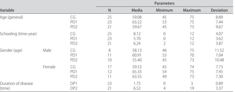
be-Table 1. Comparison of the demographic variables and the duration of PD in the groups.
Variable
Parameters
N Media Minimum Maximum Deviation
Age (general) CG
PD1 PD2
25 23 21
59.08 63.22 59.67
45 53 45
75 75 73
8.89 7.44 9.67 Schooling (time-year) CG
PD1 PD2
25 23 21
8.12 5.70 6.24
0 0 2
12 12 12
4.07 3.62 3.87
Gender (age) Male CG
PD1 PD2
8 11 10
58.13 60.91 55.40
46 53 45
75 70 73
11.52 7.04 10.48
Female CG
PD1 PD2
17 12 11
59.53 65.33 63.55
45 54 49
74 75 73
7.73 7.45 7.30 Duration of disease
(time) DP1DP2 2321 1.756.52 04 193 0.893.37
PD1: group with PD up to 3 years; PD2: group with PD for 3 years or more; CG: control group.
Table 2. Distribution of the media of the motor functions (Motor) and media of the scores separately which compose Sector III of the UPDRS among the patients.
Symptom Groups N Media* Minimum value Maximum value Deviation ANOVA
Tremor PD1
PD2 2321 5.13
a
5.52a 1.000.00 11.0012.00 2.913.87 0.71
Rigidity PD1
PD2 2321 5.57
a
6.67a 1.001.00 13.0014.00 3.033.26 0.25
Bradykinesia PD1
PD2 2321 12.74
a
15.86a 1.002.00 26.0026.00 6.126.09 0.09
Motor PD1
PD2 2321 28.26
a
35.19a 12.008.00 56.0057.00 12.4112.68 0.07
tween the two groups of patients. In the WCST test (fail-ure to maintain the set) similar results among the groups were veriied. In analyzing the inductive reasoning skills there was signiicant diference between the media of the CG and the PD groups. However, when comparing the two PD groups no diference was observed.
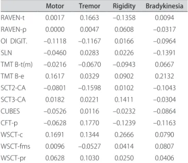
When the correlation between motor symptoms of PD - rigidity, tremor and bradykinesia- and each EF test given were studied, weak or no association between the cognitive modalities and the results of the motor function in UPDRS - sector III were observed (Table 4).
DISCUSSION
For many years, PD was described as a movement dis-order with a tendency to neglect the mental dysfunction associated with the disease1,3. Recently there has been a
systematic concern with the cognitive and behavioral as-pects of the neurodegenerative disease. In this way the neuropsychological evaluation of the EF has been the ob-ject of growing interest of researchers. However, there still remain many doubts about the functioning of EF in neurodegenerative diseases and PD in particular.
he individuals of this study are characterized by a
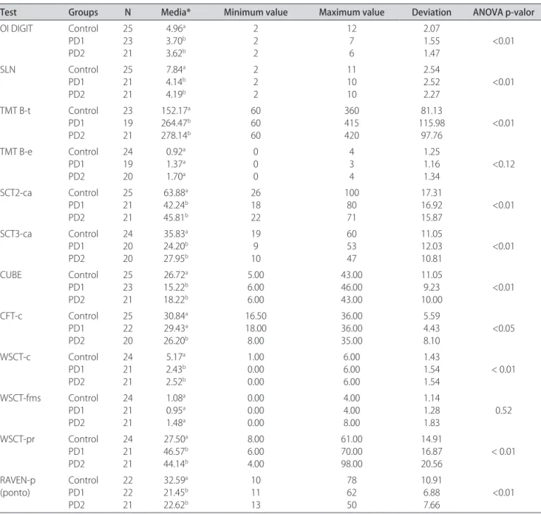
Table 3. Comparison among the results obtained from the tests of the diferent modalities for executive function among the groups studied.
Test Groups N Media* Minimum value Maximum value Deviation ANOVA p-valor OI DIGIT Control
PD1 PD2 25 23 21 4.96a 3.70b 3.62b 2 2 2 12 7 6 2.07 1.55 1.47 <0.01 SLN Control PD1 PD2 25 21 21 7.84a 4.14b 4.19b 2 2 2 11 10 10 2.54 2.52 2.27 <0.01
TMT B-t Control PD1 PD2 23 19 21 152.17a 264.47b 278.14b 60 60 60 360 415 420 81.13 115.98 97.76 <0.01 TMT B-e Control
PD1 PD2 24 19 20 0.92a 1.37a 1.70a 0 0 0 4 3 4 1.25 1.16 1.34 <0.12 SCT2-ca Control PD1 PD2 25 21 21 63.88a 42.24b 45.81b 26 18 22 100 80 71 17.31 16.92 15.87 <0.01 SCT3-ca Control PD1 PD2 24 20 20 35.83a 24.20b 27.95b 19 9 10 60 53 47 11.05 12.03 10.81 <0.01 CUBE Control PD1 PD2 25 23 21 26.72a 15.22b 18.22b 5.00 6.00 6.00 43.00 46.00 43.00 11.05 9.23 10.00 <0.01 CFT-c Control PD1 PD2 25 22 20 30.84a 29.43a 26.20b 16.50 18.00 8.00 36.00 36.00 35.00 5.59 4.43 8.10 <0.05 WSCT-c Control PD1 PD2 24 21 21 5.17a 2.43b 2.52b 1.00 0.00 0.00 6.00 6.00 6.00 1.43 1.54 1.54 < 0.01 WSCT-fms Control PD1 PD2 24 21 21 1.08a 0.95a 1.48a 0.00 0.00 0.00 4.00 4.00 8.00 1.14 1.28 1.83 0.52 WSCT-pr Control PD1 PD2 24 21 21 27.50a 46.57b 44.14b 8.00 6.00 4.00 61.00 70.00 98.00 14.91 16.87
20.56 < 0.01 RAVEN-p
(ponto) Control PD1 PD2 22 22 21 32.59a 21.45b 22.62b 10 11 13 78 62 50 10.91 6.88 7.66 <0.01
variation in regards to the level of education among the groups. It is known that intellectual experience in work activities could decrease the diiculties in carrying out neuropsychological tests in the subjects with little school-ing14. In the experimental groups, the duration of PD was
not associated with the severity of the parkinsonian mo-tor symptoms. his efect might have occurred because of the selection of patients since those older than 75 and/ or with severe tremor, bradykinesia, rigidity, postural in-stability and depression were excluded. his could have caused limitation in this study.
Neuropsychological evaluation of the EF showed low scores for PD patients in both stages with no statistical difference in the experimental groups suggesting that from the beginning of the disease the individuals with PD already show diiculty in EF. Few studies have addressed the question of cognitive reserve in patients with PD. Greater performance in cognitive tests in PD patients in the initial phase associated with a high level of cognitive reserve has been described such as in other neurodegen-erative diseases15. hese results are in agreement with the
indings of Foltynie et al.16, which also noted alterations in
the cognitive domain in patients initiating PD symptoms, or without dementia and with light motor manifestations. his is also in agreement with the study of Muslimovic et al.17, which showed cognitive deicit mainly in the
memo-ry domain areas and EF in the early phase of PD.
Fluid intelligence
In the investigation of inductive reasoning or luid in-telligence through the Raven test, signiicant deicit for the experimental groups was observed when compared to the CG, conirming works by Duncan et al.18, who found
correlations between frontal dysfunctions and diiculty in luid intelligence tasks. his also is in agreement with the indings of Prabhakaran et al.19, who mapped the
ce-rebral areas activated while a person is resolving items of the Raven test using functional MRI. his showed which areas of the cerebral areas were activated while the sub-ject solved problems using simple reasoning perceptual processes, analytic problems and problems of perceptual comparison (task control).
Operational memory
he tests used to evaluate operational memory (work-ing memory), SLN and inverse order digits, in DP1 as well as DP2 groups presented greater diiculty than the con-trol group. Dubois and Pillon20 reported that the patients
with PD presented alterations in operational memory when performing task which require short-term mem-ory; inhibition of an interference of a stimuli; digital se-quencing or special organization. Our indings are also in agreement with these studies related by Starkstein and
Merello21, which showed operational memory deicit in
PD in tests which require correct coordination of two tasks simultaneously. he recent work of Beato et al.22,
identiied that patients with PD presented inferior per-formance to the control group in working memory tasks and levodopa therapy presents a positive efect on spatial operational memory.
Inhibitory control
Attention encompasses orientation and mental con-centration directed by a task and inhibition of compet-itors. Distraction, interference of tasks by stimuli com-ing from the ambient, increases attention diiculty for individuals with frontal lobe disorders. In this study, the evaluation of inhibitory control indicated low scores for PD patients in regards to normal standards. In the study of Osternack-Pinto23 similar indings were found. PD
pa-tients had low scores in attention control tasks which re-quire good operational memory and inhibitory control to resist interference of stimuli competing for attention. In the same study, low scores were also found in SCWT and TMT B tests in PD patients.
Planning
In the investigation of planning the tests showed a dif-ference in the PD groups and the CG conirming the data found in literature3. Planning requires that an
individu-al have the capacity to evindividu-aluate individu-alternatives, make
choic-Table 4. Association measures among the means of the Motor scores in section III of UPDRS or scores separate from section III and results of tests of executive functions modalities studied.
Motor Tremor Rigidity Bradykinesia RAVEN-t 0.0017 0.1663 –0.1358 0.0094 RAVEN-p 0.0000 0.0047 0.0608 –0.0317 OI DIGIT. –0.1118 –0.1167 0.0166 –0.0964 SLN –0.0460 0.0283 0.0226 –0.1391 TMT B-t(m) –0.0216 –0.0670 –0.0943 0.0667 TMT B-e 0.1617 0.0329 0.0902 0.2132 SCT2-CA –0.0801 –0.1598 0.0102 –0.1043 SCT3-CA 0.0182 0.0221 0.1411 –0.0304 CUBES –0.0526 0.0116 –0.0232 –0.0864 CFT-p –0.0628 0.1770 –0.1239 –0.1163 WSCT-c 0.1691 0.1344 0.2666 0.0790 WSCT-fms 0.0096 –0.0527 0.0414 0.0807 WSCT-pr 0.0628 0.1030 0.0250 0.0406
es and study ideas necessary for carrying out the plan. In the qualitative analysis of the performance of PD patients in the CFT test, the majority of the patients evaluated in the study had diiculty in visual and perceptual organiza-tion, diiculty in starting a sequence, diiculty in choos-ing, rejecting and adopting alternative thought and con-duct courses. he complexity was so great for these pa-tients that many quit the task.
Cognitive lexibility
he WCST, which originally was developed to eval-uate the ability of abstraction reasoning and to change from one line of thought to another, is cited as sensitive for operational and cognitive lexibility. In the investiga-tion of cognitive lexibility with the use of WCST (com-pleted category and persevering response) a signiicant statistical diference between the CG and the PD groups was found. In studies carried out by Taylor, Saint-Cry and Lang cited by Piovezan24, the use of WCST showed
signif-icant diferences among the number of categories: there were persevering responses and a large number of errors to reach the irst category, which suggest less ability in PD patients to make a plan of action when given a task24.
Sim-ilar indings were found in the article of Sobreira et al.25
which evaluated the EF in PD and found scores below the average in the following tests: WCST, inverse digit order thus proving an involvement in the EF of these patients.
he PD patients scored lower when compared to the CG in all tests which evaluated EF, in those tests which needed quickness and motor skills (Cube, CFT, SCWT, TMT B) in their fulillment as well as those that did not need them (SLN, inverse order digits, WCST, Raven).
One of our objectives was to verify if cognitive dys-function is associated with the motor symptoms of PD when studied separately. We did not ind evidence of as-sociation between tremor, rigidity and bradykinesia in the scores of the tests which evaluated EF. Possibly, these iso-lated symptoms are not predictive factors for the preva-lence of cognitive deicits. his is partially in agreement with the indings of Piovezan24, which did not ind
corre-lation between executive deicits and scores for the scales Hoehn-Yahr and UPDRS. Also in agreement with the lit-erature, Graham and Sagar26 and Mohr et al.27, indicated
that such defects are part of a greater cognitive decline or which alternatively have these restrictions in a subgroup of patients and/or did not occur in the initial phase of the disease in the group studied.
In conclusion, the patients with PD had deicits in all tests which evaluated the modalities of EF when com-pared to the control group. here was little diference in the tests which evaluated executive cognitive domain be-tween the PD groups. he cardinal motor symptoms of the disease, when individually studied, were not
correlat-ed to ED and probably do not have prcorrelat-edictive value for the development of future cognitive incapability or dementia.
REFERENCES
1. Kummer A, Teixeira A. Neuropsychiatry of Parkinson’s disease. Arq Neurop-siquiatr 2009;67:930-939.
2. Pillon B, Czernecki V, Dubois B. Dopamine and cognitive function. Curr Opin Neurol 2003;16(Suppl 2):S17-S22.
3. Emre M. What Causes mental dysfunction in Parkinson’s disease? Mov Dis-ord 2003;18(Suppl):S63-S71.
4. Caixeta, L. Demências. São Paulo: Lemos-Editorial, 2004:146-147. 5. Javoy-Agid F, Agid Y. Is the mesocorticaldopaminergic system involved in
PD? Neurology 1980;30:1326.
6. Pillon B, Czernecki V, Dubois B. Dopamine and cognitive function. Curr Opin Neurol 2003;16(Suppl 2):S17-S22.
7. Braak H, Del Tredici K, Rüb U, de Vos RA, Jansen Steur EN, Braak E. Staging of brain pathology related to sporadic Parkinson’s disease. Neurobiol Aging 2003;24:197-211.
8. Parenté R. Retraining cognition: techniques and applications. Maryland: As-pen Publishers, 1996.
9. Lang AE, Lozano AM. Parkinson’s disease: first at two parts. N Eng J Med 1998;339:1044-1053.
10. Spreen O, Strauss E. A Compendium of neuropsychological tests: admin-istration, norms, and commentary. 2nd Ed. New York: Oxford University Press,1998.
11. Folstein MF, Folstein SE, Mchugh PR. Mini-mental state: a practical method for grading the cognitive state of patients for the clinician. J Psychiatr Res 1975;12:189-198.
12. Katz S, Downs TD, Cash HR, Grotz RC. Progress in the development of the in-dex ADL. Gerontologist 1970;1:20-30.
13. American Psychological Association - APA. Presidential Task Force on the as-sessment of age-consistent memory decline cognitive and dementia: guide-lines for the evaluation of dementia and age-related cognitive decline. Am Psychol 1998;53:1298-1303.
14. Osternack-Pinto K. [Análise Comparativa das funções neuropsicológicas de portadores de doença de Parkinson em estágios inicial e avançado: uma de-terminação de padrões para diagnóstico em população brasileira]. Thesis, University of São Paulo. São Paulo, 2006.
15. Sánchez JLMD, Rodriguez M, Carro J. Inluence of cognitive reserve on neu-ropsychological functioning in Parkinson’s disease. Acta Neuropsychiatrica 2002;14:207-215.
16. Foltynie T, Brayne CE, Robbins TW, Barker RA. The cognitive ability of an in-cident cohort of Parkinson’s patients in the UK: the Campaign study. Brain 2004;127:550-560.
17. Muslimovic D, Post B, Speelman JD, Schmand B. Cognitive proile of patients with newly diagnosed Parkinson disease. Neurology 2005; 65:1239-1245. 18. Duncan J, Emslie H, Williams P, Johnson R, Freer C. Intelligence and the frontal
lobe: the organization of goal-directed behavior. Cogn Psychol 1996;30:257-303. 19. Prabhakaran V, Smith JAL, Desmond JE, Glover GH, Gabrieli JDE. Neural
sub-stances of luid reasoning: an fMRI study of neocortical activation during perfor-mance of the Raven’s progressive matrices test. Cogn Psychol 1997; 33:43-63. 20. Dubois B, Pillon B. Cognitive deicits in Parkinson’s disease. J Neurol 1997; 24:2-8. 21. Starkstein SE, Merello M. Psychiatry and cognitive disordens in Parkinson’s
disease. Cambridge University Press 2002:88-113.
22. Beato R, Levy R, Pillon B, et al. Working memory in Parkinson’s disease pa-tients: clinical features and response to levodopa. Arq Neuropsiquiatr 2008; 66:147-51.
23. Osternack-Pinto K. [Análise Comparativa das funções neuropsicológicas de portadores de doença de Parkinson em estágios inicial e avançado: uma de-terminação de padrões para diagnóstico em população brasileira]. Thesis, University of São Paulo. São Paulo, 2006.
24. Piovezan MR. [Avaliação cognitiva em pacientes portadores de doença de Parkinson idiopática]. Dissertation, Federal University of Paraná. Curitiba, 2006. 25. Sobreira EST, Pena MCS, Filho THS, et al. Executive functions in Parkinson’s
disease. Dement Neuropsychol 2008;2:206-209.
26. Graham JM, Sagar HJ. A data-driven approach to the study of heterogeni-ty in idiophatic Parkinson’s disease: identiication of three distinct subheterogeni-types. Mov Disord 1999;14:10-20.