Arq. Bras. Oftalmol. vol.79 número5
Texto
Imagem
Documentos relacionados
Purpose: To evaluate subfoveal choroidal thickness (SFCT ) changes after in- travitreal bevacizumab (IVB) therapy for central serous chorioretinopathy (CSC) using enhanced
Comparison of central corneal thickness measurements by Pentacam, noncontact specular microscope, and ultrasound pachymetry in normal and post-LASIK eyes.. Corneal thickness
Purpose: The aim of the present study was to use enhanced depth imaging optical coherence tomography (EDI-OCT ) to investigate choroidal changes in patients with cone dystrophy
Purpose: This study was conducted to evaluate the relationships of inner/outer segment (IS/OS) junction disruption, macular thickness, and epiretinal membrane (ERM) grade
This study investigates the repeatability of the closed system FVA CS test in control subjects and in patients with glaucoma, cataracts and good Snellen visual acuity, or
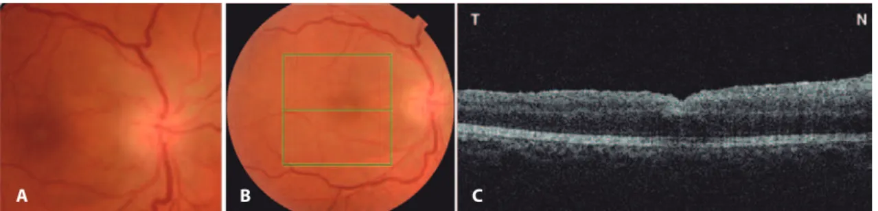
Fundus examination of the right eye revealed an oval and gray inferotemporal optic pit and two choroid colobomas (one was 2-disc size and an anteriorly located one was 8-disc
Anterior segment inflammation usually occurs within 12-48 h after surgery, and the symptoms include decreased visual acuity, increased intraocular pressure, corneal edema,
Cribriform adenoid cystic carcinoma (ACC) is the most common malig- nant epithelial tumor of the lacrimal gland and minor salivary glands; however, its occurrence in the
