Effect of trolox C on cardiac
contracture induced by
hydrogen peroxide
1Laboratório de Fisiologia Cardiovascular, Departamento de Fisiologia, Instituto de Biociências, Universidade Federal do Rio Grande do Sul, Porto Alegre, RS, Brasil
2Departamento de Quimica Analitica y Fisicoquimica, FFB, Universidad de Buenos Aires, Buenos Aires, Argentina A. Belló-Klein1, A.R. Oliveira1,
M.F.S. Miranda1, M.C. Irigoyen1, P.I. Homem-de-Bittencourt Jr.1, S. Llesuy2 and A.A. Belló1
Abstract
Hydrogen peroxide (H2O2) perfused into the aorta of the isolated rat
heart induces a positive inotropic effect, with cardiac arrhythmia such as extrasystolic potentiation or cardiac contractures, depending on the dose. The last effect is similar to the “stone heart” observed in reperfusion injury and may be ascribed to lipoperoxidation (LPO) of the membrane lipids, to protein damage, to reduction of the ATP level, to enzymatic alterations and to cardioactive compounds liberated by LPO. These effects may result in calcium overload of the cardiac fibers and contracture (“stone heart”). Hearts from male Wistar rats (300-350 g) were perfused at 31oC with Tyrode, 0.2 mM trolox C, 256 mM H2O2 or trolox C + H2O2. Cardiac contractures (baseline elevation of
the myograms obtained) were observed when hearts were perfused with H2O2 (Tyrode: 5.9 ± 3.2; H2O2: 60.5 ± 13.9% of the initial value);
perfusion with H2O2 increased the LPO of rat heart homogenates
measured by chemiluminescence (Tyrode: 3,199 ± 259; H2O2: 5,304 ±
133 cps mg protein-1 60 min-1), oxygen uptake (Tyrode: 0.44 ± 0.1;
H2O2: 3.2 ± 0.8 nmol min-1 mg protein-1) and malonaldehyde (TBARS)
formation (Tyrode: 0.12 ± 0; H2O2: 0.37 ± 0.1 nmol/ml). Previous
perfusion with 0.2 mM trolox C reduced the LPO (chemilumines-cence: 4,098 ± 531), oxygen uptake (0.51 ± 0) and TBARS (0.13 ± 0) but did not prevent the H2O2-induced contractures (33.3 ± 16%). ATP
(Tyrode: 2.84 ± 0; H2O2: 0.57 ± 0) and glycogen levels (Tyrode: 0.46
± 0; H2O2: 0.26 ± 0) were reduced by H2O2. Trolox did not prevent
these effects (ATP: 0.84 ± 0 and glycogen: 0.27 ± 0). Trolox C is known to be more effective than α-tocopherol or γ-tocopherol in reducing LPO though it lacks the phytol portion of vitamin E to be fixed to the cell membranes. Trolox C, unlike vitamin A, did not prevent the glycogen reduction induced by H2O2. Trolox C induced a
positive chronotropic effect that resulted in higher energy consump-tion. The reduction of energy level seemed to be more important than LPO in the mechanism of H2O2-induced contracture.
Correspondence
A.A. Belló
Laboratório de Fisiologia Cardiovascular
Departamento de Fisiologia Instituto de Biociências, UFRGS Rua Sarmento Leite, 500 90050-170 Porto Alegre, RS Brasil
Fax: 55 (051) 316-3166 E-mail: [email protected]
Research supported by FINEP, CAPES, CNPq, FAPERGS and PROPESP-UFRGS.
Received July 15, 1996 Accepted August 18, 1997
Key words
•Trolox C
•“Stone heart”
•Free radicals
Introduction
Bianchini and Belló (1) demonstrated a positive inotropic effect with cardiac arrhyth-mia induced by hydrogen peroxide (H2O2) by using whole perfused rat hearts. The in-jection of 0.15 ml of 128 mM H2O2 into the aorta of the isolated rat heart (perfused at constant pressure) induces cardiac contrac-ture (“stone heart”) (2) that is partially an-tagonized by nifedipine (3) or more effi-ciently by 0.1 mM indomethacin (4).
The mechanisms that seem to be respon-sible for these cardiac effects may be attrib-uted to lipoperoxidation (LPO) of membrane lipids (5), to protein damage, to reduction of the ATP level induced by H2O2, to enzy-matic alterations (6) and to cardioactive com-pounds such as prostaglandins and leukotri-enes liberated by LPO (4). These mecha-nisms may result in cytoplasmic calcium overload inducing cardiac contracture.
During the LPO process there is singlet oxygen formation that accelerates this oxi-dation, thus increasing the changes in mem-brane permeability.
Tocopherols are considered to be singlet oxygen quenchers (7,8). Trolox C, the polar portion of vitamin E, is water soluble and is reported to be more active than α- and γ -tocopherols and may interfere with the chain reaction of lipid peroxidation by reacting directly with peroxyl and alkoxyl radicals (9).
The aim of the present study was to test the hypothesis that trolox C can protect the rat heart from the oxidative stress induced by hydrogen peroxide.
Material and Methods
Male Wistar rats (300-350 g) were ob-tained from the Central Animal House of the Universidade Federal do Rio Grande do Sul. The animals were housed in plastic cages (47 x 34 x 18 cm, three animals per cage) lined with sawdust changed every 48 h, in
air-conditioned quarters and had free access to tap water and pelleted food (Purina, Nutripal, Porto Alegre, RS, Brazil).
Animals were killed by a blow to the head and the chest was opened. The heart was carefully dissected from its connections and the aorta retroperfused with Tyrode so-lution of the following composition: 120 mM NaCl, 5.4 mM KCl, 1.8 mM MgCl2, 1.25 mM CaCl2, 27 mM NaHCO3, 2.0 mM NaH2PO4, 1.8 mM Na2SO4 and 11.1 mM glucose, pH = 7.4. This solution was main-tained at 31oC, gassed with O
2:CO2 (95%/ 5%) and used for perfusion at a constant pressure of 8.02 kPa.
Four groups (six hearts each) were stud-ied: a) control: hearts perfused with Tyrode for 30 min; b) trolox: hearts perfused with Tyrode for 10 min and with Tyrode plus trolox C (0.2 mM; Aldrich Chemical Com-pany, Milwaukee, WI) for another 20 min; c) H2O2: hearts perfused with Tyrode for 10 min and with Tyrode plus 256 mM H2O2 (Perhidrol, Merck, Darmstadt, Germany) for another 20 min; d) trolox and H2O2: hearts perfused with Tyrode plus trolox C (0.2 mM) for 10 min and Tyrode plus H2O2 (256 mM) for another 20 min.
The hydrogen peroxide dose was selected according to a previously established dose-concentration curve (2). The trolox dose employed was the same as used by Wu et al. (10).
In order to record the heart beats, the apex of the left ventricle was attached to a microdisplacement myograph transducer by means of a heart clip. Using a lifter, the load was increased until the heart worked at the Lmax level. The coronary flow (drops/min) was recorded using a piezoelectric drop counter. The results obtained during the last minute of the experiment are reported as percent of the values obtained over a period of 1 min measured between the 9th and 10th min after the beginning of perfusion.
of the myograms obtained at 10 min of per-fusion (just before change of the perper-fusion fluid), and 20 min later. Contracture was considered to occur when the relaxation phase was significantly reduced and was deter-mined by measuring the baseline elevation of the myograms and reported as percent of the value measured at 10 min of perfusion. Heart rate was measured from the myograms and the values obtained in the last minute of the experiment are reported as percent varia-tion of the values obtained between the 9th and 10th minute of perfusion.
The transducers were attached to a re-corder. All recording devices were from Narco Bio-System (Houston, Texas).
At the end of the experiments, a slice (±200 mg) obtained from the left ventricle was used for glycogen determination ac-cording to the method of Van Handel (11). Each remaining heart was then homogenized in an ultra-Turrax blender using 1 g of tissue in 5 ml 140 mmol/l potassium chloride con-taining 20 mmol/l phosphate buffer, pH = 7.4. The protein of the homogenate was measured by the method of Lowry et al. (12). The suspension (approximately 1 mg of pro-tein/ml) was added to 3 mmol/l tert-butyl hydroperoxide (Sigma Chemical Company, St. Louis, MO) and assayed for chemilumi-nescence (13) in an LKB Rack Beta liquid scintillation spectrometer model 1215 (LKB-Produkter AB, Bromma, Sweden). Chemilu-minescence is reported as counts per second per mg protein of the homogenate.
Aliquots were used for malonaldehyde determination according to the technique of Buege and Aust (14) for thiobarbituric acid reactive substances (TBARS).
Aliquots were also used for oxygen up-take determination (nmol min-1 mg protein-1) using an oxymeter with a Clark electrode purchased from FQ/IQUIFIB/MADEIC, Buenos Aires, Argentina.
In order to determine the adenosine-5’-triphosphate (ATP) level, the hearts of 16 rats (300-350 g) were perfused (N = 4 per
group) in the same manner as the groups previously reported with Tyrode, 0.2 mM trolox C, 256 mM H2O2 and trolox C fol-lowed by H2O2. After perfusion, the hearts were homogenized and used for enzymatic determination of ATP (Sigma Diagnostics), according to Adams (15).
Results are reported as means ± SEM. Statistical evaluation was performed by two-way ANOVA followed by the Scheffé t-test or the Student t-test for independent samples
using the SPSS-PC software (SPSS, Inc., Chicago, IL). P<0.05 was taken to be signifi-cant.
Results
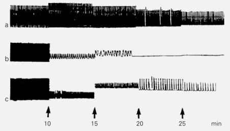
Figure 1 shows typical myograms ob-tained from hearts perfused with the differ-ent solutions. Perfusion with H2O2 induced cardiac contractures, i.e., a 60% decrease in the relaxation phase with respect to the myograms observed at 10 min of perfusion (60.5 ± 13.9). Previous perfusion with 0.2 mM trolox C did not significantly reduce the contracture induced by H2O2 (33.3 ± 16.1) (Fcal = 1.64). A small nonsignificant in-crease in the relaxation phase was demon-strable when hearts were perfused only with
a
b
c
10 15 20 25 min
Figure 1 - Typical records obtained from hearts perfused with trolox C (0.2 mM) (a), H2O2
(256 mM) (b) and trolox C followed by H2O2 (c). The figures show myograms for the last
H2O2 and previous perfusion with trolox C did not inhibit this effect (Table 3).
Discussion
The H2O2 dose used to induce cardiac contractures in the present study was high, far above the possible concentration level that would be observed in the ischemia-reperfusion phenomenon. The low tempera-ture used (31oC) could explain, in part, the high dose employed, but the most important fact is that perfusion was performed at con-stant pressure. H2O2 is so potent a coronary constrictor that it almost completely blocks coronary flow within the first 5 s. To avoid coronary constriction, hearts were perfused at a constant flow. In this condition, contrac-tures similar to those obtained in this study could be induced by doses as low as 0.25 mM. This H2O2 concentration would occur in the ischemia-reperfusion injury (16).
H2O2 can react with iron ions resulting in hydroxyl radical formation. This radical can initiate the LPO of membrane lipids. The lipoperoxide production may change mem-brane permeability leading to the formation of hydrophilic “pores” (5).
The arachidonic acid present in cell mem-branes can be liberated by LPO and form various cardioactive compounds. In particu-lar, it can be converted into prostaglandins or leukotrienes (17). Fatty acids or their oxidation products may act as ionophores. The energy loss and the presence of “pores” result in intracellular calcium overload that blocks muscle relaxation.
The overall results indicate that H2O2 induced LPO and cardiac contracture and that trolox C (0.2 or 2 mM) was effective in preventing the H2O2-induced LPO but not the contractures.
During LPO there is singlet oxygen for-mation that increases this oxidative process (7). Tocopherols and trolox C are known to be singlet oxygen quenchers capable of re-acting with LPO products. In our
observa-Chemiluminescence (cps/mg protein) x 10
3 6
5
4
2 3
1
0
22 44 66
. Trolox ! H2O2 A Trolox + H2O2 Time (min)
0.2 mM trolox C. The experiments were repeated with perfusion with 2 mM trolox C but the results were almost the same, i.e., trolox C did not block the cardiac contractures induced by H2O2 (data not shown).
Figure 2 shows the kinetic profile (77 min) of chemiluminescence initiated by tert-butyl hydroperoxide in homogenates of hearts perfused with 0.2 mM trolox C, 256 mM H2O2 and trolox C followed by H2O2.
Table 1 shows the results obtained by various methods used to study LPO. H2O2 injection increased oxygen uptake, TBARS levels and chemiluminescence of heart ho-mogenates. In the hearts previously perfused with 0.2 mM trolox C, all these phenomena were reduced, indicating LPO reduction.
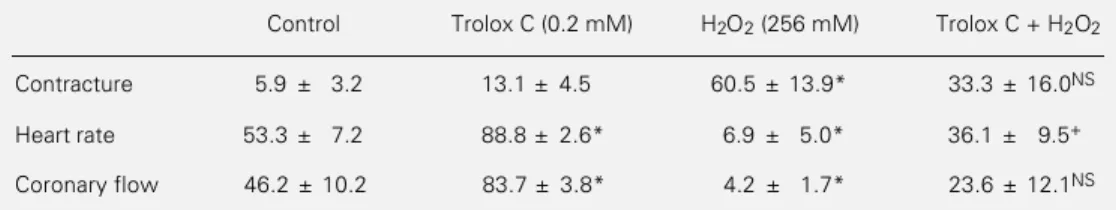
Table 2 shows the contracture, heart rate and coronary flow (% of the initial values) of the different groups. It should be pointed out that trolox C increased the heart rate and coronary flow but did not prevent the contractures and the coronary constriction induced by H2O2.
H2O2 reduced the heart glycogen concen-tration and trolox C did not prevent this reduc-tion (Table 3). The ATP level was reduced by
Figure 2 - Kinetic profile of chemiluminescence initiated by tert-butyl hydroperoxide (counts per second per mg protein) from homogenates of hearts perfused with trolox C (0.2 mM), with hydrogen peroxide (256 mM) or with trolox C followed by hydrogen peroxide.
tion, the reductions in oxygen uptake, TBARS levels and chemiluminescence all suggest that singlet oxygen and/or products of the LPO were quenched or scavenged by trolox C. However, the present study indicates that effective protection by trolox C against H2O2 -induced lipid peroxidation is not associated
per se with effective protection against H2O2 -induced contracture.
Schraufstatter et al. (6) observed that H2O2 can activate a nuclear enzyme, poly (ADP-ribose) polymerase, that uses NAD as sub-strate. The activation reduces the level of ATP necessary for the ionic pumps. H2O2 can also inhibit glycolysis, reducing the ATP stores necessary to maintain the fundamen-tal ionic difference between the two cell membrane sides. Moreover, we observed that glycogen and ATP levels were reduced by H2O2 and trolox C was unable to prevent this reduction. On the other hand, vitamin A was more effective in protecting the heart
Table 1 - Different methods to measure lipoperoxidation.
Data are reported as means ± SEM for 6 hearts in each group. *P<0.05 compared to the respective control (Student t-test); NS, not different from the control group. +P<0.05compared to H
2O2 group (ANOVA, Scheffé
test). The control group was perfused with Tyrode only.
Control H2O2 (256 mM) Trolox C (0.2 mM) Trolox C + H2O2
TBARS (nmol/ml) 0.12 ± 0.02 0.37 ± 0.10* 0.23 ± 0.10NS 0.13 ± 0.03NS+
Oxygen uptake 0.44 ± 0.15 3.21 ± 0.80* 0.44 ± 0.15NS* 0.51 ± 0.07NS+
(nmol min-1 mg protein-1)
Chemiluminescence 3,199 ± 259 5,304 ± 133* 3,324 ± 358NS* 4,098 ± 531NS
(cps mg protein-1 60 min-1)
Table 2 - Contracture, heart rate and coronary flow (% of initial value) of the different groups.
Data are reported as means ± SEM for 6 hearts in each group. *P<0.05 compared to the respective control; NS, not different from the H2O2 group. +P<0.05 compared to H2O2 group (ANOVA, Scheffé t-test).
Control Trolox C (0.2 mM) H2O2 (256 mM) Trolox C + H2O2
Contracture 5.9 ± 3.2 13.1 ± 4.5 60.5 ± 13.9* 33.3 ± 16.0NS
Heart rate 53.3 ± 7.2 88.8 ± 2.6* 6.9 ± 5.0* 36.1 ± 9.5+
Coronary flow 46.2 ± 10.2 83.7 ± 3.8* 4.2 ± 1.7* 23.6 ± 12.1NS
Table 3 - Glycogen content (g%) and ATP values (µmol/g dry tissue) in hearts perfused with differ-ent solutions.
Data are reported as means ± SEM for 4 hearts in each group. *P<0.05 compared to the respective control (Student t-test).
Glycogen ATP
Control 0.46 ± 0.06 2.84 ± 0.05
Trolox C (0.2 mM) 0.18 ± 0.01* 2.27 ± 0.42
H2O2 (256 mM) 0.26 ± 0.01* 0.57 ± 0.44*
Trolox C + H2O2 0.27 ± 0.03* 0.84 ± 0.24*
from the oxidative stress induced by H2O2, by reducing the contractures and LPO and by maintaining the glycogen level (Table 2) (18). The contracture was probably prevented by vitamin A by means of energy conserva-tion.
depression of the contractile function (“stun-ning”). Trolox C, unlike vitamin A, increased the heart rate (18). This may result in greater energy consumption, reducing the energy available for the ionic pumps, leading to an elevation of intracellular calcium. The in-creased intracellular calcium level (that may be responsible for the contracture) can acti-vate calcium-dependent phospholipases with subsequent release of fatty acids from mem-branes and activation of the arachidonic acid cascade. It is known that cardiac contractures can be induced by prostaglandins (19). The calcium overload results in cardiac stiffen-ing known as “stone heart” or cardiac con-tracture.
It can also be seen in Table 2 that trolox C
increased coronary flow and heart rate, which in turn also increased energy consumption. The vasodilation induced by trolox C is a result that merits further investigation.
Trolox C does not have the phytol por-tion of vitamin E that fixes this vitamin to the cell membrane (20). This localization is stra-tegic to protect the cell membrane against lesions induced by oxidative stress. But the reduction of the energy level seemed to be more important than LPO in the mechanism of cardiac H2O2-induced contractures.
Acknowledgments
We thank Tânia Regina Gattelli Fernandes for technical assistance.
References
1. Bianchini A & Belló AA (1988). Hydrogen peroxide effects on the contractile force and coronary flow of the rat isolated heart. Medical Science Research, 16: 1265-1266.
2. Belló AR & Belló AA (1988). Cardiac con-tracture induced by hydrogen peroxide. Medical Science Research, 16: 1149-1150.
3. Belló AR & Belló AA (1989). Effect of nifedipine on cardiac contracture induced by hydrogen peroxide. Medical Science Research, 17: 237-238.
4. Belló AR & Belló AA (1989). Indomethacin antagonizes cardiac contracture induced by hydrogen peroxide. Medical Science Research, 17: 627-628.
5. Blokha VV, Kagan VY, Sitkovskii MV, Danilov VS, Kols OR & Koslov YP (1972). Peroxidation of lipids and conduction of excitation in frog muscles. Biofizika, 17: 549-551.
6. Schraufstatter IU, Hyslop PA, Hinshaw DB, Spaag RG, Sklar LA & Cochrane CG (1986). Hydrogen peroxide induced injury of cells and its prevention by inhibitors of poly (ADP-ribose) polymerase. Proceed-ings of the National Academy of Sciences, USA, 83: 4908-4912.
7. Halliwell B & Gutteridge JMC (1989). Free Radicals in Biology and Medicine. 2nd edn. Clarendon Press, Oxford.
8. Kaiser S, Di Maschio P, Murphy M & Sies H (1990). Physical and chemical scaveng-ing of sscaveng-inglet molecular oxygen by toco-pherols. Archives of Biochemistry and Bi-ophysics, 277: 101-108.
9. Sies H & Murphy ME (1991). Role of toco-pherols in the protection of biological sys-tems against oxidative damage. Journal ofPhotochemistry and Photobiology. B, Biology, 8: 211-224.
10. Wu TW, Hashimoto N, Au Jx, Wu J, Mickle DAG & Carey D (1991). Trolox pro-tects rat hepatocytes against oxyradical damage and the ischemic rat liver from reperfusion injury. Hepatology, 13: 575-580.
11. Van Handel E (1965). Estimation of glyco-gen in small soft tissue. Analytical Bio-chemistry, 11: 256-265.
12. Lowry OH, Rosebrough NJ, Farr AL & Randall RJ (1951). Protein measurement with the Folin phenol reagent. Journal of Biological Chemistry, 193: 265-275. 13. Llesuy SF, Milei J, Gonzalez-Flecha BS &
Boveris A (1990). Myocardial damage in-duced by doxorubicin: hydroperoxide-ini-tiated chemiluminescence and morpholo-gy. Free Radical Biology and Medicine, 8: 259-264.
14. Buege JA & Aust SD (1978). Microsomal lipid peroxidation. Methods in Enzymol-ogy, 52: 302-309.
15. Adams H (1963). Adenosine-5’-triphos-phate determination with phosphoglycer-ate kinase. In: Bergmeyer H (Editor), Methods of Enzymatic Analysis. Academ-ic Press, New York, 539-543.
16. Brunetto AF (1995). Efeitos mecânicos, lipoperoxidação e entrada de Ca2 +
induzidos por peróxido de hidrogênio em miocárdio de rato. Doctoral thesis, Depar-tamento de Fisiologia, Universidade Fed-eral do Rio Grande do Sul, Porto Alegre, Brasil.
17. Basu DK & Karmazyn M (1987). Injury to rat hearts produced by an exogenous free radical generating system. Study into the role of arachidonic acid and eicosanoids. Journal of Pharmacology and Experimen-tal Therapeutics, 242: 673-685.
18. Belló-Klein A, Oliveira AR, Brunetto AF, Irigoyen MC, Llesuy S & Belló AA (1994). Effect of vitamin A on cardiac contracture induced by hydrogen peroxide. Medical Science Research, 22: 411-413. 19. Karmazyn M, Leung CKH & Dhalla NS
(1979). Prostaglandin actions and interac-tions on isolated perfused rat hearts. Ca-nadian Journal of Physiology and Pharma-cology,57: 1275-1282.