Electrophysiological evidence for
the presence of NR2C subunits of
N
-methyl-D-aspartate receptors in rat
neurons of the nucleus
tractus solitarius
Departamento de Fisiologia, Faculdade de Medicina de Ribeirão Preto, Universidade de São Paulo, Ribeirão Preto, SP, Brasil
V. Baptista, W.N. Ogawa, J.F. Aguiar and W.A. Varanda
Abstract
The nucleus tractus solitarius (NTS) plays an important role in the control of autonomic reflex functions. Glutamate, acting on N -methyl-D-aspartate (NMDA) and non-NMDA ionotropic receptors, is the major neurotransmitter in this nucleus, and the relative contribution of each receptor to signal transmission is unclear. We have examined NMDA excitatory postsynaptic currents (NMDA-EPSCs) in the subpostremal NTS using the whole cell patch clamp technique on a transverse brainstem slice preparation. The NMDA-EPSCs were evoked by stimulation of the solitary tract over a range of membrane poten-tials. The NMDA-EPSCs, isolated pharmacologically, presented the characteristic outward rectification and were completely blocked by 50 µM DL-2-amino-5-phosphonopentanoic acid. The I-V relationship of the NMDA response shows that current, with a mean (± SEM) amplitude of -41.2 ± 5.5 pA, is present even at a holding potential of -60 mV, suggesting that the NMDA receptors are weakly blocked by extracellular Mg2+ at near resting membrane potentials. This weak
block can also be inferred from the value of 0.67 ± 0.17 for parameter δ obtained from a fit of the Woodhull equation to the I-V relationship. The maximal inward current measured on the I-V relationship was at -38.7 ± 4.2 mV. The decay phase of the NMDA currents was fitted with one exponential function with a decay time constant of 239 ± 51 and 418 ± 80 ms at a holding potential of -60 and +50 mV, respec-tively, which became slower with depolarization (e-fold per 145 mV). The biophysical properties of the NMDA receptors observed in the present study suggest that these receptors in the NTS contain NR2C subunits and may contribute to the synaptic signal integration. Correspondence
W.A. Varanda
Departamento de Fisiologia FMRP, USP
Av. Bandeirantes, 3900 14049-900 Ribeirão Preto, SP Brasil
Fax: +55-16-633-0017 E-mail: [email protected]
V. Baptista was the recipient of a FAPESP fellowship, W.N. Ogawa was the recipient of a CAPES fellowship and the research of W.A. Varanda was supported by FAPESP.
Received March 3, 2004 Accepted October 1, 2004
Key words
•Nucleustractus solitarius •Electrophysiology •Synapse
•NMDA receptors •NR2C subunits
The nucleus tractus solitarius (NTS) is the primary site for a variety of peripheral sensory inputs including those of cardiovas-cular, respiratory, gustatory, and gastrointes-tinal origins (1). Most of the afferent fibers involved in the control of cardiovascular and respiratory functions terminate around the
visceral afferent fibers (6,7) and that it acts on N-methyl-D-aspartate (NMDA) and non-NMDA ionotropic receptors in second-or-der neurons within the NTS (5,8). The rela-tive role of NMDA and non-NMDA recep-tors in synaptic transmission in the NTS is not well understood. Several lines of evi-dence indicate that non-NMDA receptors play the predominant role in synaptic trans-mission within the NTS (9) while activation of NMDA receptors may modulate the auto-nomic signal transmission by depolarizing second order neurons subjected to high fre-quency or convergent stimulation (5). The specific type of NMDA receptor determines the extent of its involvement in the synaptic transmission process. NMDA receptors con-sist of hetero-oligomers of the NR1, NR2A-NR2D and NR3A, NR3B subunits (10,11). The NR1 subunit, expressed ubiquitously in the central nervous system (CNS), confers the essential functions of the NMDA recep-tors. In contrast, other subunits show more limited expression and confer a functional diversity (11). The NMDA receptor function depends on agonist binding and membrane potential, representing a unique feature among the ligand-gated ion channels. The voltage dependence of the NMDA receptors is mainly due to a voltage-dependent block by extracellular Mg2+ (12,13) and this
prop-erty dominates their physiological role (11-13).
To gain insight into the glutamatergic transmission in the subpostremal NTS, we have analyzed the NMDA postsynaptic cur-rents evoked by solitary tract stimulation. A transverse slice preparation of the medulla oblongata containing the subpostremal NTS was obtained from 30- to 35-day-old Wistar rats of either sex and used in the experi-ments. The animals were anesthetized with Nembutal (50 mg/kg, ip). Following decapi-tation and craniotomy, the brain and upper cervical spinal cord were removed and sub-merged in ice-cold (2-3ºC) artificial cere-brospinal fluid (aCSF), pH 7.35-7.4,
equili-brated with carbogen (95% O2, 5% CO2).
The aCSF contained 122 mM NaCl, 2.5 mM KCl, 1.0 mM MgCl2, 2.0 mM CaCl2, 25 mM
NaHCO3, 1.25 mM NaH2PO4, and 25 mM
glucose, with osmolality of 305-310 mOsm/ kg.H2O, and was continuously gassed with
carbogen. After dissection, the brainstem was glued with cyanoacrylate glue to an L-shaped agar block (4% agar in aCSF) and placed on the stage of a vibrating tissue slicer (MA756, Campden Instruments, Le-icester, England). Two transverse slices (300 µm) containing the area postrema were ob-tained from each animal and incubated in aCSF for 60 min at 32ºC. A single slice was transferred to the recording chamber on the stage of an upright microscope (E600 Nikon Inc., Tokyo, Japan) and held in place with a nylon net mounted on a platinum wire. The chamber was continuously perfused with aCSF at a rate of 2-3 ml/min, driven by gravity. All drugs were applied at known concentrations with the perfusion solution. Experiments were performed at room tem-perature (23-25ºC). Strychnine was pur-chased from Sigma (St. Louis, MO, USA), DL-2-amino-5-phosphonopentanoic acid (DL-AP5), 6,7-dinitroquinoxaline-2,3 dione (DNQX), trans -2-carboxy-5,7-dichloro-4-phenylaminocarbonyl amino-1,2,3,4-tetrahy-droquinoline (L-689-560) and bicuculline methochloride were purchased from Tocris Cookson Inc. (Ellisville, MO, USA). All other salts were purchased from Sigma. Ef-forts were made to minimize the number of animals used and their suffering in accor-dance with the Guide for the Care and Use of Laboratory Animals of the Faculty of Medi-cine of Ribeirão Preto, USP.
Patch pipettes were pulled from borosili-cate glass tubing (Sutter Instrument Co., Novato, CA, USA) on a P-97 puller (Sutter Instrument Co.) and fire polished on a microforge (MF-83; Narishige, Tokyo, Ja-pan). The internal solution was 130 mM CsF, 10 mM NaCl, 1 mM MgCl2, 3 mM
adjusted to 7.3 with CsOH, and osmolality of 295-305 mOsm/kg.H2O. The reference
electrode was an Ag/AgCl wire connected to the extracellular solution via an agar bridge (2.5% in the internal solutions). When filled with the above solution the pipettes had a resistance of 4-8 MΩ. Junction potentials were of the order of 10 mV and these were taken into account in the results shown. Cells were approached by the ‘blind patch’ method and seal resistances in excess of 5 GΩ were obtained prior to entering the whole-cell con-figuration. The average access resistance, was 18.4 ± 0.4 MΩ (N = 40 cells) and was corrected by 70-80%. Recordings were ob-tained with an EPC-7 (List Medical, Darm-stadt, Germany) patch clamp amplifier. Whole-cell currents and voltages were lowpass filtered at 3 kHz (8 pole Bessel filter -LPF8; Warner Instruments Corp., Hamden, CT, USA) digitized at 10 kHz by a computer driven A/D converter (Digidata 1200B; Axon Instruments, Foster City, CA, USA), and stored on the hard disk using the pClamp6 software (Axon Instruments). Data were an-alyzed off-line using the MiniAnalysis pro-gram (Synaptosoft, New Jersey, NJ, USA), Clampfit or Axoscope (Axon Instruments). Synaptic responses of the NTS neurons were evoked by electrical stimulation (15 V, 50100 µs, 0.20.5 Hz, stimulus isolation unit -DS2A - Digitimer Ltd., Garden City, Eng-land) delivered by a twisted pair platinum electrode (100 µm in diameter) positioned on the ipsilateral solitary tract under visual control. The I-V relationship of NMDA re-ceptors-excitatory postsynaptic currents (NMDAR-EPSCs) was fitted by the follow-ing equation:
Eq. 1
where V is the holding potential, VR is the
reversal potential, gmax the maximal
conduc-tance measured at holding potentials between +10 and +50 mV, a represents the
dissocia-tion constant in the absence of transmem-brane voltage, δ is the fraction of the mem-brane voltage at the blocking site and gives the voltage dependence of Mg2+ binding,
and R, T, z, and F have their usual meaning. The decay time constants or the rise time, taken as the time from the onset to the peak of the response of NMDA receptor currents, as a function of membrane potential, was fitted by Equation 2:
Eq. 2
where A and B are constants. The pooled data are reported as the mean ± SEM and statistical significance between values (P < 0.05) was determined by the Student t-test.
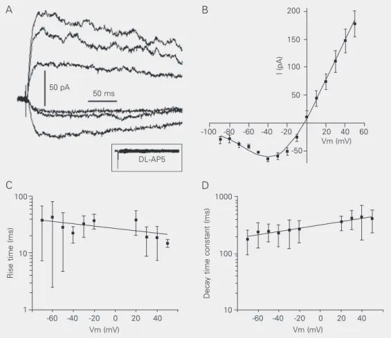
The NMDA-evoked EPSCswere isolated by perfusing the preparation with 5 µM DNQX, 5 µM strychnine and 50 µM bicu-culline to block non-NMDA, glycine and GABAA receptors, respectively (Figure 1A).
-fold per 145 mV.
The I-V relationship of the NMDA re-sponse has a characteristic “J” shape with a region of negative slope conductance for voltages more negative than -40 mV, where Mg2+ block of the NMDA receptor channel
is evident (Figure 1B, N = 5). In order to estimate the sensitivity of the NMDA recep-tor to Mg2+ block, the I-V curves obtained
from 5 subpostremal NTS neurons were fit-ted by Equation 1 (Figure 1B). This equation
assumes a model where Mg2+ ions block the
open channel at a site within the membrane electrical field (16-18). The reversal poten-tial obtained from the fit was -2.1 ± 7.3 mV. The maximal slope conductance
Eq. 3
derived from the linear range of the I-V relationships (from +10 to +50 mV) was 3.4
Rise time (ms)
100
10
1
Decay time constant (ms)
1000
100
10 50 pA
50 ms
DL-AP5
I (pA)
200
150
100
50
-100 -80 -60 -40 -20 20 40 60 Vm (mV) -50
-60 -40 -20 0 20 40 -60 -40 -20 0 20 40
Vm (mV) Vm (mV)
Figure 1. N-methyl-D-aspartate (NMDA) receptor-mediated currents recorded from secondary neurons of the nucleus tractus solitarius of the rat. A, Pharmacological isolation of the NMDA excitatory postsynaptic currents by perfusion with bicuculline (50 µM), strychnine (5 µM) and DNQX (5 µM). Holding potentials varied from -90 to +50 mV in 10-mV steps. The traces shown correspond to the following voltages: -70 (lowest trace), -30, 0, +20, +40, and +50 mV (uppermost traces). Addition of DL-2-amino-5-phosphonopentanoic acid (DL-AP5, 50 µM) to the bath completely abolished the synaptic currents (inset). B, I-V relationship of the NMDA responses showing the characteristic “J” shape, with a region of negative slope conductance. The reversal potential estimated from the fit was -2.1 ± 7.3 mV and peak inward current occurred near -40 mV (-38.7 ± 4.2 mV). C, Plots of excitatory postsynaptic current rise time against the membrane potential. Note that the rise time tends to be faster with depolarization. D, Plot of the decay time constant against the membrane potential. The decay time constants are dependent on membrane voltage, being prolonged by depolarization in an exponential manner (increasing e-fold per 145 mV). The average at -70 mV differed significantly from that at +50 mV (P < 0.05, Student t-test).
A B
± 0.6 nS (N = 5). The parameter δ in Equa-tion 1 gives the fracEqua-tion of membrane poten-tial acting at the binding site or, in other words, the voltage dependence of the Mg2+
block. We found a value of δ equal to 0.7 ± 0.2 (N = 5), and a value of a equal to 10 ± 4 mM (N = 5) suggesting a weak Mg2+ block
and that the Mg2+ blocking site is about 70%
of the way across the membrane electrical field from the outside (but not necessarily 70% of the distance across the membrane, if the electrical field is not constant) (16). With the average values of a and δ obtained from the fitting with Equation 1, the KD for Mg2+
in the NMDA receptor can be calculated by the following equation (16):
KD = a*eV
δzF/RT Eq. 4
We found a KD equal to 299 ± 37 µM (N = 5)
at a holding potential of -60 mV, a value relatively high, confirming the suggestion of a weak block of the NMDA receptors by Mg2+. It has been shown (19) that NMDA
receptors containing NR2A or NR2B sub-units have a much steeper voltage-depend-ence (δ ~1) and pass maximal inward current at voltages close to -25 mV. On the other hand, receptors containing NR2C or NR2D subunits show a weaker voltage-dependence (δ ~0.7) and pass maximal inward current at about -35 mV. The maximal inward current obtained from our I-V curve occurred at -38.7 ± 4.2 mV. Thus, these two parameters, δ and the voltage giving maximal inward
current, suggest that the subpostremal NTS neurons contain subunits of the NR2C or NR2D type. The current response of NMDA receptors containing the NR2D subunit de-cays very slowly, with time constants of the order of 1 s; NMDA receptors made up of NR2A subunits have fast kinetics, with de-cay time constants around 50 ms, and those containing either the NR2B or NR2C
sub-unit have decay time constants around 250-280 ms (20). In the present study, the decay phase of NMDA current was fitted with a single exponential function, with decay time constants of 239 ± 50 ms at a holding poten-tial of -60 mV, and of 418 ± 80 ms at +50 mV. Taken together, the values of δ, the voltage at which the inward current is maxi-mal and the decay time constants, suggest the presence of NMDA receptors containing NR2C subunits in the subpostremal NTS. Because the intensity of NMDA receptor block by Mg2+ is relatively weak, this
References
1. Beckstead RM & Norgren R (1979). An autoradiographic examina-tion of the central distribuexamina-tion of the trigeminal, facial, glossopha-ryngeal and vagal nerves in the monkey. Journal of Comparative Neurology, 184: 455-472.
2. Loewy AD (1990). Central autonomic pathway. In: Loewy AD & Spyer KM (Editors), Central Regulation of Autonomic Functions. Oxford, New York.
3. Barraco R, El-Ridi M, Ergene E, Parizon M & Bradley D (1992). An atlas of the rat subpostremal nucleus tractus solitarius. Brain Re-search Bulletin, 29: 703-765.
4. Grabauskas G & Bradley RM (2003). Frequency-dependent proper-ties of inhibitory synapses in the rostral nucleus of the solitary tract.
Journal of Neurophysiology, 89: 199-211.
5. Bonham AC & Chen C (2002). Glutamatergic neural transmission in the nucleus tractus solitarius: N-methyl-D-aspartate receptors. Clini-cal and Experimental Pharmacology and Physiology, 29: 497-502. 6. Doyle MW & Andresen MC (2001). Reliability of monosynaptic
sensory transmission in brain stem neurons in vitro. Journal of Neurophysioloy, 85: 2213-2223.
7. Talman WT, Perrene MH & Reis DJ (1980). Evidence for L-gluta-mate as the neurotransmitter of baroreceptor afferent nerve fibers.
Science, 209: 813-815.
8. Aylwin ML, Horowitz JM & Bonham AC (1997). NMDA receptors contribute to primary cisceral afferent transmission in the nucleus of the solitary tract. Journal of Neurophysiology, 77: 2539-2548. 9. Yen JW, Chan JYH & Chan SHH (1999). Differential roles of NMDA
and non-NMDA receptors in synaptic responses of neurons in nucleus tractus solitarius of the rat. Journal of Neurophysiology, 81: 3034-3043.
10. Nishi M, Hinds H, Lu H, Kawata M & Hayashi Y (2001). Motoneuron specific expression of NR3B, a novel NMDA-type glutamate
recep-tor subunit that works in a dominant-negative manner. Journal of Neuroscience, 21: 1-6.
11. Dingledine R, Borges K, Bowie D & Traynelis SF (1999). The gluta-mate receptor ion channels. Pharmacological Reviews, 51: 7-60. 12. Nowak L, Bregestovski P, Ascher P, Herbert A & Prochiantz A
(1984). Magnesium gates glutamate-activated channels in mouse central neurones. Nature, 307: 462-465.
13. Mayer ML, Westbrook GL & Guthrie PB (1984). Voltage-dependent block by Mg2+ of NMDA responses in spinal cord neurones. Nature,
309: 261-263.
14. Anchisi D, Scelfo B & Tempia F (2001). Postsynaptic currents in deep cerebellar nuclei. Journal of Neurophysiology, 85: 323-331. 15. O’Brien JA, Isaacson JS & Berger AJ (1997). NMDA and non-NMDA
receptors are co-localized at excitatory synapses of rat hypoglossal motoneurons. Neuroscience Letters, 227: 5-8.
16. Woodhull AM (1973). Ionic blockage of sodium channels in nerve.
Journal of General Physiology, 61: 687-708.
17. Neher E & Steinbach JH (1978). Local anaesthetics transiently block currents through single acetylcholine-receptor channels. Journal of Physiology, 277: 153-176.
18. Acher P & Nowak L (1988). The role of divalent cations in the N-methyl-D-aspartate responses of mouse central neurones in cul-ture. Journal of Physiology, 399: 247-266.
19. Kuner T & Schoepfer S (1996). Multiple structural elements deter-mine subunit specificity of Mg2+ block in NMDA receptor channels.
Journal of Neuroscience, 16: 3549-3558.