Rev Bras Ter Intensiva. 2009; 21(4):461-464
Tumoral pulmonary mass secondary to
Schistosoma mansoni
infection resembling
neoplasia: case report
Massa tumoral secundária a infecção por Schistosoma mansoni
simulando neoplasia de pulmão: relato de caso
CASE REPORT
INTRODUCTION
Patients with chronic schistosomiasis may have pulmonary involvement featuring a variable range symptoms and radiological indings.(1) he lungs
may be involved due to anomalous eggs migration via portal system into the pulmonary artery system (via porto-systemic anastomosis) and less common-ly by adult worms migration. here are extensive parenchyma involvement cases, as well as others with predominant arteritis, with pulmonary hyperten-sion and cor pulmonale.
Lung parenchyma involvement is mainly characterized by granu-lomatous reaction to Schistosoma mansoni eggs. The granulomas have no preferential location, and may be found in all lung segments and pleura. Inter-alveolar thickening and connective tissue fibrosis were also reported.(2) Lesions from dead worms are rarer, and characterized by
ne-crotic areas surrounded by intensive exudation, usually reabsorbed and involved by cicatricial tissue.(3,4)
A case involving a young female patient with atypical pulmonary schisto-somiasis faking neoplasia is presented. Aspects regarding diferential diagnosis are discussed.
Cláudio Dornas de Oliveira1,
Glaucio de Oliveira Nangino2,
Paulo César Correia3, Carlos
Vechio Salomão3, Marcelo Alencar
Resende4, Leonardo da Cruz
Peixoto4, Maurício Buzelini Nunes5
1. Physician of Santa Casa de Belo Horizonte – Belo Horizonte (MG), Brazil.
2. Physician of Santa Casa de Belo Horizonte – Belo Horizonte (MG), Brazil.
3. Physician of Santa Casa de Belo Horizonte – Belo Horizonte (MG), Brazil.
4. Physician of Santa Casa de Belo Horizonte – Belo Horizonte (MG), Brazil.
5. Physician of Santa Casa de Belo Horizonte – Belo Horizonte (MG), Brazil.
ABSTRACT
Patients with chronic Schisto-soma mansoni infection may feature a range of pulmonary symptoms and radiological findings. Eggs, and rare-ly adult worms, may passiverare-ly enter the pulmonary circulation, usually via the portal system, where they may cause pulmonary inflammation, fi-brosis, hypertension and cor pulmo-nale. A 25-year-old patient who lived in a schistosomiasis endemic area with a pulmonary mass suggestive of malignancy underwent exploratory thoracotomy. The mass was adher-ent, with no resection possibility. The lung-biopsy specimen
evalua-tion showed several granulomas with
Schistosoma mansoni eggs and
hy-perplasic connective tissue with no sign of malignancy. The patient had respiratory failure and hypotension immediately post-surgery. Specific treatment (praziquantel) and pred-nisone were given. The patient had pneumonia and septic shock. The patient was given antibiotics, vaso-pressors, mechanical ventilation and hemodialysis with no improvement, and subsequently died 28 days after the surgery.
Keywords: Schistosomiasis; Schistosoma mansoni; Neoplasias/secondary; Lung diseases, parasitic; Case reports
Received from Santa Casa de Belo Horizonte – Belo Horizonte (MG), Brazil.
Submitted on September 7, 2009 Accepted on November 28, 2009
Author for correspondence:
Cláudio Dornas de Oliveira Santa Casa de Belo Horizonte- CTI adulto, 2º andar
Av. Francisco Sales, 1111- Santa Eigênia
CEP: 31150-221 - Belo Horizonte (MG), Brazil.
462 Oliveira CD, Nangino GO, Correia PC, Salomão CV, Resende MA, Peixoto LC et al.
Rev Bras Ter Intensiva. 2009; 21(4):461-464
CASE REPORT
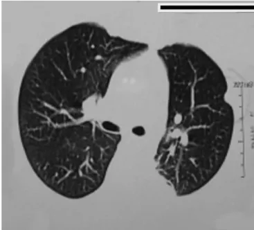
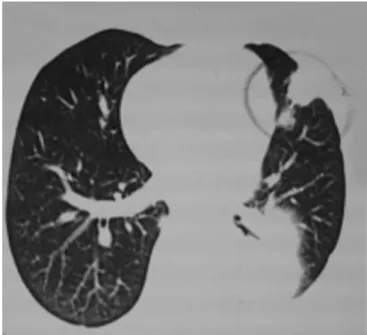
his was a 25 years old female patient who lived in endemic schistosomiasis area and was referred to a tho-rax surgery service with dysphagia, weight loss and exer-tion dyspnea. here was no relevant history of previous diseases or comorbities. he physical examination evi-denced severe malnutrition, asymmetrical thorax expan-sion and bronchial sounds at the lower left hemithorax. Additional tests showed: chest X-ray: heterogeneous hypo-transparency at lower left lobe (Figure 1); radio-graphic exam with contrast and upper digestive endos-copy: signs of esophageal lower third extrinsic compres-sion; chest CT: heterogeneous mass taking the upper the left lower lobe basal lateral and basal posterior areas, with adjacent pleural thickening (Figure 2). Presence of two similar images, but smaller, on the upper lingula segment and anterior left upper lobe (Figures 3 and 4); increased left liver lobe (non-oriented examination); spirometry: moderate restrictive respiratory disorder; bronchoscopy: extrinsic left basal bronchia compression; bronchial bi-opsy: non-speciic bronchitis.
Surgical approach was decided. Enteral nutrition support was started, as well as preoperative evaluations. he patient underwent exploratory thoracotomy. A large mass was found, invading the parietal pleura, left lung hilum, aorta, diaphragm and left atrium, with no resec-tion possibility. A biopsy was performed and the patient was referred for post-operative follow-up at the intensive care unit. She required mechanical ventilation and va-soactive amines from the admission. She coursed with severe restrictive ventilatory disorder, and refractory
hypoxemia. he histopathology revealed thickened and ibrosed pleura, pulmonary parenchyma with ibrosis and several granulomas containing Schstosoma mansoni eggs; no lesions suggesting neoplasia were found (Figure 5). he patient underwent praziquantel 50 mg/kg single dose treatment plus prednisone 1 mg/kg/day. Echo-Dop-pler and the Swan-Ganz catheter evidenced pulmonary hypertension. Lower limbs duplex scan was negative for venous thromboembolism.
Subsequently she developed pulmonary infection and
Figure 1 – Posterior-anterior chest X-ray: heterogeneous hypo-transparency at left lower lobe.
Figure 2 – Chest CT: heterogeneous mass at left lower lobe basal posterior part.
Tumoral pulmonary mass secondary to Schistosoma mansoni 463
Rev Bras Ter Intensiva. 2009; 21(4):461-464 septic shock. Antibiotics, vasoactive amines, ventilatory
support and hemodyalisis, were given with no improve-ment. Subsequently she died, 28 days after the surgery.
DISCUSSION
From a radiological point of view, chronic schisto-somiasis pulmonary parenchyma changes are commonly described as: difused iniltrate, focal opacities and mi-cro-nodules.(5) Pulmonary nodes secondary to
schisto-somiasis are rare, and pose a diferential diagnosis with Figure 5 – Pathology – fragment of lung: schistosomotic granuloma, with inlammatory iniltrate surrounding the lesion, and connective hyperplasia.
Figure 4 – Chest CT: nodular image at left upper lobe upper portion, adjacent to the pleura.
lung neoplasia, and are frequently deined only after exploratory thoracotomy.(6-8) he term ‘pseudotumoral
schistosomiasis presentation’ was irst used in 1975 de-scribing a pulmonary node in a patient who died, being a autopsy indings granulomatous reaction, Schistosoma mansoni eggs, and pulmonary arterioles obliteration.(9)
The presence of a large mass associated to Schis-tosoma infection (bilharziasis) was described in 1953 in Cairo.(10) The gross presentation during the
sur-gery suggested lung neoplasia, requiring pneumec-tomy. Similarly, the histology showed fibrotic and thickened pleural tissue with cicatricial granulomas, fibrosed lung tissue, Schistosoma eggs surrounded by histiocytes, eosinophils and fibroblasts. The authors pointed out that, as first hypothesis, the lung mass producing these pathology findings was secondary to schistosomiasis. The schistosomiasis pseudo-neoplasic forms represent anomalous response to the parasite eggs. Hyperplastic connective tissue formations de-velop around the granulomatous formations, as a host reaction.
It is proposed that, in schistosomiasis endemic areas, pulmonary schistosomiasis is considered a diferential diagnosis for complex structures, as pulmonary masses.
RESUMO
Indivíduos infectados com Schistosoma mansoni na fase crônica da doença podem apresentar comprometimento pul-monar com sintomatologia e alterações radiológicas variáveis. Os pulmões podem ser acometidos pela migração anômala de ovos do sistema porta para o sistema arterial pulmonar (atra-vés de anastomoses porto-sistêmicas) e menos comumente por migrações ectópicas de vermes adultos. Há casos com extenso comprometimento parenquimatoso e outros com predomínio de arterites, com hipertensão pulmonar e cor pulmonale. Pa-ciente jovem, residente em área endêmica de esquistossomo-se, com massa pulmonar sugestiva de neoplasia foi submeti-da a toracotomia exploradora sem possibilisubmeti-dade de ressecção da massa. Exame histopatológico mostrou vários granulomas esquistossomóticos e hiperplasia do tecido conjuntivo, sem sinais de neoplasia. Evoluiu com insuiciência respiratória e instabilidade hemodinâmica no pós-operatório imediato. Re-cebeu tratamento especíico (praziquantel) associado a pred-nisona. A paciente cursou com infecção pulmonar e choque séptico. Recebeu antibioticoterapia, aminas vasoativas, suporte ventilatório e tratamento hemodiálitico sem melhora. Evoluiu para óbito 28 dias após cirurgia.
464 Oliveira CD, Nangino GO, Correia PC, Salomão CV, Resende MA, Peixoto LC et al.
Rev Bras Ter Intensiva. 2009; 21(4):461-464
REFERENCES
1. Schwartz E. Pulmonary schistosomiasis. Clin Chest Med. 2002;23(2):433-43. Review.
2. Bogliolo L. [Schistosomiasis mansoni. Pathology]. Rev Bras Malariol Doencas Trop. 1959;11:359-424. Portuguese. 3. Barbato EC. [Schistosomal pneumopathy and chronic cor
pulmonale]. Arq Bras Cardiol. 1953;6(3):195-305. Portu-guese.
4. Sami AA. Pulmonary manifestations of schistosomiasis. Dis Chest. 1951;19(6):698-705.
5. Rocha RL, Pedroso ERP, Rocha MOC, Lambertucci JR, Greco DB, Ferreira CE. Forma pulmonar crônica da es-quistossomose mansoni: avaliação clínico-radiológica. Rev Soc Bras Med Trop. 1990;23(2):83-9.
6. Fatureto MC, Correia D, Silva MBO, Barra MFC, Silva
AV, Tarquinio DC, et al. Nódulo pulmonar esquistosso-mótico simulando neoplasia: relato de caso. Rev Soc Bras Med Trop. 2003;36(6):735-7.
7. Lambertucci JR, Silva LCS, Queiroz LC. Pulmonary nodu-les and pleural efusion in the acute phase of schistosomiasis
mansoni. Rev Soc Bras Med Trop. 2007;40(3):374-5.
8. Ryan ET, Aquino SL, Kradin RL. Case records of the Mas-sachusetts General Hospital. Case 29-2007. A 51-year-old man with gastric cancer and lung nodules. N Engl J Med. 2007;357(12):1239-46.
9. Akoun G, Huchon G, Rolland J, Barrière L, Marsac J, Ronco P, et al. [Letter: Pseudotumoral pulmonary schis-tosomiasis due to Schistosoma mansoni]. Nouv Presse Med. 1975;4(33):2408. French.