Parotid Incidentaloma Identi
fi
ed by Positron
Emission/Computed Tomography: When to
Consider Diagnoses Other than Warthin Tumor
Carolina Bothe
1Alejandro Fernandez
2Jacinto Garcia
1Montserrat Lopez
1Xavier León
1Miquel Quer
1Joan Lop
11Department of Otorhinolaryngology, Hospital Sant Pau, Barcelona, Spain
2Department of Nuclear Medicine, Hospital de Sant Pau, Barcelona, Barcelona, Spain
Int Arch Otorhinolaryngol 2015;19:112–115.
Address for correspondence Carolina Bothe, MD, Department of Otorhinolaryngology, Hospital Sant Pau, Avda. Sant Antoni Mª Claret, 167, Barcelona 08025, Spain (e-mail: [email protected]).
Introduction
It is not unusual tofind unexpected hypermetabolic foci as a result of scanning using whole-body positron emission/com-puted tomography (PET/CT). Known asincidentalomas, such
findings are not necessarily related to the tumor or disease being studied. Parotid gland incidentalomas (PGIs) are defined
as new focal intraglandular deposits of radiotracer in patients without prior history of parotid disease.1These deposits are most commonly due to benign lesions such as Warthin tumor (WT),1–5 but they can also be caused by metastatic lymph
nodes or malignant tumors.6
The parotid gland is the salivary gland that most commonly develops tumors, most of which are benign. After pleomorphic Keywords
►
parotid gland
►
adenolymphoma
►
PET scan
►
cigarette smoking
Abstract
Introduction
Parotid gland incidentalomas (PGIs) are unexpected hypermetabolic foci
in the parotid region that can be found when scanning with whole-body positron
emission/computed tomography (PET/CT). These deposits are most commonly due to
benign lesions such as Warthin tumor.
Objective
The aim of this study was to determine the prevalence of PGIs identi
fi
ed in
PET/CT scans and to assess the role of smoking in their etiology.
Methods
We retrospectively reviewed all PET/CT scans performed at our center in
search of PGIs and identi
fi
ed smoking status and standardized uptake value (SUVmax) in
each case. We also analyzed the database of parotidectomies performed in our
department in the previous 10 years and focused on the pathologic diagnosis and
the presence or absence of smoking in each case.
Results
Sixteen cases of PGIs were found in 4,250 PET/CT scans, accounting for 0.4%.
The average SUVmax was 6.5 (range 2.8 to 16). Cytology was performed in
fi
ve patients;
it was benign in four cases and inconclusive in one case. Thirteen patients had a history
of smoking. Of the parotidectomies performed in our center with a diagnosis of Warthin
tumor, we identi
fi
ed a history of smoking in 93.8% of those patients.
Conclusions
The prevalence of PGIs on PET/CT was similar to that reported by other
authors. Warthin tumor is frequently diagnosed among PGIs on PET/CT, and it has a
strong relationship with smoking. We suggest that a diagnosis other than Warthin
tumor should be considered for PGIs in nonsmokers.
received August 22, 2014 accepted
November 14, 2014 published online December 29, 2014
DOI http://dx.doi.org/ 10.1055/s-0034-1397334. ISSN 1809-9777.
Copyright © 2015 by Thieme Publicações Ltda, Rio de Janeiro, Brazil
Original Research
adenoma, WT is the second most prevalent parotid tumor. It presents as a slow-growing mass in the tail of the gland, and is usually asymptomatic. It generally occurs between the sixth and seventh decades of life, predominantly in men. It has a strong association with smoking, which is therefore consid-ered a risk factor for its development.7–10WT can be
multi-centric or bilateral, and it rarely becomes malignant.
To our knowledge, only two previous studies have re-ported the prevalence of PGI, and both were performed in Asia.2,11 Furthermore, clinical features such as the role of smoking have not been considered in their etiology. The present study aimed to determine the prevalence of parotid incidentalomas in18F-fluorodeoxyglucose (18F-FDG) PET/CT and evaluate the presence of smoking in this group of patients. We also reviewed the database of parotidectomies performed in our hospital to assess the relationship between WT and smoking.
Materials and Methods
Patients
We conducted a retrospective review of the reports of PET/CT performed in 4,250 patients in our hospital between June 2009 and February 2013. We identified 16 cases of parotid focal uptake in patients without known disease of the parotid glands. The group consisted of 10 men and 6 women, 48 to 88 years of age. In most cases, PET-CT was indicated as an extension study of diagnosed neoplasms or to confirm locoregional relapse after treatment. In two cases, it was indicated to rule out giant cell arteritis.
We reviewed the history of smoking in the 16 cases: 13 had smoked or were current smokers. We identified additional studies performed on the parotid masses,finding that only 5 patients underwent cytologic analysis byfine needle aspira-tion cytology (FNAC). In 8 of the 16 cases, the lesion was located in the right parotid gland, in 7 cases it was located on the left, and in 1 case it was bilateral. Two patients had more than one deposit within the same gland.
We analyzed the database of parotidectomies performed in our department in the previous 10 years and focused on the pathologic diagnosis and the presence or absence of smoking in each case.
18F-Fluorodeoxyglucose Positron Emission/Computed
Tomography
Combined 18F-FDG whole-body PET/CT (Gemini TF, Philips, Amesterdam) with 64-slice CT was performed. Standard patient preparation included: 6-hour fasting, hydration, and serum glucose level of less than 150 mg/dL before tracer injection. Patients were asked to rest quietly in supine position, and vesical evacuation was done before the acquisition of images.
A dose of 3.7 MBq/kg of 18F-FDG was intravenously in-jected. Low-dose CT and PET from the base of the skull to the proximal thighs were performed, with an additional acquisi-tion of head and neck images. The total examinaacquisi-tion time was
20 minutes. An abnormal PET-CTfinding was defined as a
significant increased uptake, higher than that of the sur-rounding normal tissue.
Results
We found PGIs in 16 patients who underwent PET/CT between June 2009 and February 2013, corresponding to 0.4% of patients undergoing PET/CT. The incidentalomas were more frequent in men (62.5%, 10 cases), and the average age at diagnosis was 68 years.
The most common indication for the test was lung cancer (7 cases, corresponding to 43.75%), followed by breast cancer (3 cases), giant cell vasculitis (1 case each of Horton disease and Takayasu arteritis), and 1 case each of locally advanced oropharyngeal cancer, gallbladder carcinoma with liver me-tastasis, peritoneal carcinomatosis of gynecologic origin, and retroperitoneal lymphoma. (See►Table 1.)
Five of the 16 patients underwent FNAC of the parotid mass. Three offive masses had an inflammatory component without a malignant component, one was considered a mixed tumor suggestive of pleomorphic adenoma, and the other was inconclusive. Of the 16 patients, only the patient with the pleomorphic adenoma underwent surgery; a superficial pa-rotidectomy was performed and the histology was confirmed. In the remaining 11 cases, FNAC was not performed but the mass was monitored by periodic clinical follow-up. Thirteen patients (81.25%) had a history of smoking. One of the three nonsmokers was the patient with the pleomorphic adenoma. The average standardized uptake value of parotid tumors was 6.51, ranging from 2.87 to 16.1. The term used to describe the lesions washyperintenseorhypermetabolic intraglandular nodule.
A review of the database of parotidectomies performed in our department revealed that 311 patients were surgically treated for parotid gland tumors in the previous 10 years and 80 cases (25.7%) were identified as WT. This was the second most frequent tumor after pleomorphic adenoma. A history of smoking was identified in 93.8% of patients with WT (75 cases). This percentage was high compared with the prevalence of smoking in pleomorphic adenomas, which was 38.8%. (See►Table 2.)
Discussion
Ourfindings show that the prevalence of PGIs on PET/CT in our institution is similar to that reported in the two previous studies in Asia. When analyzing the presence of cigarette smoking in our patients with PGIs, we found it was a common factor in 81.25% of the patients.
In our study, FNAC was performed infive cases. Three were negative for malignancy without typing the lesion. One case was a pleomorphic adenoma and the other had a nondiag-nostic smear. The remaining patients did not undergo addi-tional testing on the PGI due to the advanced stage of the primary tumor, the absence of symptoms, or the unlikelihood of metastasis based on clinical and tomographic features.
In the largest series published to date, Wang et al found 58 PGIs in 19,333 patients, representing a prevalence of 0.3%.11 They obtained histologic confirmation in 51 patients, 41 with benign lesions (mainly represented by pleomorphic adeno-mas and WT) and 10 with malignant tumors. In a smaller
sample, Horiuchi et al detected four cases of abnormal parotid FDG uptake in 1,872 PET scans, with an estimated prevalence of 0.2%; all four cases were diagnosed with WT.2Other series of cases also found WT as the main cause of PGI.1,3
As PGIs are likely benign lesions, metastasic disease should not be suspected initially without histopathologic confirmation, even in patients with known malignancy elsewhere.1,4,5Nevertheless, these lesions warrant further radiologic and histopathologic correlations. Ultrasonogra-phy and FNAC are useful tools to characterize parotid masses, avoiding erroneous staging of primary malignan-cies. The objective of cytologic diagnosis of parotid masses is the differential diagnosis between benign and malignant lesions.12 Complex histopathology and heterogeneity of cellular patterns of salivary gland tumors explain why typing of primary salivary malignancies by FNAC is a challenge. The accuracy of FNAC depends on the experience of the clinician who performs the procedure and the pathologist who evaluates cytologic material.12
We found the prevalence of smoking in parotid tumors surgically treated in our hospital was significantly higher in patients with WT than in patients with other tumors (p<0.001). More than 90% of patients with WT were smokers, and cigarette use in patients with other benign and malignant neoplasms was lower, 38 and 55.4%, respec-tively. Several other authors have studied the relation between WT and smoking. According to their publications, between 80 and 94% of patients with WT smoke,7–9,13and
the risk for development of WT correlates with the level and duration of smoking.7,9Sadetzki et al found a remarkably high odds ratio (15.3) for developing WT in the presence of smoking.9
Conclusion
Based on the high prevalence of WT as PGI on PET/CT and the strong relationship with smoking, we suggest diagnosis other than Warthin tumor should be considered for PGI in
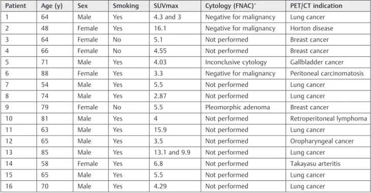
Table1 Data of 16 patients with parotid incidentalomas detected by PET/CT
Patient Age (y) Sex Smoking SUVmax Cytology (FNAC) PET/CT indication
1 64 Male Yes 4.3 and 3 Negative for malignancy Lung cancer
2 48 Female Yes 16.1 Negative for malignancy Horton disease
3 64 Female No 5.1 Not performed Breast cancer
4 66 Female No 4.55 Not performed Breast cancer
5 71 Male Yes 4.03 Inconclusive cytology Gallbladder cancer
6 88 Female Yes 3.3 Negative for malignancy Peritoneal carcinomatosis
7 54 Male Yes 5.5 Not performed Lung cancer
8 74 Male Yes 2.87 Not performed Lung cancer
9 79 Female No 5.5 Pleomorphic adenoma Breast cancer
10 81 Male Yes 4 Not performed Retroperitoneal lymphoma
11 63 Male Yes 15.9 Not performed Lung cancer
12 65 Male Yes 3.5 Not performed Oropharyngeal cancer
13 85 Male Yes 13.1 and 9.9 Not performed Lung cancer
14 58 Female Yes 6.8 Not performed Takayasu arteritis
15 65 Male Yes 5.5 Not performed Lung cancer
16 70 Male Yes 4.29 Not performed Lung cancer
Abbreviations: FNAC,fine needle aspiration cytology; PET/CT; positron emission/computed tomography; SUVmax, standardized uptake value.
Table 2 Data of parotidectomies performed in our department in the previous 10 years
Histologic diagnosis Number of cases (%) Smoking status (%)
Pleomorphic adenoma 134 (43.1%) 52 (38.8%)
Warthin tumor 80 (25.7%) 75 (93.8%)
Malignant tumors 56 (18%) 31 (55.4%)
Other diagnosis 41 (13.2%) 17 (41.5%)
Total 311 (100%) 175 (56.3%)
International Archives of Otorhinolaryngology Vol. 19 No. 2/2015
nonsmokers. Further studies with histologic diagnosis and larger samples are warranted to confirm our hypothesis.
References
1 Lee SK, Rho BH, Won KS. Parotid incidentaloma identified by com-bined 18F-fluorodeoxyglucose whole-body positron emission tomog-raphy and computed tomogtomog-raphy:findings at grayscale and power Doppler ultrasonography and ultrasound-guidedfine-needle aspira-tion biopsy or core-needle biopsy. Eur Radiol 2009;19(9):2268–2274
2 Horiuchi M, Yasuda S, Shohtsu A, Ide M. Four cases of Warthin’s tumor of the parotid gland detected with FDG PET. Ann Nucl Med 1998;12(1):47–50
3 Klijanienko J, Petras S, De Bosschere L, Paulmier B, Le Tourneau C, Rodriguez J. False-positive FDG PET/CT uptake in Warthin tumor in head and neck oncological patients confirmed by afine needle aspiration. Diagn Cytopathol 2012;40(3):282–284
4 Enomoto A, Nakahara H, Uchihashi T, Tsuji H, Hamada S. Fluo-rodeoxyglucose-positive Warthin tumor in a neck node mimick-ing metastasis in primary intraosseous left posterior mandibular cancer staging with positron emission tomography/computed tomography. J Oral Maxillofac Surg 2011;69(7):2052–2054
5 Iwai T, Baba J, Shibasaki M, et al. 18F-fluorodeoxyglucose-positive Warthin tumor in a contralateral cervical lymph node mimicking
metastasis in tongue cancer staging with PET/CT. J Craniofac Surg 2012;23(5):e507–e509
6 Coronado Poggio M, Couto Caro RM, Rodado Marina S, Martín Curto LM. 18F-FDG PET/TAC Semiology. Rev Esp Med Nucl 2008; 27(4):284–304, quiz 305–306
7 Peter Klussmann J, Wittekindt C, Florian Preuss S, Al Attab A, Schroeder U, Guntinas-Lichius O. High risk for bilateral Warthin tumor in heavy smokers—review of 185 cases. Acta Otolaryngol 2006;126(11):1213–1217
8 Pinkston JA, Cole P. Cigarette smoking and Warthin’s tumor. Am J Epidemiol 1996;144(2):183–187
9 Sadetzki S, Oberman B, Mandelzweig L, et al. Smoking and risk of parotid gland tumors: a nationwide case-control study. Cancer 2008;112(9):1974–1982
10 Reddy VM, Thangarajah T, Castellanos-Arango F, Panarese A. Conservative management of Warthin tumour. J Otolaryngol Head Neck Surg 2008;37(5):744–749
11 Wang HC, Zuo CT, Hua FC, et al. Efficacy of conventional whole-body 18 F-FDG PET/CT in the incidentalfindings of parotid masses. Ann Nucl Med 2010;24(8):571–577
12 Zbären P, Schär C, Hotz MA, Loosli H. Value offine-needle aspira-tion cytology of parotid gland masses. Laryngoscope 2001;111(11 Pt 1):1989–1992
13 Chedid HM, Rapoport A, Aikawa KF, Menezes AdosS, Curioni OA. Warthin’s tumor of the parotid gland: study of 70 cases. Rev Col Bras Cir 2011;38(2):90–94