UNIVERSIDADE DE LISBOA
FACULDADE DE CIÊNCIAS
DEPARTAMENTO DE BIOLOGIA ANIMAL
Studying laminins in skeletal muscle development:
Regulators of muscle stem cells and synaptic organizers
Inês Curado Batista Antunes
Mestrado em Biologia Evolutiva e do Desenvolvimento
Dissertação orientada por:
Professora Doutora Sólveig Thorsteindóttir
Doutora Andreia Marcelino Nunes
“Destiny is a funny thing. You never know how things are going to work out. But if you keep an open mind and open heart, I promise you will find your own destiny someday.”
Agradecimentos/ Acknowledgments
Estes agradecimentos são a última peça deste puzzle quebra-cabeças que foi esta tese! Foram mais complicados de escrever do que pensei, não porque não tenha a quem agradecer, mas porque tenho uma quantidade brutal de pessoas sempre dispostas a ajudar-me, e para as quais não estava a conseguir escrever palavras à altura dos agradecimentos merecidos (e nem sei se estão à altura, depois dizem-me )
Começo por agradecer à professora Solveig que me deixou entrar neste projecto fantástico, que desde o momento inicial eu disse que ia ser meu! Agradecer a coragem que teve em colocar o seu laboratório nas mãos da pessoa mais desastrada e despistada que há registo. Por ter acreditado em mim, pela motivação e incentivo constante, e pela paciência para as minhas crises existenciais constantes. Trabalhar neste laboratório foi sempre um prazer, mesmo nos dias mais negros (e se tais dias houve!), aqui encontrei um óptimo ambiente para se trabalhar e não só Obrigada pela oportunidade que me deu de passar de uma novata a brincar aos laboratórios e poder ter evoluído até onde evoluí. Mais experiências estão à nossa espera, com tanto ou mais sucesso!
Agradecer (e muito!) à Andreia! Sem ti esta tese não teria sido possível. E não, não é um clichê! Estiveste, e estás, lá sempre para mim, nas dúvidas mais parvas, nos momentos de nervosismo e ansiedade, sempre ali a manter a minha sanidade mental, quando eu já não achava possível que ela existisse. Um ombro para quando os meus olhos insistiam em derramar lágrimas :p Todas as maratonas de longas horas no laboratório, e as prolongadas sessões no confocal, onde passamos toda a discografia pop dos anos 90! És uma pessoa extraordinária, e ter-te conhecido já faz valer este ano e todas as suas peripécias. És a melhor companheira de bancada que alguém pode pedir, sempre atenta e disponível, e com uma paciência que desafia os limites do universo. De tudo o que esta tese me deu, e muitas coisas estão nessa lista, a tua amizade é das mais valiosas! Obrigada por me teres adoptado Ah e o Danny claro, o cão mais fofinho do mundo!!! (Sabias que eu ia ter que falar dele não é verdade?) Este agradecimento parece-me poucozinho, mas são tantas e tantas e tantas e tantas coisas pelas quais te tenho que agradecer, que não havia palavras suficientes. Fica só um obrigada com reticências que acumulam tudo!
À Ana agradeço a sua presença sempre incondicional, não só neste ano, mas especialmente este ano. Por todas as vezes que abandonaste o teu trabalho para me apoiar. Por todas as vezes que correste para o comboio porque ficaste à espera que me despachasse. Por todos os sábados, domingos e feriados que vieste comigo para a fcul, não porque tivesses que o fazer, mas para me fazeres companhia. Oficialmente a minha tech support girl, sempre lá para me ajudar com os desafios da formatação! Pela paciência que tiveste em ouvir-me deambular sobre laminina, embriões, ratinhos…etc…etc… Por seres uma amiga para todas as ocasiões!
Aos meus companheiros, colegas e amigos que o mestrado me ofereceu: Ritinha por teres sido o meu apoio incondicional durante o primeiro ano de tese, mas não só. Por teres oferecido a tua casa vezes e vezes sem conta, pelas vezes em que me levavas o computador porque as minhas costas já estavam a desistir, por todo o tempo que passamos quando ainda eramos proto-cientistas. Depois fugiste para o Algarve, achas bem!!? Se eu pudesse agradecia-te com um jantar de sushi, mas sabes que isso não consigo comer, por mais que tentes :p Agradecer ao André Mesquita por ser a personificação de um velhinho rabugento, mas com o sentido de humor que como se costuma dizer, é um gosto adquirido, só para os fortes. Obrigada por todo o companheirismo destes dois anos, por estares sempre a dizer que eu constato o óbvio, mas também por me ajudares sempre que tinha que ir ao IGC e nunca fazia ideia de onde eram os edifícios naquele labirinto! À Mariana Ferreira, a minha primeira amiga BEDiana, já desde as aulas de excel na licenciatura, onde percebeste rapidamente a minha inimizade com as tecnologias :p
e que gosta imenso de me pregar sustos quando estava concentrada a abrir ovos… Actualmente a minha guia turística do IMM!
Agradecer também à Teresa e ao Rodrigo que insistem sempre em sugerir sushi cada vez que combinamos um jantar, e só depois se lembram que eu sou a ovelha negra que não gosta de comida não cozinhada!
Não menos importante, agradecer aos elementos do grupo BD que me receberam muito bem. Ao Luís pela imensa ajuda que me deu na microscopia e tratamento, pelas horas gastas no confocal todo XPTO, essencialmente pelo espirito de entre-ajuda que sempre demonstrou. Ao André pela sua boa disposição, pelas piadas à hora de almoço, e pela ajuda que me deu a tratar dos meus ratinhos, quando eu não podia! À Marta pela sua disponibilidade e eficiência a ajudar-me sempre que precisei, um obrigada! À Prof. Gabriela pela sua boa disposição, e por fim, à Patricia, que apesar de só a ver de tempos a tempos no lab, traz sempre uma alegria nova ao ambiente!
Agradeço também aos meus amigos fculianos, Patricia, Luís Dias, Célia, Manel, Filipa, Mariana, começamos jovens e inocentes a licenciatura e hoje estamos todos em áreas diferentes mas com a pipoca queimada em comum
À Rita Graça, uma amiga de longa data, que vive a 20minutos de mim, mas co-habitamos em fusos horários diferentes, e apesar de só nos vermos quase que sazonalmente, eu sei que estás sempre a um telefonema de distância e não hesitas para me ajudar. Temos que melhorar a nossa resolução de ano novo de nos vermos mais frequentemente!
E por fim, mas que foram os primeiros sempre desde a idade das fraldas, à minha família que me apoiou sempre nesta aventura louca do curso superior. Aos tios e primos do coração. À minha avó Lurdes, que é um pilar daquilo que sou hoje, parte integrante e essencial do caminho que construí até aqui.
E claro, aos meus Pais que me aturaram até hoje, que me apoiaram sempre incondicionalmente. Por terem sempre acreditado em mim, e por permitirem que continuasse a estudar até querer, sem nunca pedir nada em troca, e quase irem à bancarrota à minha custa. Eu sei que trabalharam e investiram muito nisso, e por tal sou imensamente grata! Espero que esta tese seja um orgulho para vocês!
Abstract
MDC1A is a crippling neuromuscular disease caused by the absence of the α2-chain of laminins 211/221, major components of basement membranes. The onset of this disease during development in
utero is marked by impaired muscle growth which correlates with a reduction in the number of
mononucleated muscle cells in the fetal muscle masses (Nunes et al., 2017). Skeletal muscle development starts during early embryogenesis, when the dermomyotomal Pax3- and/or Pax7-positive muscle precursors cells are induced to enter the myogenic program and subsequently delaminate from the dermomyotome to form the myotome. Later on, when the dermomyotome dissociates, Pax3- and/or Pax7-positive muscle stem cells are released, some of which differentiate into myoblasts and fuse with myotomal cells or with each other, forming the primary myofibers during primary myogenesis that occurs between E11.5 and E14.5. The primary myofibers later serve as a scaffold for the formation of secondary myofibers and secondary myoblasts fuse with both primary and secondary myofibers to increase their size. Motor axons enter the muscle masses in parallel with primary myogenesis, but it is during secondary myogenesis (between E14.5 until birth) that the nerve contacts the muscle, and proper innervation is essential for normal fetal muscle development. During mid-secondary myogenesis, the Pax7-positive muscle stem cells become closely associated with the myofibers and their basement membrane. Laminin 211 and 221 are assembled around the adult myofiber and synaptic endplate, respectively, and are known to play important roles both in myofiber and neuromuscular junction development.
In this thesis we aimed to contribute to the study of the fetal myogenesis defect in dyW mice in two ways:
First, we asked what cell types produce laminins during fetal myogenesis. We performed a detailed analysis of laminin production and assembly in fetal muscles at stages preceding the onset of MDC1A. We found that mononucleated cells, including Pax7-positive cells, are a major source of laminins at the beginning of secondary myogenesis, but during later stages of secondary myogenesis, myofibers also express laminin genes. This suggests that Pax7-positive muscle stem cells play a major role in constructing the laminin microenvironment in the fetal muscles. We then used the Myf5cre-NICD
mouse model (Mourikis et al., 2012) where Pax7-positive cells are unable to differentiate and thus do not form muscle fibers. We found that Pax7-positive cells at E14.5 produce and assemble laminins 211, 411 and 511, but at E17.5, the capacity to produce laminins is greatly diminished as they only produce laminin 511. These results indicate that Pax7-positive cells at E17.5 require the presence of myofibers to produce and assemble laminins 211 and 411.
Second, since it is known that fetal myogenesis depends on innervation, we used the dyW mouse
model, which has a mutation in the Lama2 gene, to assess the role of laminin 211/221 during neuromuscular junction development. We found that laminin 521 is closely assembled around the synapse, while laminins 421 is present, but does not seem to be in direct contact with the synapse. In the wildtype, laminin 221 has a distribution like laminin 421. However, our spatial analysis showed that α2-laminin deficient synapses tend to have a less clustered organization compared to wildtype ones and display a deficient AChR patterning. Based on these results, we hypothesize that laminin 221 might play a crucial role in NMJ development and that this may contribute to the onset of the fetal myogenesis defect in dyW mice.
Together, our results provide new insights into laminin production and assembly during fetal muscle development and provide new indications into the mechanism underlying disease onset during development in utero in a mouse model for MDC1A.
Keywords: Skeletal muscle; Laminins; Pax7-positive muscle stem cells; neuromuscular junction; Merosin-deficient congenital dystrophy type 1A
Resumo alargado
O desenvolvimento do músculo, ou miogénese, é um processo bastante conservado entre os vertebrados. Todos os músculos-esqueléticos do tronco e dos membros são provenientes dos sómitos, estruturas epiteliais que se formam em ambos os lados do tubo neural. Os sómitos são posteriormente padronizados em diferentes compartimentos que darão origem a diferentes linhagens celulares. A porção mais ventral do sómito perde a sua estrutura epitelial e forma o esclerótomo, fonte de células precursoras do esqueleto axial. A porção mais dorsal, o dermamiótomo, permanece epitelial e é constituído pelos precursores miogénicos (MPCs) e os percursores da derme, entre outros.
Os músculos-esqueléticos iniciam o seu desenvolvimento quando os progenitores no dermamiótomo, que expressam os factores de transcrição Pax3 e/ou Pax7 são induzidos a activar o programa de diferenciação miogénica, controlado pelos factores regulatórios da miogénese (MRF), nomeadamente Myf5, MyoD, Mrf4 e Miogenina.
O dermomiótomo encontra-se dividido em três compartimentos distintos: (1) dermomiótomo dorsomediano (2) dermomiótomo central e (3) dermomiótomo ventrolateral. O desenvolvimento dos músculos epaxiais inicia-se no ratinho por volta de E8.5 com a formação do miótomo através da adição das células do dermamiótomo quando estas delaminam do dermomiótomo e povoam a zona ventral ao dermamiótomo para constituir o miótomo. As células precursoras musculares no dermamiótomo, ou células musculares estaminais, que passam a expressar os MRFs, entram no miótomo como mioblastos, mas no miótomo acabam por diferenciar-se em miócitos. O miótomo cresce nos estádios subsequentes com a adição progressiva de células estaminais musculares que diferenciam. Com o início da dissociação do dermamiótomo a E10.5, os progenitores que não se diferenciam acabam por migrar para as massas constituídas por miócitos. Entre E11.5 e E14.5, alguns destes progenitores diferenciam-se em mioblastos primários que fundem com os miócitos para formar as fibras primárias- miogénese primária. Durante os estádios subsequentes até ao nascimento, outra porção de células estaminais que se diferencia em mioblastos, desta vez secundários, que se fundem entre si para formas as fibras secundárias, mas também fundem com as fibras primárias. Esta fase é responsável pelo aumento do tamanho das massas musculares, quer em número de fibras quer no tamanho das mesmas. O sistema de inervação do músculo, mais especificamente a formação das junções neuromusculares (NMJs), sinapses especializadas que se formam entre o músculo e o nervo, desenvolve-se em paralelo com a miogénese. O primeiro contacto entre músculo e nervo antecede o início da miogénese secundária. Por esta altura, já existe uma pré-padronização da distribuição dos receptores de acetilcolina (AChR) no músculo, que será posteriormente remodelada. Dado que a miogénese e a inervação são processos interdependentes para o correcto funcionamento do músculo, estes processos requerem uma comunicação estruturada entre o músculo e o nervo.
Durante a miogénese secundária (por volta de E16.5) as células musculares estaminais, positivas para Pax7, migram para o espaço existente entre a fibra muscular e a membrana basal. Esta localização é mantida pelas células estaminais musculares que não se diferenciam durante a miogénese in utero e que constituem a população de células satélite, as células estaminais musculares adultas. Dado que estas se encontram em contacto directo com a membrana basal, a matriz extracelular adquire um papel crucial na regulação do comportamento destas células. Os diferentes elementos que constituem a membrana basal, tais como colagénio, perlecano e laminina permitem que as células estaminais musculares reconheçam o microambiente que as envolve. Além do microambiente que providencia à fibra e às células estaminais musculares durante o desenvolvimento do músculo esquelético, a membrana basal é um componente essencial no desenvolvimento das NMJs.
De entre os vários componentes da membrana basal, as lamininas são dos componentes mais bem estudados. As lamininas são trímeros, que apresentam uma estrutura cruciforme ou em T com três
cadeias: alpha (α), beta (β) e gamma (γ). Actualmente são conhecidas 16 isoformas diferentes denominadas com base na sua constituição. Por exemplo, a laminina 211 é constituída pelas cadeias α2, β1 e γ1. As lamininas ligam-se principalmente a dois tipos de receptores no músculo: (1) integrinas, receptores transmembranar compostos por duas sub-unidades alpha (α) e beta (β); (2) distroglicano, que se liga intracelularmente à distrofina. Durante a miogénese secundária, as principais isoformas presentes no músculo e nas NMJ são, respectivamente, 211, 411, 511 e 221, 421 e 521. Porém, no músculo adulto, a isoforma que permanece a volta das miofibras é a 211, enquanto nas NMJs adultas continuam presentes as isoformas 221, 421, 521, todas elas cruciais para o desenvolvimento e correcto funcionamento do sistema neuromuscular. As lamininas são determinantes desde cedo no desenvolvimento do músculo-esquelético durante a formação do miótomo através do controlo do balanço entre proliferação e diferenciação das células do dermamiótomo. Em estádios mais tardios do desenvolvimento fetal, as lamininas são parte integrante do microambiente das fibras e das células estaminais musculares que parece ser determinante para o crescimento normal das massas musculares. Em paralelo, as lamininas desempenham papel igualmente preponderante durante o desenvolvimento das junções neuromusculares
As células estaminais musculares localizadas entre a membrana basal e a fibra representam no músculo adulto a principal fonte da capacidade regenerativa. Para que a reserva de células estaminais musculares não se esgote é necessário garantir que exista um equilíbrio entre a proporção de células que se mantêm quiescentes, as células que são activadas e as células que se diferenciam no momento da regeneração. A membrana basal que hospeda estas células representa um elemento determinante em distintas vias de sinalização que operam no sentido de instruir as células a manterem-se quiescentes, a activar, a proliferar ou diferenciar. A sinalização Notch destaca-se como reguladora deste processo. Quando abolida, as células estaminais musculares diferenciam-se precocemente sem a necessária proliferação que permite manter a população e desta forma a população acaba por esgotar-se. A Distrofia muscular congénita merosina negativa (MDC1A) é um tipo de distrofia causado por mutações no gene LAMA2 que levam à perda das lamininas 211 e 221 da membrana basal das fibras e junções neuromusculares, respectivamente. Esta doença é caracterizada por fraqueza muscular, neuropatia, dificuldades respiratórias, entre outros sintomas. Neste estudo, usámos o modelo de ratinho dyW como modelo de estudo para a MDC1A. Estudos recentes do nosso laboratório demonstraram que no ratinho, o desenvolvimento da MDC1A inicia-se in utero entre E17.5 e E18.5. O início desta distrofia é demarcado por uma diminuição significativa no número de células positivas para Pax7, em paralelo com uma diminuição do crescimento do músculo fetal.
O trabalho realizado nesta tese teve como objectivo compreender melhor como é que as células musculares constroiém o seu microambiente e de que forma alterações no microambiente tanto das células musculares como das junções neuromusculares influencia o crescimento do músculo fetal.
Numa primeira abordagem, avaliámos a capacidade das células musculares, tanto as células estaminais musculares como as fibras, de produzirem e montarem as matrizes de laminina. Os nossos resultados demonstram que numa fase inicial da miogénese secundária, as células estaminais musculares são as principais produtoras de laminina no músculo e montam as suas matrizes de laminina mesmo na ausência das fibras. Durante fases mais tardias da miogénese secundária, as fibras passam a expressar os diferentes genes de laminina e a sua presença parece ser importante para que as matrizes de laminina sejam mantidas no microambiente das células estaminais musculares. Desta forma, este trabalho revela que as células musculares desempenham papéis diferentes na construção das matrizes de laminina em fases distintas da miogénese secundária e que as células estaminais e as fibras são interdependentes na construção das matrizes de laminina.
Numa segunda abordagem, esta tese teve como objectivo compreender em maior detalhe o papel da laminina 221 durante a formação das NMJs e compreeender a sua influência no início/progressão da
MDC1A. Para tal, estudámos o desenvolvimento das NMJs durante a miogénese secundária em ratinhos
dyW. Os nossos resultados revelam que enquanto as lamininas α2 e α4 não aparentam ter um contacto directo com sinapse a E15.5, as lamininas α5 apresentam uma proximidade com a sinapse. Esta dinâmica não parece estar alterada na ausência da laminina α2 (211/221). Contudo, a nossa análise do desenvolvimento das NMJ na ausência de laminina 221, ainda que preliminar, sugere que a distribuição dos receptores de acetilcolina está alterada e que há uma tendência para que os receptores se encontrem mais dispersos ao longo do músculo na ausência de lamininas α2. Estes resultados apontam para um papel das lamininas na agregação dos receptores junto da fenda sináptica.
Em suma, o trabalho desenvolvido ao longo desta tese realça a complexidade das dinâmicas de produção e construção das matrizes de laminina durante a miogénese secundária. Os dados desta tese exemplificam igualmente a diversidade de microambientes aos quais as células estaminais estão sujeitas durante diferentes fases da miogénse secundária. Esta tese analisa em particular o papel das lamininas α2 durante o desenvolvimento das NMJs e fornece novas evidências acerca da influência da inervação do músculo no início da MDC1A.
Palavra-chave: Músculo-esquelético; Lamininas, Células estaminais Pax7+; Junção neuromuscular, MDC1A.
Table of contents
Agradecimentos/ Acknowledgments I
Abstract III
Resumo alargado V
List of figures and tables XI
List of abbreviations and acronyms XII
Chapter 1 - Introduction 1
Myotome formation 2
Primary and Secondary Myogenesis 4
Extracellular Matrix 5
Laminins 6
Laminins in Skeletal Muscle 8
Laminins in Neuromuscular Junctions 8
Satellite Cells 10
Merosin-deficient congenital muscular dystrophy type 1A (MDC1A) 12
Aims of this thesis 12
Chapter 2 – Materials and Methods 15
Embryo collection 16
Genotyping 16
Cryosectioning 16
In situ probe production 16
a. Transformation 16
b. Primary culture 16
c. Linearization 17
d. Transcription 17
In situ hybridization 17
a. RNA whole-mount in situ hybridization 17
b. In situ hybridization on sections 18
c. Immunohistochemistry over in situ hybridization 18
Immunohistochemistry 18
a. Whole-Mount immunohistochemistry DMSO/Methanol Method 18
b. Whole-Mount immunohistochemistry – thick sections 19
c. Immunohistochemistry on 30 and 100µm sections 19
Immunohistochemistry on Myf5Cre-NICD fetuses 19
Real time quantitative RT-qPCR 19
Statistical analysis 20
Data analysis of dyW muscle masses 20
a. Coordinate acquisition 20
b. Data visualization 21
c. Quadrat analysis and Variance-to-Mean Ratio 21
d. G function 23
e. Ripley’s K function 24
Chapter 3 - Results 27
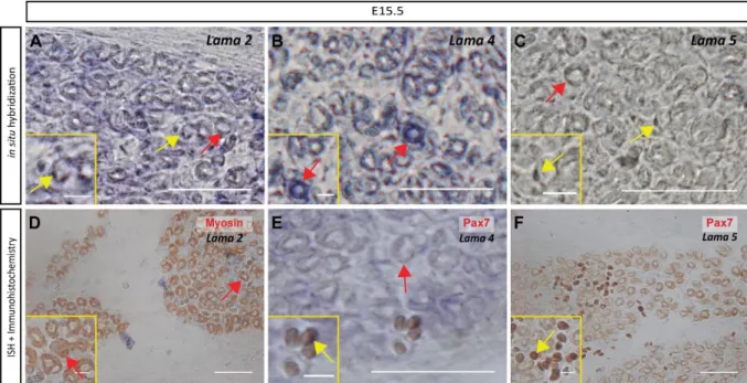
Both myofibers and mononucleated cells contribute to the production of Lama2, Lama4 and Lama5
mRNA 28
A stage-dependent role for mononucleated cells in the synthesis of laminin 30 Number of synapses increases in neuromuscular junctions lacking laminin 211 and 221 32 The absence of α2 laminin does not interfere with the distribution of α4 and α5 laminin in the synapse
36
Chapter 4 - Discussion 39
Laminin assembly dynamics in fetal muscles: mononucleated cells and myofibers working
together 40
The muscle stem cell population: a diverse population with different niches 42 Absence of α2-laminins appears to lead to alterations in acetylcholine receptor clustering 43
Does innervation influence muscle stem cell dynamics? 43
Final considerations 44
Chapter 5 - References 47
Annexes 55
Annex I – Protocols 56
Annex II – Solutions and Reagents 58
Annex III – Antibodies 64
List of figures and tables
Figure 1 - The main phases of skeletal muscle development throughout time, terminology used to designate cells and their major markers.
Figure 2 - Myotomal myogenesis.
Figure 3 - Primary and secondary myogenesis.
Figure 4 - Laminin trimer structure and laminin isoforms. Laminins are cruciform or T-shaped heterotrimers.
Figure 5 - The neuromuscular junction & the laminin distribution within the synaptic cleft. Figure 6 - Neuromuscular synapse formation in the mouse.
Figure 7 - Satellite cell fate decisions. Figure 8 - The Notch signaling pathway.
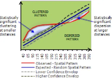
Figure 9 - Schematic representation of Ripley’s K function results.
Figure 10 - Both myofibers and mononucleated cells express Lama2, Lama4 and Lama5 genes at E15.5, but expression in mononucleated cells appears stronger.
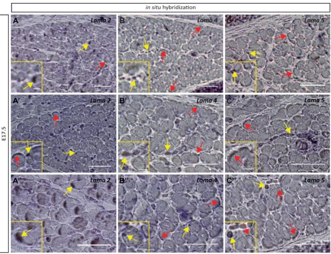
Figure 11 - Both myofibers and mononucleated cells still express Lama2, Lama4 and Lama5 genes at E17.5.
Figure 12 - Characterization of laminin assembly in the presumptive epaxial muscle area in the absence of myofibers at E14.5.
Figure 13 - Laminin assembly in the presumptive epaxial muscle area decreases as development proceeds.
Figure 14 – E15.5 dyW-/- fetuses display differences concerning the synaptic number and size.
Figure 15 - Spatial analysis of the distribution of synapses in wildtypeE15.5 longissimus muscle. Figure 16 – Spatial analysis of the distribution of synapses in dyW-/- E15.5 longissimus muscle. Figure 17 - Laminin distribution in the NMJ at E15.5.
Figure 18 – Schematic representation depicting the main alteration in neuromuscular junction development in α2-laminin-deficient muscles at E15.5.
Figure 19 – Schematic representation of Pax7-positive cells possible laminin niches at E14.5 and E17.5. Figure 20 – The diversity of laminin niches in the heterogeneous population of Pax7-positive cell.
Tables:
Table 1 – Laminin nomenclature and composition. Table S1 – Laminin probes
Table S2 – Variance to mean ratio analysis.
Table S3 – Nearest-neighbour distance analyses – G Function – R score and statistical significance, Z-test – associated.
List of abbreviations and acronyms
AChR – Acetylcholine ReceptorBM – Basement Membrane
DGC- Dystrophin-associated Glycoprotein Complex DML – Dorsomedial Lip
DNA- Deoxyribonucleic Acid
ECM – Extracellular Matrix
FACS - Fluorescence-Activated Cell Sorting
FGF – Fibroblast Growth Factors
JAK-STAT - Janus Kinase/Signal Transducers and Activators of Transcription LAMA2-CMD – Laminin α2 Congenital Muscular Dystrophy
MDC1A – Merosin-deficient Congenital Muscular Dystrophy Type 1A
MHC – Myosin Heavy Chain
MPC – Myogenic Precursors Cells
MRF – Myogenic Regulatory Factor
mRNA – messenger Ribonucleic Acid
NICD – Notch Intracellular Domain NMJ – Neuromuscular Junction
RNA - Ribonucleic Acid
Shh – Sonic hedgehog
VLL – Ventrolateral Lip
VMR – Variance-to-Mean Ratio
Chapter 1 - Introduction
Chapter 1
Introduction
Introduction
Muscle development occurs through a process conserved among amniotes (Picard 2002). In the vertebrate embryo, all trunk and limb muscles arise from the somites, transient embryonic structures originated from the paraxial mesoderm (Brent & Tabin 2002; Pu et al., 2015; Chal & Pourquié, 2017). The specification of somites results from a dynamic molecular process, involving a segmentation clock, which generates pulses of Notch, FGF and Wnt signalling (Andrade et al., 2005).
Once formed the somites are patterned into different compartments that will give rise to distinct cell lineages (Brent & Tabin, 2002). The most ventral part undergoes an epithelial to mesenchymal transition and will form the sclerotome, the source of the axial skeleton and tendons. On the other hand, the most dorsal part of the somite will remain epithelial forming the dermomyotome, which contains myogenic progenitors (MPCs) (Brent & Tabin 2002; Dumont et al., 2015b; Chal & Pourquié, 2017), as well as dermis progenitors (Cinnamon et al., 1999; Scaal & Christ, 2004; Chal & Pourquié, 2017), among others.
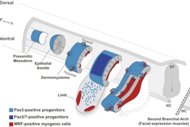
Skeletal muscle development is initiated with the specification and differentiation of myoblasts. In response to specific signals, dermomyotomal cells, expressing Pax3 and/or Pax7 and precursors of muscle stem cells (see Figure 1 for terminology), activate the myogenic program which is controlled by specific transcription factors, the myogenic regulatory factors (MRFs), Myf5, MyoD, Mrf4 and Myogenin (Brent & Tabin, 2002; Buckingham & Rigby, 2014).
Figure 1 - The main phases of skeletal muscle development throughout time, terminology used to designate cells and their major markers. The muscle stem cells express one or both of the transcription factors Pax3 and Pax7. As they start
expressing the myogenic regulatory factors (MRFs), Myf5, MyoD, Mrf4 and Myogenin, they first become committed myoblasts and then differentiate. The differentiated myoblasts – myocytes – express specific muscle fiber proteins, such as myosin, dystrophin and desmin. These myocytes fuse giving rise to multinucleated muscle cells – the myotubes. A second wave of differentiation gives rise to the secondary myotubes that contribute to muscle growth. Adapted from Deries and Thorsteinsdóttir, 2016; Nunes, 2017.
Myotome formation
All epaxial and hypaxial skeletal muscle are derived from the dermomyotome (Brent & Tabin, 2002, Thorsteinsdóttir et al., 2011). The dermomyotome can be regionalized in three main domains. The
epaxial dorsomedial lip (DML) the central dermomyotome, and the hypaxial ventrolateral lip (VLL) (Eloy-Trinquet & Nicholas, 2002, Brent & Tabin, 2002).
The formation of the myotome (Figure 2) is a crucial step of skeletal muscle formation (Gros et al., 2004). In amniotes, it starts when the cells from the DML are induced to express Myf5 (Pownall & Emerson, 1992, Brent & Tabin, 2002) and downregulate Pax3 (Williams & Ordahl, 1994) (E8.5) (Biressi et al., 2007). Myf5 and MyoD are the first MRFs expressed in the myogenic program and are determination factors, i.e. they set the myogenic fate (Kablar et al., 2003). Mrf4 can also act as a determination factor (Kassar-Duchossoy et al., 2004), while Myogenin (and also Mrf4) act later in the myogenesis program, as differentiation factors (Kablar & Rudnicki, 2000; Tajbakhsh & Buckingham, 2000; Kablar et al., 2003; Buckingham & Rigby, 2014).
Figure 2 - Myotomal myogenesis. All epaxial and hypaxial skeletal muscles are derived from the dermomyotome. The
dermomyotome can be compartmentalized into three domains, the epaxial dorsomedial lip (DML), the central dermomyotome and the hypaxial ventrolateral lip (VLL). The cells from the epaxial DML activate the expression of myogenic regulatory factors (MRF) Myf5 and MyoD. Those cells migrate to the underlying space giving rise to the myotome. The epaxial myotome gives rise to the epaxial muscles (transversospinalis, longissimus, iliocostalis and levatores costarum), while the hypaxial myotome originates the intercostal and abdominal muscles. More cells are progressively added to the myotome as myoblasts that either differentiate into myocytes or fuse with the existing ones. The myotome grows both dorso-ventrally and medio-laterally and myogenesis proceeds as a rostro-to-caudal wave of differentiation. From Buckingham and Rigby, 2014.
Myf5 and MyoD-positive myoblasts (see Figure 1) migrate to the underlying space giving rise to the third somitic compartment – the myotome - which continues to grow due to the ongoing entry of cells. The myotome is also a compartmentalized structure, as the epaxial myotome that originates from the cells migrating from the DML, and the hypaxial myotome develops from cells delaminating from the VLL. At certain axial levels, the hypaxial myotome does not form, but cells delaminate and migrate long distances to form the muscles of the limbs, tongue and diaphragm (Deries et al., 2016).
More and more cells are progressively added to the myotome as myoblasts where they either differentiate into new myocytes or fuse with the existing myocytes to form first bi- and then
multinucleated myotubes, thus contributing to myotome growth (Relaix et al., 2005; see Figure 1 for terminology). Consequently, the myotome grows medio-laterally and myogenesis proceeds as a rostral-to-caudal wave of maturation (Gros et al., 2004).
Eventually, the central dermomyotome loses its epithelial structure and the Pax3- and/or Pax7-positive muscle stem cells enter the underlying, myotomal space. These cells can either activate Myf5 or MyoD and differentiate or keep proliferating, providing a reserve cell population for muscle growth during development (Buckingham & Rigby, 2014).
The myotome is a transient structure that transforms into the epaxial (deep back) muscles between E11.5 and E12.5 in the mouse (Deries et al., 2010, 2012) thus losing its segmented organization. This process includes translocation, re-orientation, elongation of existing myotomal myocytes (Deries et al., 2010). Concomitantly, the dissociation of the dermomyotome brings in MPCs of which some differentiate into myoblasts that drive primary myogenesis in the epaxial muscle masses (Deries et al., 2010).
Primary fibers of the epaxial muscle masses cleave into four muscle masses, corresponding to the transversospinalis, iliocostalis, longissimus and levatores costarum all clearly distinguishable at E15.5 (Vallois, 1922). Each newly formed muscle includes populations of Pax3- and Pax7-positive muscle stem cells intermingled with them, which contribute to their future growth and development (Deries et al., 2010).
Primary and Secondary Myogenesis
Post-myotome myogenesis is marked by the formation of myofibers which occurs in two main phases: embryonic or primary myogenesis, from E11.5 to E14.5, and fetal or secondary myogenesis, spanning from E14.5 to postnatal development (Patton et al., 1997; Biressi et al., 2007; Figure 3).
Primary myogenesis provides the basic muscle pattern of the body. It involves the differentiation of a subset of Pax3- and/or Pax7-positive muscle stem cells into primary myoblasts that will contribute to formation of the primary myofibers (Figure 1 and Figure 3), upon which the muscle grows during secondary myogenesis (Messina et al., 2010).
Figure 3 - Primary and secondary myogenesis. Post-myotome myogenesis is marked by the formation of myofibers. It is
divided in two distinct phases: (A) Primary or embryonic myogenesis (from E11.5 to E14.5) and (B) Secondary or fetal myogenesis (from E14.5 to birth). Primary myogenesis occurs simultaneous with the entry of nerves into the muscle masses (Hurren et al., 2015), and is essential to form the basic muscle pattern of the body, upon which the muscle grows during secondary myogenesis. The increment in muscle mass occurs during secondary myogenesis, with a differentiation wave of secondary myoblasts that fuse with each other at the site of the innervation of the primary myofiber, giving rise to the secondary myofibers. From Nunes, 2017.
In the trunk, primary myofibers are generated by the fusion of primary myoblasts with the myotomal myocytes forming the primary myotubes, while in the limbs, diaphragm and tongue, differentiated myoblasts fuse with each other to form the primary myotubes. The primary myotubes will eventually attach to cartilaginous structures, such as the vertebrae of the trunk, and thus become the scaffold on which myogenesis will proceed (Deries et al., 2010).
Secondary myogenesis is responsible for the increment in muscle mass, both in the number of myofibers as well as their size (Biressi et al., 2007). Secondary myofibers form through a wave of differentiation of Pax7-positive cells into secondary myoblasts that will start to fuse with each other at the site of innervation of the primary myofiber, forming the secondary myofibers. In the absence of functional innervation, the formation of secondary muscle fibers is impaired. (Wigmore & Evans, 2002). Secondary myoblasts then also fuse with all existing myofibers, increasing their size (Biressi et al., 2007).
In early development, MPCs express either Pax3 or Pax7 or both. However, as development proceeds, Pax3-positive cells decline in frequency (Deries et al., 2010) and, at least in the case of epaxial muscles, become restricted to the ventrolateral edge of the differentiated muscle masses. From E14.5 onwards, expressing muscle stem cells are found in large numbers, which shows a role of Pax7-positive cells in driving fetal stages of muscle growth including the freshly started secondary myogenesis (Biressi et al., 2007; Hutcheson et al., 2009). By this time, the fetal muscle stem cells only express Pax7 (Hutcheson et al., 2009) and become closely associated with differentiating muscle fibers at E15.5. Between E16.5 and E18.5 the basement membrane (or basal lamina) which has been assembled around the muscle fibers, comes to enclose the Pax7-positive cells underneath it. The location underneath the basement membrane is maintained during later stages of myogenesis and is a landmark of adult muscle stem cells, firstly termed satellite cells due to this characteristic location (Mauro, 1961; Zammit & Beauchamp, 2001). It is believed that satellite cells are direct descendants of the Pax3 and/or Pax7-positive cells of the dermomyotome (Kassar-Duchossoy et al., 2005, Relaix et al., 2005).
Extracellular Matrix
Skeletal muscle development is understood today as a complex process that goes much beyond myoblast differentiation and fusion. The impact of transcription factors and signalling pathways in determining the course of events is now well recognized. However, there is a third element that adds complexity and tinkering to this developmental process, namely the extracellular matrix (ECM). Soon after somites develop, a basement membrane is laid down around it (Duband et al., 1987, Ostrovsky et al., 1988), playing a key role in maintaining the undifferentiated epithelial state of the dermomyotome (Bajanca et al., 2006).
The importance of the ECM is illustrated by the wide range of syndromes that arise form genetic abnormalities regarding several of its components (Frantz et al., 2010). Mutations affecting the expression of ECM components and receptors are associated with developmental arrest affecting several different tissues, and in some cases embryonic lethality (Poschl et al., 2004; Yurchenco et al., 2011).
The structural and scaffolding role of the ECM is undeniable. However, it is a highly dynamic network in constant remodelling. The ECM is composed of glycoproteins such as collagens, fibronectin, laminins, tenascins, and polysaccharides like glycosaminoglycans and proteoglycans (Frantz et al., 2010), and can be characterized as two biochemically and morphologically separate entities: the interstitial matrix as observed for example in connective tissue (Laurila & Leivo, 1993), and the pericellular matrix, which includes basement membranes, which is in close contact with cells and has a different molecular composition.
Interstitial collagens, often secreted by fibroblasts, are the most abundant fibrous proteins within the interstitial matrix, constituting the main structural element providing tensile strength, regulating cell adhesion, migration and therefore often directing tissue development (Rozario & DeSimone, 2010). Interstitial matrices also often contain fibronectin, another fibril-forming matrix protein, which plays a crucial role in mediating cell attachment, polarization and tissue compartmentalization (Rozario & DeSimone, 2010; Thorsteinsdottir et al., 2011).
Basement membranes are evolutionarily ancient macromolecular structures first described in muscle as a “membranaceous sheath of the most exquisite delicacy” (Bowman, 1840), present in every tissue (LeBleu et al., 2007). BMs are a network of several large glycoproteins and proteoglycans which line epithelial and endothelial cells and surround muscle, nerve, and fat cells (Durbeej, 2010; Yurchenco et al., 2011). The main components of BMs are type IV collagen, laminin, nidogen/entactin, perlecan and agrin. (LeBleu et al., 2007; Yurchenco et al., 2011; Thorsteinsdóttir et al., 2011).
BMs are first assembled in the pre-implantation embryo and soon after in Reichert’s membrane (Wartiowaara et al., 1979; Leivo et al., 1980; Thorsteinsdóttir, 1992). They require the presence of laminin 111 and 511, which are already expressed at E4.5 (Miner, 2004). Both collagen type IV and laminins are able to self-assemble in a calcium dependent way and they constitute the basic networks of the BM, (Murray & Edgar, 2000). Components such as nidogen and perlecan play a decisive role in stabilization of the structure by bridging the two networks (LeBleu et al., 2007).
BMs have a structural role, whereby through direct cell-ECM interaction, they give cells an accurate understanding of their surroundings. In addition, BMs can also modulate cell behaviour by binding (or repelling) molecules, such as paracrine factors, thus creating areas of high (or low) concentrations of these factors. Thus BMs are not only providers of mechanical support but also play active roles in developmental and regenerative processes by modulating key cellular responses such as proliferation, polarization, migration, differentiation, survival and apoptosis (Frantz et al., 2010; Rozario & DeSimone, 2010; Thorsteinsdóttir et al., 2011).
In skeletal muscle, these roles are vividly illustrated through the processes of myogenesis and synaptogenesis (Patton et al., 1997; Sanes, 2003; Rogers & Nishimune, 2017).
Laminins
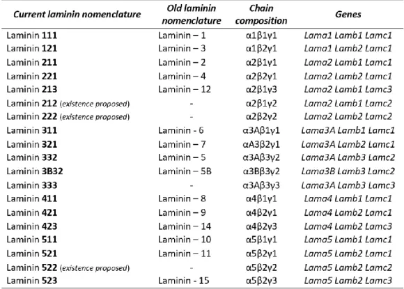
Laminins are the major and best studied BM components (Aumailley, 2013). They are cruciform or T-shaped heterotrimers consisting of one α, one β and one γ chain (Aumailley et al., 2005; Durbeej, 2010; Domogatskaya et al., 2012), forming at least 16 known isoforms (Durbeej, 2010). The current nomenclature is based on their chain composition, as for example, laminin 211 is composed of an α2, β1 and γ1 chain (Aumailley et al., 2005; Figure 4, Table 1). The individual chains are joined through the long coiled-coil domains to produce a molecule with one long arm and up to three short arms (Lebleu et al., 2007; Durbeej, 2010; Aumailley, 2013).
Laminins can be classified according to their domain composition and fall into polymerizing and non-polymerizing groups (Yurchenco, 2015). Their polymerization is an important contributor to basement membrane assembly (Cheng et al., 1997; Hohenester & Yurchenco, 2013). The biological role of laminins is mediated mostly by their interaction with cell surface receptors that link laminin matrices to intracellular signalling pathways. The major receptors are integrins, and certain non-integrin receptors such as dystroglycan (Durbeej, 2010).
Integrins are transmembrane heterodimeric receptors composed of an α and a β subunit, involved in a multitude of functions. The majority of integrins are involved in cell-ECM adhesion (Burkin & Kaufman, 1999; Barczyk et al., 2010). The predominant laminin-binding integrin receptors are α1β1,
α2β1, α3β1, α6β1 and α7β1 (Nishiuchi et al., 2006; Scheele et al., 2007; Yurchenco et al., 2011). Integrin α7β1 binds strongly to laminins 211 and 221, while α6β1 has more affinity to laminins 511 and 521 (Nishiuchi et al., 2006).
Dystroglycan is a non-integrin laminin receptor, part of the dystrophin glycoprotein complex (DGC), which is composed of two subunits, encoded by a single gene (Dag1). Dystroglycan binds to laminin (extracellularly) and dystrophin (intracellularly) linking the muscle fiber cytoskeleton to the basement membrane (Durbeej, 2010; Domogatskaya et al., 2012). Laminins can also bind to two other non-integrin receptors, the heparin sulfates and sulfated glycolipids (Hohenester & Yurchenco, 2013).
Figure 4 - Laminin trimer structure and laminin isoforms. Laminins are cruciform or T-shaped heterotrimers. They are
constituted by one α, one β and one γ chain that assemble to form at least 16 known isoforms, with one long arm and up to three short arms. Laminins are denominated based on their chain composition, as for example, laminin 211 is composed by α2, β1 and γ1 chain. From Domogatskaya et al., 2012.
It has already been shown that laminins play a crucial role in tissue morphogenesis, beginning to be expressed in the preimplantation embryo in the mouse, during implantation and throughout organogenesis into the postnatal period (Miner, 2004; Durbeej, 2010). Laminins have often specific cell type-specific functions in processes such as adhesion, differentiation, migration and resistance to apoptosis (Domogatskaya et al., 2012), with vital roles in many physiological functions in all types of muscle, in the nervous system, as well as in skin, kidney, the digestive system and vasculature (Durbeej, 2010).
Table 1 - Laminin nomenclature and chain composition. From Thorsteinsdóttir et al., 2011
Laminins in Skeletal Muscle
The influence of laminins on skeletal muscle begins early in embryo development during the assembly of the dermomyotome basement membrane which has a role in preventing precocious myogenesis (Bajanca et al, 2006). Another basement membrane is assembled soon after in the interface between the myotome and the sclerotome (Tosney et al., 1994; Bajanca et al., 2004).
The laminin isoform content in skeletal muscle changes throughout development, postnatal time and adulthood (Patton et al., 1997; Nunes et al., 2017). In the myotome, laminins 211 and 511 are found. During primary myogenesis, essentially no assembled laminins are present, but later on, at the onset of secondary myogenesis (E14.5), laminins 211, 411 and 511 are assembled around myotubes (Patton et al., 1997; Nunes et al., 2017). The adult expression of laminins is a subset of those made during development. The α2- and γ1- laminin chains are expressed ubiquitously in the myofiber as well as in the synaptic basement membranes. α5- and α4-chain containing laminins initially have a widespread pattern, but become restricted to the neuromuscular junction perinatally, where β2 is also present. β1 is excluded from the NMJ, but is present in the myofiber basement membrane thus occupying 99% of the muscle basement membranes (Patton et al., 1997). In conclusion, laminin 211 is the major isoform surrounding the muscle fibers in the adult, while laminin 221, 421 and 521 becomes characteristic of the neuromuscular junction area (Patton et al., 1997; Patton, 2000; Yurchenco, 2015). Both of them are crucial for myofiber function and survival (Vachon et al., 1996; Holmberg & Durbeej, 2013).
Laminins in Neuromuscular Junctions
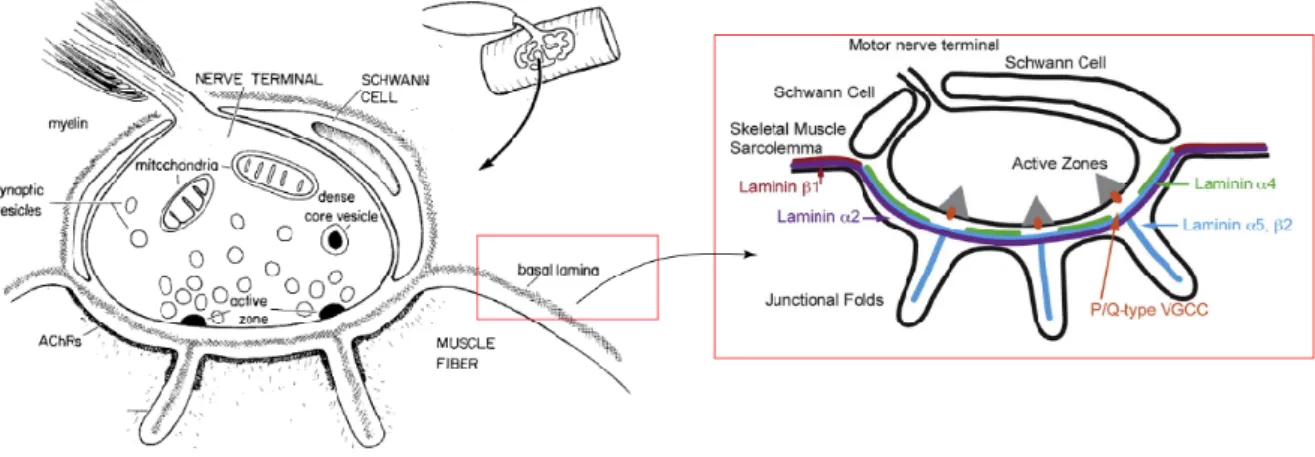
The specialized junction that forms between the motor neuron and skeletal muscle fibers is known as the neuromuscular junction (NMJ) (Witzemann, 2006; Rogers & Nishimune, 2017). Synapse formation requires a complex interplay between the pre- and post-synaptic region (Figure 5). There is an interchange of information that assures the proper maturation of the neuromuscular junction (Noakes et al., 1995; Sanes & Lichtman, 1999).
Figure 5 - The neuromuscular junction & the laminin distribution within the synaptic cleft. The neuromuscular junction
is a specialized synapse between the motor neuron and myofiber. Acetylcholine receptors in the myofiber membrane distribute in a prepattern before innervation, however this initial distribution is remodeled after the nerve contacts the muscle. Laminins are muscle-derived synaptic organizers crucial for the development and maturation of the NMJ. The synaptic basal lamina is constituted by laminin 221, 421 and 521. Adapted from Hall & Sanes, 1993, and Patton, 2001.
The first contact between nerves and myogenic cells occurs before the beginning of secondary myogenesis, with acetylcholine and its receptor (AChR) being the main communication system of the neuromuscular system (Witzemann, 2006; Hurren et al., 2015).
The distribution of acetylcholine receptors on muscle fibers is prepatterned, even before the contact with a motor neuron and subsequent contact between the neuron and the myofiber reinforces this pattern (Figure 6; Grady et al., 2005). Multiple innervations are eliminated to form a single synaptic contact, and by E17.5 the synaptic site and endplate pattern in fully established (Grady et al., 2005). The process of innervation by motor neurons requires a guidance system allowing the communication between them and their target, the endplate. Laminins are known to play a role in this process (Patton, 2003; Sanes, 2003). As mentioned before, the basement membrane envelops the entire muscle fiber, as well as the synaptic cleft (Sanes, 2003).
Figure 6 - Neuromuscular synapse formation in the mouse. The post-synaptic area of the neuromuscular junction is
characterized by the presence of aneural AChR clusters, i.e. which are clusters of AChR in the myofiber membrane that formed without nerve contact. When innervation occurs, there is a reorganization of the AChR clusters in the membrane of myofibers, with the receptors becoming concentrated in the synaptic region while primitive clusters in non-synaptic areas disappear. From Wu et al., 2010.
Laminins are muscle-derived synaptic organizer that promote pre- and post-synaptic differentiation by an autocrine mechanism (Nishimune et al., 2008). This process depends largely on the aggregation of dystroglycan in the post-synaptic membrane, known to bind to agrin, a nerve-derived synaptic organizer (Witzemann, 2006; Nishimune et al., 2008; Rogers & Nishimune, 2017). Laminins are crucial for the proper development and maturation of the NMJ and the synaptic basement membrane contains laminins 221, 421, 521 (Patton et al., 1997; Patton, 2000). Laminins 421 and 521 have a determinant role in organizing active zones and endplate structures in NMJs (Patton, 2000). These two laminins are responsible for the correct alignment of the pre- and post-synaptic structures, and the specific presence of the laminin β2 chain is crucial for motor neuron adhesion, inhibition of neurite outgrowth, ultimately acting as a stop signal (Noakes et al., 1995; Nishimune et al., 2008). Mouse models lacking laminin-α2 chain display partial detachment of motor neuron terminals from the endplate, have demyelination of motor axons and Schwann cell infiltration into the synaptic cleft (Patton, 2000). Furthermore disruption between laminins and their receptors (integrins and/or dystroglycan), due to aging or injury, results in defects at the NMJ. A fragmented distribution of AChR and the loss of precise apposition of the pre- and post-synaptic apparatus are the major phenotypes (Noakes et al., 1995; Patton et al., 2001). Ultimately, breaking this structural connection leads to a progressive degeneration of the neuromuscular system.
Satellite Cells
Vertebrate skeletal muscle has an exquisite regenerative capacity that maintains the neuromuscular system healthy and functioning (Chargé & Rudnicki, 2004). Satellite cells, which are localized between the basement membrane and the muscle fiber, (Mauro, 1961) are muscle stem cells (Tajbakhsh, 2009). They are derived from the Pax3- and Pax7- expressing cells of the central dermomyotome, and represent the principal regenerative source of the skeletal muscle in postnatal life (Kassar-Duchossoy et al., 2005; Relaix et al., 2005).
Muscle regeneration is characterized by a degenerative phase triggered by the disruption of the myofibers allowing the entry of macrophages (Chargé & Rudnicki, 2004), and a regenerative phase which is accomplished by satellite cell activation, proliferation, differentiation into myoblasts and fusion of these myoblasts with each other or the pre-existing fibers (Chargé & Rudnicki, 2004; Kuang et al., 2007; Sambasivan et al., 2011).
Upon injury, satellite cells are activated in response to both intrinsic and extrinsic signals. They proliferate for a while after which some downregulate Pax7, upregulate Myf5 and MyoD, enter the myogenic program and differentiate into myoblasts which either fuse with each other giving rise to new multinucleated myofibers or fuse with existing ones. After that, satellite cells which did not enter the myogenic program, re-enter quiescence again and become nested underneath the basal lamina (Schultz & Jaryszak, 1985).
Recent studies have shown that the satellite cell population is heterogeneous, with 90% of Pax7+/Myf5+ cells, known to be more prone to differentiation, and about 10% of Pax7+/Myf5-. The latter is believed to be a more stem population crucial to restore and maintain the satellite cell pool after regeneration (Kuang et al., 2007).
The reservoir of muscle stem cells is maintained and expanded mainly through asymmetric and symmetric divisions, respectively (Figure 7). The ratio of these two types of divisions is extensively influenced by the satellite cell niche. The niche that is essentially asymmetric, with the cells contacting with two opposing environments, the basement membrane and the myofiber membrane, leads to an asymmetric distribution of proteins, for example integrin α7β1 on the basal side, near the basement membrane, and β-catenin on the apical side, near the myofiber (Dumont et al., 2015a).
Figure 7 - Satellite cell fate decisions. (A) Satellite cells are located under the basement membrane, juxtaposed to the muscle
fiber, in a quiescent state. Satellite cells are a heterogeneous population that in response to both intrinsic and extrinsic signals are activated and re-enter the cell cycle. Then they proliferate and some of them upregulate the MRFs Myf5 and MyoD. (B) When activated, the satellite stem cells can undergo two different types of divisions. The symmetric divisions allow for the expansion of the stem cell pool. The asymmetric divisions maintain the stem cell pool while generating committed myogenic progenitors. The committed cell proceeds in the myogenic program, giving rise to myoblasts that will fuse to form new myofibers. From Dumont et al., 2015a.
Several signalling pathways are known to play roles in the return to quiescence (e.g. Le Grand & Rudnicki, 2007; Shea et al., 2010; Chakkalakal et al., 2012). However, Notch signalling (Figure 8) has emerged as a master regulator of satellite cell state. Notch signalling occurs when Delta1-like ligand and the Notch receptor are present on adjacent cell surfaces (Conboy & Rando, 2002). Similarly with lateral inhibition, also controlled by Notch, when an asymmetric cell division occurs, the daughter cell committed to differentiate will express high levels of delta1, whereas the other one expresses Notch3 receptor. The cell expressing Notch will receive a notch signal from the Delta1-expressing cells and the subsequent release of the Notch intracellular domain (NICD) to the nucleus, promotes the expression of Pax7 (Bröhl et al., 2012) and the return to quiescence, (Kuang et al., 2007) maintaining their proliferative capacity. Depletion of Notch receptor will lead to spontaneous uncontrolled differentiation resulting in the satellite cell pool depletion (Vasyutina et al., 2007), while forced expression of notch intracellular domain (NICD) maintains Pax7-positive cells in an undifferentiated state
Figure 8 - The Notch signaling pathway. Notch
signaling is activated when a notch-ligand (Delta-like, Jagged) in the neighbor cell contacts with the notch receptor in the receiving cell. This connection induces a proteolytic event and the notch receptor is cleaved. This cleavage releases the notch intracellular domain (NICD), which is translocated to the nucleus and funcions as a transcription factor that, together with other cofactors, activates the transcription of Notch target genes. From Anderson et al., 2014
thus blocking myogenic differentiation (Mourikis et al., 2012).
The unequivocal necessity of Pax7-positive satellite cells in adult regenerative myogenesis, does not rule out the importance of the contribution of other cell types to the regeneration process. These other cell types can definitely modulate the behaviour and survival of the Pax7-positive satellite cells (Sambasivan et al., 2011). For example, the presence of nerves have been shown to influence satellite cells since after denervation, satellite cell numbers decline dramatically, impairing the regenerative capacity of muscles (Jejurikar et al., 2002).
Merosin-deficient congenital muscular dystrophy type 1A (MDC1A)
Merosin-deficient congenital muscular dystrophy type 1A (MDC1A) is a type of congenital muscular dystrophy, also known as laminin-α2 CMD (LAMA2-CMD). It is a neuromuscular disease resulting from a mutation in the laminin-α2 gene (LAMA2) (Tomé et al., 1994). MDC1A is characterized by severe muscle weakness and neonatal hypotonia, as well as delayed motor milestones and neuropathies (Gawlik & Durbeej, 2011), representing about 40% of cases of CMD in Europe (Allamand & Guicheney, 2002).
As mentioned above, laminin 211 is the main isoform in the basement membrane surrounding muscle fibers, while 221 is mostly concentrated at the neuromuscular junction. (Patton et al., 1997; Nunes et al., 2017). The absence of the α2 chain and consequently the laminin 211 and 221 trimers, is thought to result in a disruption between the basement membrane and the muscle fiber cytoskeleton, leading to a structural instability and consequently increased muscle fiber degeneration (Vachon et al., 1996).
There are several mouse models for studying laminin-α2 deficiency, the one recommended as a MDC1A model by Cure CMD (www.curecmd.org) being the dyW/dyW mouse, which expresses a small
amount of truncated α2 chain (Gawlik & Durbeej, 2011). Using the dyW
/dyW model, Nunes et al. showed for the first time that the onset of the disease occurs prenatally and in the absence of any muscle fiber damage. Rather, they found that E18.5 dyW/dyW muscles display significantly fewer Pax7-positive muscle cells and an overactivation of the JAK-STAT signalling compared to same stage wild-type muscles (Nunes et al., 2017). One way to explain these results is that the absence of laminin 211/221 somehow shifts the balance of muscle stem cell division to asymmetric divisions, which could lead to an early depletion of the stem cell pool.
It is imperative to gain a deeper and more detailed understanding of the functional mechanisms underlying the communication between muscle cells and their basement membrane and how mutation affecting this communication lead to disease, such as muscular dystrophies. It is important to comprehend the way in which the different players involved in skeletal muscle formation regulate each other’s development. What are the key players coordinating the formation of a functional musculoskeletal system? In what way do these players communicate with each other? A better understanding of how this communication occurs, and how it goes wrong in disease, will allow us to envision new and precise therapies targeting the key events involved in the onset of diseases such as muscular dystrophies. Such an approach may be more effective since it targets the disease early, at an earlier stage of development.
Aims of this thesis
As mentioned above, laminin matrices have been found to play crucial roles during skeletal muscle development. Here we aim to build on the results obtained in our group and characterize the fetal myogenesis defect in the dyW/dyW mouse in more detail. We propose to first analyse how normal fetal
fetal muscle masses express the Lama2, Lama4 and Lama5 genes, and studying the laminin assembly dynamics around the muscle stem cells and myofibers during secondary myogenesis. Knowing how this occurs in normal fetuses will contribute to understanding what goes wrong in dyW/dyW fetuses. Secondly, we know that the development of a functional musculoskeletal system depends on innervation. Thus we will study the development of NMJs in the absence of α2-laminin, aiming to determine if the NMJs are somehow altered in the dyW/dyW mouse model, and if so, in what way.
Chapter 2 – Materials and Methods
Chapter 2
Materials and
Methods
Materials and methods
Embryo collection
a. Wild-type fetuses (E15.5 - E18.5) obtained from crossing of outbred CD-1 mice (Jackson Laboratories), were used to assess the distribution of laminin variants during normal myogenesis. The pregnancies were dated through observation of the copulation plugs, with the morning of the plug staging the embryonic day 0.5 (E0.5). Pregnant females were anesthetized with an anaesthetic drug – isoflurane (2-chloro-2-(difluoromethoxy)-1,1,1-trifluoro-ethane) - and confirmed unconscious with loss of pinch toe and reflexes. They were sacrificed by cervical dislocation. Fetuses were removed from the uterus in cold phosphate buffered saline (PBS), then beheaded and processed for cryosectioning and immunohistochemistry.
b. dyW mice have a LacZ-neo cassette inserted in the Lama2 gene, and homozygous animals produce a small amount of a truncated laminin α2 protein lacking the N-terminal LN domain (Engvall YEAR). To obtain homozygous dyW-/- mutants and wildtype controls, heterozygous
dyW were crossed.
c. Myf5Cre/+:R26Rstop-NICD-nGFP/+ mice express the notch intracellular domain (NICD) under the
control of the Myf5 promotor, which enables the constitutive activation of NICD in muscle stem cells that have activated Myf5 gene (Mourikis et al., 2012).
Genotyping
The fetuses from heterozygous dyW crossings were genotyped with the following primers: 5’
ACTGCCCTTTCTCACCCACCCTT 3’, 5’ GTTGATGCGCTTGGGACTG 3’ and 5’
GTCGACGACGACAGTACTGGCCTCAG 3’. (Annex I – P1)
All procedures involving mice were performed under two approved protocols: (3/2016) from the Animal Welfare Body of the Faculty of Sciences, University of Lisbon.
Cryosectioning
Fetuses staged between E15.5 and E18.5 were processed for cryosectioning, and fixed in 4% paraformaldehyde (PFA) in phosphate buffered saline (PBS) or 2% (fixative for in situ hybridization) FISH. They were washed and incubated for 48h in a sucrose gradient: solution 1 (0.12M phosphate buffer with 4% sucrose), solution 2 (0.12M phosphate buffer with 15%). Finally, the fetuses were washed and incubated in solution 3 (0.12M phosphate buffer with 15% sucrose and 7.5% gelatine) for 3h at 37°C and frozen in small aluminium boats on the surface of dry ice-chilled isopentane. Sections were made with a Leica CM1860 UV Cryostat.
In situ probe production
a. Transformation
One microliter of Lama2, Lama4 and Lama5 plasmid (1-10ng) was added to 50µL of DHα5 competent E. coli bacteria. The resulting solutions were incubated on ice for 30min, subjected to heat shock during 35s at 42°C without shaking and immediately transferred to ice for 2min. Nine hundred and fifty microliters of pre-warmed lysogeny broth (LB) medium without ampicillin were added to the bacteria tubes, which were incubated at 225rpm at 37°C for 1h. After that, 100µL of each tube was plated in the pre-warmed selective plates. The plates remained inverted overnight at 37°C.
b. Primary culture
Four tubes per plasmid with 4mL of LB medium treated with ampicillin (100µg/mL; selection plates) pre-warmed at 37°C were inoculated during 8h at 300rpm with isolated colonies picked up from
the plates. After that, 1mL of culture was used to perform a confirmation Mini-Prep (Annex I – P2), in order to choose the best colony and verify if the transformation was efficient.
Subsequently, we proceeded to a secondary culture, where 500µL of primary culture were incubated in 100mL of LB medium with ampicillin, at 37°C for 16h in autoclaved Erlenmeyer flasks. The plasmid of interest was then extracted using a JetStar - The Novel Plasmid Purification System (Annex I – P3), and diluted in 30µL of TE and stored at -20°C.
c. Linearization
Ten micrograms of the plasmid were digested for 2h30 at 37°C with 5 units of the proper restriction enzyme, see SI - Table A.
The plasmid linearization was confirmed through gel electrophoresis (1% agarose). DNA was precipitated with 300µL of DEPC water and 800µL of isopropanol at -20°C for 2h, followed by a 13000rpm centrifugation at 4°C during 45min. The pellet was washed in 1200µL of ethanol (EtOH) 100% followed by a 15min centrifugation in the previous conditions. The supernatant was discarded and a second EtOH wash for 5min was performed. The DNA pellet was resuspended in 30µL of TE buffer.
d. Transcription
One microgram of linearized DNA was incubated for 2h30 at 37°C with 2µL of transcription buffer 10x, 2µL of DIG labelling mix, 1µL of DTT, 0.5µL of RNAsin, 1µL of RNA polymerase and the necessary amount of DEPC/nuclease free water to make up the 19.5µL volume. RNA presence was confirmed through gel electrophoresis (2% agarose), and the RNA proceeded to precipitation with 2.5µL of LiCl 4M, 75µL of EtOH 100% and 1µL of EDTA 0.5M pH = 8.0, leaving the solution at -20°C overnight. After centrifugation and removal of the supernatant, the pellet was washed by a gradient of ethanol. The pellet was resuspended in 30µL of DEPC water and measured in Nanodrop for concentration and purification ratios. The probe was diluted in hybridization solution (hybmix, see Annex II) at a concentration of 1µg/mL and stored at -20°C.
In situ hybridization
To determine mRNA expression pattern of Lama2, Lama4 and Lama5, fetuses from E15.5 to E18.5 were fixed in 4% PFA for in situ hybridization as previously described (Bajanca et al., 2004).
a. RNA whole-mount in situ hybridization
We began by testing two possible approaches of the in situ hybridization protocol. Initially, we tested the efficiency of RNA whole-mount in situ hybridization adapted for mouse fetuses from Cepko/Tabin lab and based on Bajanca et al. 2004.
Fetuses were collected and dissected, leaving only the back musculature, in PBS DEPC 1x, and fixed in fresh 4% PFA/PBS DEPC at 4°C overnight. They were washed in PBT and dehydrated in a methanol gradient, to be stored at -20°C.
The in situ hybridization protocol begins by a methanol to PBT gradient rehydration. The fetuses were bleached with 6% H2O2 and treated with proteinase K (10µg/mL). Then incubated in 2mg/mL glycine for 10min and fixed in a solution of 0.2% glutaraldehyde and 4% PFA in PBT. Finally they were washed in a gradient of PBT:Hybmix and incubated overnight at 70°C in the probe solution. In the next day, fetuses were washed in pre-warmed hybmix at 70°C.
From this step onwards, all washes were performed at room temperature, starting with long washes (2 x 30min) in TBST. For the blocking step, fetuses were left incubating for 1h in a solution of TBST +