Dissertação de Mestrado
Characterization of axon-Schwann cell interactions
implicated in neuronal energy metabolism
Ana Carolina Temporão Marques Filipe
Mestrado Integrado em Bioengenharia – Biotecnologia Molecular
2018
Supervisor
Roman Chrast, PhD
Departamento de Neurociência e Departamento de Neurociência Clínica, Instituto Karolinska, Estocolmo, Suécia
Co-supervisor
Filipa Bouçanova, MSc
Departamento de Neurociência e Departamento de Neurociência Clínica, Instituto Karolinska, Estocolmo, Suécia
Co-supervisor
Joana Paes de Faria, PhD
Instituto de Investigação e Inovação em Saúde, Universidade do Porto, Porto, Portugal Instituto de Biologia Molecular e Celular, Universidade do Porto, Porto, Portugal
Caracterização de interações axónio-célula de Schwann
implicadas no metabolismo de energia neuronal
Characterization of axon-Schwann cell interactions
implicated in neuronal energy metabolism
Ana Carolina Temporão Marques Filipe
Supervisor: Doutor Roman Chrast
Co-supervisor: Filipa Bouçanova
Co-supervisor: Doutora Joana Paes de Faria
Faculdade de Engenharia da Universidade do Porto, Portugal
Instituto de Ciências Biomédicas Abel Salazar da Universidade do Porto, Portugal
Instituto Karolinska, Suécia
Instituto de Investigação e Inovação em Saúde, Portugal
Instituto de Biologia Molecular e Celular, Universidade do Porto, Porto, Portugal
FEUP | ICBAS – I
Agradecimentos
Há cinco anos, a semente,
Moldada pelos grãos de uma terra fértil,
Brotava e espreitava curiosa
O encanto da luz do dia.
Penetrando a terra, as raízes revigoravam
E importavam nutrientes da água saciante
Para o caule ascender com empenho e diversão.
E ascendeu.
Cada pedaço de luz absorveu,
E cada instante de vida saboreou.
Primaveras com rebentos,
E verões abrasadores.
Outonos com brisas
E Invernos com nórdicos ventos.
Percurso belo, imortal,
Que agora é saudoso.
Terra, a família.
Água, as amizades.
Luz, o curso.
A todos vós…
Obrigada pela vida.
Eis a flor.
FEUP | ICBAS –
III
Author Contributions
Ana Temporão performed the studies of glutamate stimulation in SC cultures; contributed
to the initial set up of the FRET-based experiments; characterized the mouse models by
genotyping, qPCR and immunohistochemistry; and wrote the manuscript.
Filipa Bouçanova designed research; supervised the studies of glutamate stimulation in
SC cultures; performed FRET-based experiments; characterized the mouse models by
genotyping, qPCR and immunohistochemistry; and guided the writing of the manuscript.
Roman Chrast designed research, provided intellectual support along the project, and
guided the writing of the manuscript.
Joana Paes de Faria guided the writing of the manuscript.
Poster presentations
Filipa Bouçanova, Ana Temporão, Enric Domènech-Estévez, Hasna Baloui, Linnéa
Nilsson, Hjalmar Brismar and Roman Chrast. Characterization of axon-Schwann cell
interactions implicated in neuronal energy metabolism.
2nd Biomedicum Young Researchers Symposium (2018; Stockholm, Sweden)
II Nordic Neuroscience (2017; Stockholm, Sweden)
FEUP | ICBAS –
V
Resumo
O sistema nervoso periférico (SNP) é composto por nervos sensoriais e motores que, respectivamente, detectam sinais internos ou externos, como dor e calor, e estimulam tecidos efectores como glândulas e músculos para realizar uma função ou reflexo. Um SNP funcional depende inteiramente da integridade da relação entre o axónio e a célula de Schwann (SC), sendo esta essencial para o próprio desenvolvimento e para a manutenção a longo prazo dos nervos periféricos. Assim, a disrupção da comunicação axo-glial pode estar envolvida na patogénese de neuropatias periféricas, como a doença de Charcot-Marie-Tooth, neuropatias diabéticas e esclerose lateral amiotrófica, que apresentam alta prevalência a nível mundial. Por essa razão, é imperativo explorar mais profundamente a fisiologia e a fisiopatologia do SNP, a fim de que novos caminhos terapêuticos se abram para modular a progressão dessas doenças. Uma potencial nova função das SCs é a de fornecer suporte metabólico e energético ao axónio subjacente. Ambas as células no SNP expressam transportadores de monocarboxilatos (MCTs) através dos quais substratos altamente energéticos, como o lactato, podem ser trocados. Embora a distribuição celular dos diferentes MCTs sugira um tráfego metabólico da SC para o axónio, a sua caracterização funcional é ainda desconhecida. Para além disso, não é claro em que medida o suporte metabólico é necessário. Será continuamente fornecido pela glia ou apenas desencadeado por necessidades energéticas mais elevadas dos axónios, como durante a atividade neuronal?
Para preencher a lacuna existente na área, nós colocamos a hipótese de que as SCs detetam a atividade neuronal via receptores de neurotransmissores, desencadeando cascatas de sinalização intracelular e potencialmente aumentando o importe/a geração de lactato. Este monocarboxilato pode então ser transferido para o axónio através de MCTs para a produção de energia.
Com este trabalho, nós mostrámos que a breve exposição ao glutamato desencadeia a ativação das vias de sinalização ERK e AKT/mTOR/S6RP em culturas primárias de SCs, principalmente quando são previamente expostas a neuregulina-1. Essas respostas intracelulares podem preceder ajustes na sua atividade metabólica. Para avaliar potenciais variações nos níveis de lactato intracelular, começámos a estabelecer um ensaio baseado em FRET usando células HEK293T e uma sonda de deteção de lactato codificada geneticamente, a Laconic. O conhecimento assim obtido será no futuro aplicado a SCs. Finalmente, observámos preliminarmente que a ablação de MCT1 ou MCT4 especificamente em SCs não parece perturbar a estrutura nem a função do SNP, o que pode dever-se a uma expressão compensatória de outros transportadores.
Palavras-chave
Sistema nervoso periférico; Célula de Schwann; Glutamato; Lactato; Transportadores de monocarboxilatos
FEUP | ICBAS –
VII
Abstract
The peripheral nervous system (PNS) is composed of sensory and motor nerves that, respectively, detect internal or external signals such as pain and heat, and stimulate effectors like glands and muscles to perform a function or reflex. A functional PNS relies entirely on the integrity of the axon-Schwann cell (SC) unit and the crosstalk between the two partners is essential for proper development and long-term maintenance of peripheral nerves. Thus, impaired SC-axon communication may be involved in the pathogenesis of peripheral neuropathies, such as Charcot-Marie-Tooth disease, diabetic neuropathies and amyotrophic lateral sclerosis, which show high prevalence worldwide. For that reason, it is imperative to explore more deeply the physiology and pathophysiology of the PNS, so that new therapeutic avenues open to modulate the progression of these diseases.
One potential novel role of SCs is to provide metabolic and energetic support to the underlying axon. Both cells in the PNS express monocarboxylate transporters (MCTs) through which high energetic substrates, such as lactate, can be exchanged. Although the cellular distribution of different MCTs suggests SC-to-axon metabolic traffic, their functional characterization is still missing. Furthermore, it is not clear to what extent metabolic support is required. Is it continuously provided by glia or only triggered by higher energy demands of axons, such as during neuronal activity?
To fill the existing gap in the field, we hypothesized that SCs detect neuronal activity via receptors for neurotransmitters, triggering intracellular signaling cascades and potentially increasing lactate import/generation. This monocarboxylate may then be transferred to the axon through MCTs for energy production.
Here, we show that short exposure to glutamate triggers the activation of ERK and AKT/mTOR/S6RP signaling pathways in primary cultured SCs, particularly when primed with neuregulin-1. These intracellular response may precede adjustments in their metabolic activity. To evaluate potential variations in intracellular lactate levels, we started to establish a FRET-based assay using HEK293T cells and a genetically encoded lactate-sensing probe, Laconic. The knowledge obtained with this will be applied to SCs in the future. Finally, we preliminarily observed that SC-specific ablation of either MCT1 or MCT4 does not seem to perturb PNS structure and function, which could be due to a compensatory expression of other transporters.
Keywords
FEUP | ICBAS –
IX
Abbreviations List
AKT – Protein kinase B
ALS – Amyotrophic lateral sclerosis
AMPA – α-amino-3-hydroxy-5-methyl-4-isoxazolepropionic acid ANLSH – Astrocyte-neuron lactate shuttle hypothesis
ARC – AR-C155858
ATP – Adenosine triphosphate BCA – Bicinchoninic acid assay BPE – Bovine pituitary extract BSA – Bovine serum albumin cKO – Conditional knockout
CMT – Charcot-Marie-Tooth diseases CNS – Central nervous system
DAPI – 4',6-diamidino-2-phenylindole, dihydrochloride DMEM – Dulbecco's modified eagle medium
DNA – Deoxyribonucleic acid DRG – Dorsal root ganglion
ECL – Enhanced chemiluminescence EDL – Extensor digitalis longus
EDTA – Ethylenediamine tetraacetic acid EGF – Epidermal growth factor
ErbB – Epidermal growth factor receptor ERK – Extracellular signal-regulated kinase FBS – Fetal bovine serum
FRET – Förster resonance energy transfer Fsk – Forskolin
Glu – Glutamate
GLUT – Glucose transporter
HEK293T – Human embryonic kidney 293 T cell line HEPES – 4-(2-hydroxyethyl)-1-piperazineethanesulfonic acid IENFD – Intraepidermal nerve fiber density
KD – Dissociation constant
KRH – Krebs-Ringer-HEPES buffer LDH – Lactate dehydrogenase LKB1 – Liver kinase B1
MAG – Myelin-associated glycoprotein MBP – Myelin basic protein
MCT – Monocarboxylate transporter mGluR – Metabotropic glutamate receptors
X – Ana Temporão | Masters Dissertation
MPZ/P0 – Myelin protein zero
mTFP – Monomeric teal fluorescent protein mTOR – Mammalian target of rapamycin NGS – Normal goat serum
NMDA – N-methyl-D-aspartic acid
NMDA-R – N-methyl-D-aspartic acid receptor NMJ – Neuromuscular junction
Nrg1 –
N
euroregulin-1OCT – Optimal cutting temperature PB – Phosphate buffer
PBS – Phosphate-buffered saline
pCMBS – 4-(Chloromercuri)benzenesulfonic acid sodium salt PCR – Polymerase chain reaction
PFA – Paraformaldehyde
PGP9.5 – Protein gene product 9.5 PI3K – Phosphoinositide 3-kinase PLL – Poly-L-Lysine
PLP – Proteolipid protein
Pmp2 – Peripheral myelin protein 2 PNS – Peripheral nervous system
qPCR – Quantitative polymerase chain reaction RNA – Ribonucleic acid
RT – Room temperature
RT-qPCR – Quantitative real-time polymerase chain reaction S6RP –S6 ribosomal protein
SC – Schwann cell
SDS – Sodium dodecyl sulphate
PAGE – Polyacrylamide gel electrophoresis shRNA – Short hairpin RNA
SLI – Schmidt-Lanterman incisure SN – Sciatic nerve
SOD1 – Superoxide dismutase 1 TBS – Tris-buffered saline TCA – Tricarboxylic acid
TESPA – 3-triethoxysilylpropylamine Ubiq – Ubiquitin-conjugating enzyme E2 WB – Western Blot
FEUP | ICBAS –
XI
Table of Contents
Agradecimentos ... I
Author Contributions ... III
Poster presentations ... III
Resumo ... V
Abstract ... VII
Abbreviations List ... IX
1. Introduction ... 1
1.1. Peripheral Nervous System: Function, Structure and Development... 1
1.2. Metabolic communication between SCs and peripheral axons ... 5
1.3. Implications of disrupted SC-axon crosstalk ... 8
1.4. Aim, Hypothesis and Strategy ... 11
2. Materials and Methods ... 13
2.1. Reagents ... 13
2.2. Glutamate stimulation of primary cultured SCs ... 14
2.3. FRET-based intracellular lactate measurements ... 15
2.4. Characterization of the mouse models ... 15
3. Results and Discussion ... 21
3.1. In vitro study of the intracellular response of SCs to neurotransmitters ... 21
3.1.1. Cell signaling cascades triggered in cultured SCs by glutamate exposure . 21
3.1.2. Monitoring fluctuations of the intracellular levels of lactate in cultured SCs in
response to glutamate ... 29
3.2. Characterization of in vivo models to study the role of MCTs in SCs ... 33
3.2.1. Validation of the mouse models ... 34
3.2.2. Analysis of the effects of MCT1 or MCT4 depletion in the PNS ... 38
4. Conclusion and Future Perspectives ... 41
FEUP | ICBAS – 1
1. Introduction
1.1. Peripheral Nervous System: Function, Structure and Development
The peripheral nervous system (PNS) contains all the nerves and ganglia that lie outside the brain and spinal cord, structures that constitute the central nervous system (CNS). While neuronal cell bodies reside within the CNS or the peripheral ganglia, the axons bundle together to build up the nerves. Some of them are composed by sensory fibers, which transmit to the CNS signals from the internal or external environment that are detected by sensory receptors (e.g. mechanoreceptors in the skin). After being processed in the CNS, the output is carried by motor neurons to produce a response upon activation of effector organs, such as muscles and glands. Therefore, the PNS functions as the bond between the CNS and the periphery of the body (Kandel, Schwartz & Jessel, 2000).
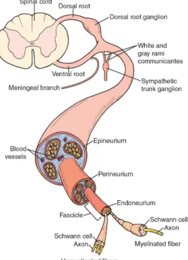
Peripheral nerves are organized in a way that three tissue compartments can be distinguished (Figure 1). Endoneurium is the innermost part, consisting of connective tissue found between individual axons within a fascicle. Bundles of multiple axons are surrounded by a sheath of fibroblasts and collagenous connective tissue, called perineurium, which functions as a blood-nerve barrier. The epineurium embeds these structures in a collagenous matrix where adipose tissue, elastin fibers and vascular endothelial cells can also be found (Jessen & Mirsky, 1999).
Figure 1. Schematic representation of the structure of peripheral nerves. As found in (Kisner & Colby,
2 – Ana Temporão | Masters Dissertation
The structure and function of peripheral nerves is ensured by the intimate interactions established between neurons and glial cells. During development, both cell types derive from the neural plate, which is later divided into neural tube and neural crest. The former differentiates into the brain and spinal cord and into the motor neurons of the PNS. Neural crest cells, in turn, migrate away to the periphery to give rise to all sensory neurons and glial cells of the PNS (Jessen & Mirsky, 2005).
Schwann cells (SCs) are the major type of peripheral glial cells and the counterpart of the oligodendrocytes in the CNS. Both cells spirally extend their cell membrane to produce myelin around axons, even though this process is differently regulated. Besides SCs, peripheral glial cells include satellite cells enveloping cell bodies in the ganglia; perisynaptic SCs at neuromuscular junctions; terminal SCs at sensory axons; olfactory ensheathing cells engulfing axons of the olfactory nerve; and enteric glial cells surrounding autonomic ganglia of the gut (Jessen, 2004).
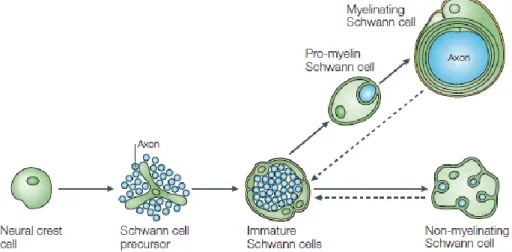
In the PNS, the maturation of neural crest cells generates SC precursors – found at the embryonic day (E) 12/13 in mouse nerves (Jessen & Mirsky, 2005). Their differentiation around E13-15 in mice gives rise to immature SCs, which are found ensheathing numerous axons of mixed caliber. A fate decision point is reached perinatally, in which immature SCs undergo a morphogenetic process known as radial sorting that leads to the formation of myelinating and non-myelinating SC that ensheath large and small diameter axons, respectively (Feltri et al., 2016).
Figure 2 – Schematic representation of the progression of Schwann cell lineage. As found in (Jessen &
Mirsky, 2005).
At the moment of decision between myelinating versus non-myelinating phenotype, SC metabolism and gene expression are tightly modulated, in particular by the axons with which they are associated. Among several neuronal growth factors, neuroregulin-1 (Nrg1) plays a major role at this stage of development, instructing the survival, migration, proliferation and differentiation of SC-lineage cells (Nave & Salzer, 2006). This occurs through the signaling of Nrg1 type III via ErbB2/ErbB3 receptors on the glial membrane, which triggers SC’s intracellular pathways that
FEUP | ICBAS –
3
target, among others, ERK and AKT (Harrisingh et al., 2004; Lyons et al., 2005; Ogata et al., 2004).Thinner axons (diameter < 1μm), which display less Nrg1 at their surface, are ensheathed together by the cytoplasm of non-myelinating SCs into Remak bundles (Griffin & Thompson, 2008; Sherman & Brophy, 2005). Since ion channels are diffusely distributed along the unmyelinated fibers, the signal transmission in Remak bundles is continuous and slow. In large caliber axons (diameter > 1μm), in turn, Nrg1 acts as a positive regulator of myelin-sheath thickness in function of axon size (Michailov et al., 2004). Each axonal segment is then enwrapped by myelin sheath provided by a single myelinating Schwann cell (Feltri, Poitelon, & Previtali, 2016). Interestingly, Nrg1 overexpression in neurons instructs non-myelinating SCs to myelinate de novo the thin axons with which they are associated (Taveggia et al., 2005).
Moreover, a notable feature of the PNS is that fully differentiated SCs retain plasticity throughout life and can readily revert to a phenotype similar to that of immature Schwann cells, a phenomenon that typically occurs in response to nerve injury (Ceci et al., 2014; Morrison et al., 1999). Nrg1 also appears to promote SC de-differentiation in injured nerves (Zanazzi et al., 2001). Although solving similar tasks, the maturation of Schwann cells and oligodendrocytes is distinct and modulated by different factors (Nave & Werner, 2014). Indeed, Nrg1/ErbB signaling is not necessary for CNS myelination to occur and oligodendrocytes only require physical association with axons at the last step of maturation (Brinkmann et al., 2008). Therefore, the regulation of myelination by neurons seems to be less pronounced in the CNS.
There are also clear differences between the CNS and the PNS with respect to the composition of myelin. The most abundant proteins of peripheral myelin are the specific glycoproteins myelin protein zero (MPZ/P0) and protein 2 (Pmp2), whereas CNS myelin is rich in proteolipid protein (PLP) (Jahn et al., 2009; Patzig et al., 2011). Moreover, MBP is not required for PNS myelination but is rate limiting for CNS myelination (Kirschner & Ganser, 1980; Readhead et al., 1987).
Myelin produced by either SCs or oligodendrocytes is repeatedly wrapped and compacted around segments of axons to electrically insulate them (Hildebrand et al., 1993; Webster, 1971). During myelination, the cytoplasmic leaflets of the glial membrane are fused together forming dark dense lines visible in electron microscopy, which alternate with intraperiod lines of the myelin sheath (Scherer & Arroyo, 2002). Nodes of Ranvier consist of the short regions that separate consecutive myelinated segments (internodes) and where action potentials are generated (Salzer, 2003). This way, myelination provides the basis for the rapid and energy efficient saltatory impulse propagation required for motor, sensory, and cognitive functions of the vertebrate nervous system (Nave & Werner, 2014).
To efficiently support this role, axons and glia form a symbiotic unit where distinct structural domains are organized (Salzer, 2003) (Figure 3). At each end of the internode, a paranodal junction separates the nodes of Ranvier from a juxtaparanodal region, where Na+ channels and K+ channels are respectively clustered (Buttermore et al., 2013). A continuous network of non-compact myelin connects the outermost (abaxonal) region of the myelin sheath,
4 – Ana Temporão | Masters Dissertation
where most glial organelles and cytosolic components are found, to the innermost (adaxonal) layer, which is in direct contact with the narrow periaxonal space that separates axons from the myelin sheath (Nave, 2010b). This channel-like system is additionally shaped by Cajal bands, positioned underneath the SC plasma membrane, and by the lumina of the paranodal loops and Schmidt-Lanterman incisures, both comprising gap-junction connections between adjacent membranes (Nave, 2010b). SLIs are exclusive structures of the PNS, consisting on local stacks of non-compacted myelin radially disposed around the axon (Nave, 2010b). Hence, although axons are almost completely surrounded by myelin, the intercellular exchange of nutrients, ions and other small molecules can take place in this system of non-compacted myelin that connects the periaxonal space to the glial soma (Nave, 2010b). However, the physiological purposes for the existence of this structure are not completely understood.
Figure 3 – Schematic representation of a myelinated axon – with the unrolled sheath of SC shown on the
right, where several compartments can be distinguished. As found in (Nave, 2010b).
Despite myelination playing a valuable role on the proper function of the vertebrate nervous system, the significance of neuron-glia interactions goes much beyond the benefits provided by myelination itself. Indeed, glial cells also play a critical role on the survival, function and regeneration of neurons, as they regulate their structural integrity, provide trophic and metabolic support, and confer neuroprotection (Samara et al., 2013).
FEUP | ICBAS –
5
1.2. Metabolic communication between SCs and peripheral axons
The complex functions and large dimension of the nervous system require fast neuronal communication. In vertebrates that is possible by means of myelination, which allows quick and efficient impulse propagation as areas of regeneration of action potentials are restricted to very short regions on the axonal surface (Nave, 2010a). However, myelination itself creates a nearly complete physical barrier that deprives axons from the free access to extracellular metabolites (Nave, 2010b). Nodal uptake of metabolites might not be sufficient to meet their energy demands, at least in fibers with longer internodes, larger caliber, or higher firing frequencies (Hirrlinger & Nave, 2014).
Axons can connect the neuronal cell body to target cells so far away that more than 99% of the neuronal mass is axonal, as it is the case for sciatic nerve fibers (Nave, 2010b). The length and volume of neurons are often further increased by axonal ramifications (Matsuda et al., 2009). Consequently, distal regions of long axons receive limited metabolic resources from the neuronal soma, thus requiring the exchange of metabolites with the surrounding space (Nave, 2010b). Indeed, the assumption that long axons may require additional metabolic support is compatible with the progressive length-dependent loss of axons observed in peripheral neuropathies (Spencer et al., 1978).
Although myelin was long thought to have a passive function in the nervous system, it is now well-recognized that the function and structural integrity of neurons depend on their continuous and reciprocal interaction with glial cells (Samara et al., 2013).
A growing body of evidence suggests that myelinating glial cells are able to provide trophic and metabolic support to axons to compensate for their physical insulation (Pellerin et al., 1998; Lee et al., 2012; Saab et al., 2013). To that end, the channel-like system of non-compacted myelin may serve as the path through which metabolites and trophic factors flow in direction to the adaxonal layer of myelin (Nave, 2010b). Notwithstanding with this potential functionality of the myelin structure, this function is presumably held by all axon-associated glial cells, since non-myelinating Schwann cells were shown to also protect the survival of sensory axons (Chen et al., 2003).
The first evidence for this glial function emerged from studies in the CNS, which gave rise to the astrocyte-neuron lactate shuttle hypothesis (ANLSH) (Pellerin et al., 1998). Blood-borne glucose represents the major energy substrate for the nervous system, being taken up by astrocytes in the CNS (Pellerin, 2003). Glucose is unlikely to be exported, since it is immediately phosphorylated by hexokinase, being instead used to produce pyruvate/lactate by glycolysis (Hirrlinger & Nave, 2014). Alternatively, astrocytes are enzymatically equipped to store it as glycogen, likely to be used under conditions of energy deprivation (Brown & Ransom, 2007; Chih et al., 2001). Lactate is then transferred to neurons and converted back to pyruvate, which undergoes oxidative metabolism in mitochondria to generate high amounts of ATP (Pellerin et al., 1998).
The ANLSH has been refined and the revised hypothesis includes the participation of oligodendrocytes in the shuttling of lactate in the CNS (Funfschilling et al., 2013; Lee et al., 2012;
6 – Ana Temporão | Masters Dissertation
Rinholm et al., 2011). Oligodendrocytes are able to import glucose either from the extracellular space or from astrocytes (with whom they are connected by connexins) to produce lactate (Rinholm & Bergensen, 2012). During myelination, they may consume it for energy production, as well as for lipid synthesis (Rinholm & Bergersen, 2014; Sánchez-Abarca et al., 2001). In mature CNS, glycolytic metabolism may yield sufficient energy to support oligodendrocyte survival and lactate is thought to be exported to the periaxonal space to metabolically support the underlying axon (Funfschilling et al., 2012; Lee et al., 2012; Rinholm & Bergersen, 2012).
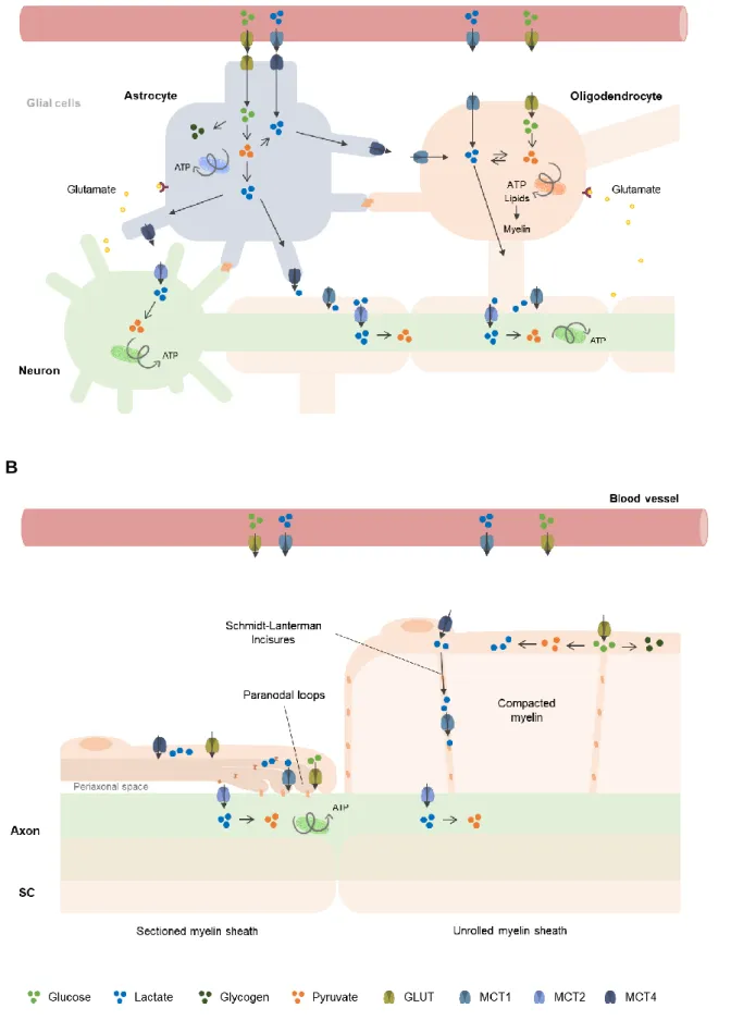
Since the ANLSH emerged, a growing amount of studies have been done to explore the energy metabolism in the CNS, which is currently believed to be distributed across the three abovementioned cellular compartments (Amaral et al., 2013). Although less work has been devoted to PNS metabolism, emerging data also points to a division of metabolic activities between SCs and neurons (Brown et al., 2012; Chen et al., 2003; Viader et al., 2011). The exchange of energy substrates in the nervous system relies on the specific expression of connexins, glucose transporters (GLUT), monocarboxylate transporters (MCTs) and enzymes according to the metabolic tasks of the cells (Hirrlinger & Nave, 2014) (Figure 4).
Regarding the CNS, glucose enters the brain parenchyma via GLUT1 at the blood-brain barrier and is taken up by astrocytes through GLUT1, but also by oligodendrocytes and neurons through GLUT1 and GLUT3, respectively (Maher et al., 1994). Similarly in the PNS, GLUT1 is present in the perineurium and in the abaxonal side of Schwann cells, and GLUT3 mediates the uptake of glucose by the axon (Jensen et al., 2014).
Lactate shuttling requires particularly high lactate dehydrogenase (LDH) activity and rapid intercellular lactate transport (Hui et al., 2017). Lactate can only be used as a source of energy if oxidized to pyruvate via lactate dehydrogenase. Neurons express the LDH isoform LDH1, which preferentially uses lactate as substrate, whereas astrocytes express mostly LDH5, typically present in lactate-producing tissues (Bishop et al., 1972; Pellerin et al., 1998). The different distribution of these isoenzymes supports the idea that astrocytes might act as lactate sources for neurons to use it as energy substrate, in agreement with the ANLSH.
FEUP | ICBAS –
7
Lactate is exchanged between cells through MCTs. These transmembrane proteins perform also the proton-coupled transport of other monocarboxylates, namely pyruvate and ketone bodies, although at much lower extent (Halestrap, 2012). Four MCT types were functionally characterized, showing different affinities for monocarboxylates: MCT2 has a high affinity; MCT1 and MCT3 show an intermediate to high affinity, and MCT4 a low affinity (Bergersen et al., 2001; Grollman et al., 2000). Their selective presence in tissues tends to be related with their glycolytic versus oxidative phenotype. In the CNS, oligodendrocytes, neurons, and astrocytes predominantly express MCT1, MCT2, and MCT4, respectively (Lee et al., 2012). This cellular distribution of MCTs appear to be correlated with their main metabolic properties, which is consistent with the lactate shuttle hypothesis (Morrison et al., 2013).Regarding the presence of MCTs in the PNS, our group has demonstrated the expression of MCT1, MCT2 and MCT4 by SCs, while MCT1 and MCT2 were found in mouse DRG neurons (Domènech-Estévez et al., 2015). A higher level of MCT1 expression was observed in maturing PNS, which suggested an increased need of monocarboxylates, presumably lactate, during that time (Domènech-Estévez et al., 2015). Regarding their spatial distribution, MCT1 was found in Schmidt-Lanterman incisures (SLIs) and in paradonal regions, both Cx32-rich structures composed by non-compacted myelin (Balice-Gordon et al., 1998; Domènech-Estévez et al., 2015). In turn, MCT4 was localized in the perinuclear and abaxonal compartments of mSCs, suggesting its presence in Cajal bands and in the outer cytoplasmic mesaxonal line (Domènech-Estévez et al., 2015). Altogether the available data indicate that each MCT isoform shows a preferred cellular distribution in CNS matching metabolic phenotype of glia and neurons (Domènech-Estévez et al., 2015). In the PNS, MCTs are expressed in different compartments of the SC-axon complex, potentially allowing lactate transport following a concentration gradient.
A schematic view of the main neuron-glia metabolic interactions in the CNS and PNS are shown in Figure 5.
8 – Ana Temporão | Masters Dissertation
A
B
Figure 5. Schematic representation of the metabolic communication between neurons and glial cells in the
FEUP | ICBAS –
9
1.3. Implications of disrupted SC-axon crosstalk
Several evidences for an axon-glia metabolic communication come from studies with animal models exhibiting axonal degeneration independently of myelin loss. Regarding the CNS, mice lacking oligodendroglia-specific genes PLP1 (Garbern et al., 2002) and CNP (Lappe-Siefke et al., 2003) did not show extensive signs of impaired myelination but presented axonal degeneration. In the PNS, mouse mutants for the myelin-associated glycoprotein (MAG) presented axonal degeneration and decreased axon caliber in sciatic nerve fibers, despite their apparently normal myelination (Li et al., 1994). Moreover, SC-specific deletion of the metabolic regulator liver kinase B1 (LKB1) led to axon degeneration as a consequence of perturbed energy homeostasis independently of dysmyelination (Beirowski et al., 2014).
Given that lactate was proposed to be a crucial fuel for metabolic support to axons, it is not surprising that disrupting MCT1-mediated transfer of lactate from oligodendrocytes led to axonal damage (Lee et al., 2012). Importantly, in an in vitro experiment, the addition of free lactate to the medium rescued MCT1 blockage and ameliorated axonal phenotype, showing that neurodegeneration was due to decreased lactate export from oligodendrocytes and not import into neurons (Lee et al., 2012).
Similarly, axonal dysfunction in central and peripheral neuropathies may occur through myelin-unrelated mechanisms such as the failure of metabolic support.
Amyotrophic lateral sclerosis (ALS) is characterized by a progressive loss of motor neurons in the brain and spinal cord, also manifesting degeneration of peripheral fibers (Riva et al., 2014). Reduced levels of MCT1 expression in oligodendroglia were observed in superoxide dismutase 1 (SOD1)-mutant mice, a model of ALS and in brain samples from ALS patients (Lee et al., 2012). Together with the failure of mitochondrial bioenergetics (Ferri et al., 2006) and perturbations in axonal transport (Marinkovic et al., 2012), the lack of glial supply of lactate to motor neurons may also potentially be involved in the pathogenesis of ALS (Beirowski, 2013). Whether MCT dysfunction compromising SC-axon metabolic coupling is implicated in ALS pathogenesis is still unknown.
Peripheral neuropathies are a common cause of morbidity in elderly populations, representing a significant economic and societal burden (Hughes, 2002). The etiology for this type of peripheral neuropathies goes from metabolic irregularities (e.g. diabetic neuropathy) and genetic mutations (e.g. Charcot-Marie-Tooth diseases) to inflammation (e.g. demyelinating polyneuropathies) and infection (e.g. leprosy) (Samara et al., 2013).
Heritable peripheral neuropathies are collectively designated as Charcot-Marie-Tooth diseases (CMT). Most of the genes mutated in CMT play a role in maintaining the structure or function of the axon-SCs complex formed by and motor/sensory neurons (Saporta & Shy, 2014). CMT neuropathies cause distal muscle weakness and atrophy, and they can be roughly divided into demyelinating (CMT1), axonal (such as CMT2) and more rare intermediate variants (Timmerman et al., 2013). As in some forms of CMT2 disease the neurodegeneration occurs in the absence of detectable changes in myelin integrity, the disruption of metabolic support of the
10 – Ana Temporão | Masters Dissertation
peripheral neurons may also be implicated in the pathogenesis of this CMT variant (Timmerman et al., 2013). This hypothesis is further supported by the observation that many types of peripheral neuropathies exhibit deficits in axonal energy, such as mitochondrial dysfunction, as a common feature (Beirowski et al., 2014; Viader et al., 2011).
In order to develop new therapeutic approaches to treat or at least improve the quality of life of people suffering from these diseases, it is crucial to explore the metabolic SC-axon communication in physiological and pathological conditions.
FEUP | ICBAS –
11
1.4. Aim, Hypothesis and Strategy
Both neurons and glia play critical roles on nervous system homeostasis, and abnormalities in their relationship are at the core of innumerous neuropathic disorders. The delivery of high-energy substrates from glial cells to neurons via MCTs may be one of the most important events underlying their communication. Although much work has been done to explore CNS metabolism, comprehensive knowledge regarding the physiological relevance of glia-axon metabolic support and the role of MCTs in the PNS is still missing.
Therefore, the aim of this master’s thesis is:
1. To explore SCs’ metabolic adaptations to neuronal cues, in particular lactate flow and production;
2. To study the role of monocarboxylate transporters in SCs in vivo.
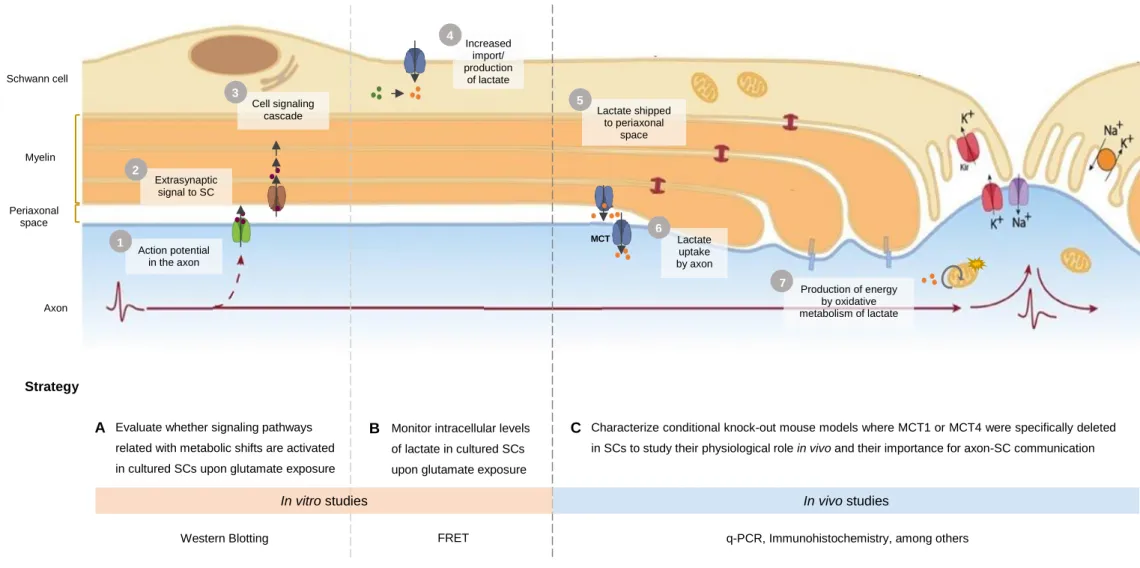
Firstly, we hypothesized that extrasynaptically released neurotransmitters interact with receptors in SCs, triggering the activation of certain signaling pathways associated with the reprogramming of their metabolic status. We believe that it would enhance lactate uptake and/or glycolytic metabolism leading to increased intracellular levels of lactate. This monocarboxylate would then be shipped to the periaxonal space and taken up by the axon through MCTs, finally undergoing oxidative metabolism to provide the energy needed for signal transmission along the axon.
To test these hypotheses, we elaborated a strategy divided into in vitro studies and in vivo approaches. First, we intended to evaluate whether signaling pathways reported to precede metabolic shifts are activated in cultured SCs in response to glutamate. To do that, we compared the phosphorylation levels of proteins involved in these pathways between stimulated and non-stimulated cultures using Western Blot. Additionally, we planned to monitor the intracellular levels of lactate in SCs upon glutamate exposure, by performing continuous FRET imaging of cultured SCs expressing a lactate-sensitive probe. Lastly, we aimed to characterize conditional knock-out mouse models where MCT1 or MCT4 were specifically deleted in SCs to study their physiological role in vivo and their importance for axon-SC metabolic communication. The validation of the mouse models and the effects of MCT depletion were assessed by immunohistochemistry and quantitative PCR, among others.
12 – Ana Temporão | Masters Dissertation Extrasynaptic signal to SC Increased import/ production of lactate Lactate shipped to periaxonal space Lactate uptake by axon Cell signaling cascade Production of energy by oxidative metabolism of lactate Action potential in the axon
Evaluate whether signaling pathways related with metabolic shifts are activated in cultured SCs upon glutamate exposure stimulation A Schwann cell Myelin Periaxonal space Axon
Monitor intracellular levels of lactate in cultured SCs upon glutamate exposure
B Characterize conditional knock-out mouse models where MCT1 or MCT4 were specifically deleted in SCs to study their physiological role in vivo and their importance for axon-SC communication
C 2 3 1 4 5 6 7 MCT Hypothesis Strategy
In vitro studies In vivo studies
Western Blotting FRET q-PCR, Immunohistochemistry, among others
FEUP | ICBAS –
13
2. Materials and Methods
2.1. Reagents
Buffers and Solutions
Lysis buffer (for protein): 80mM TrisHCl (Duchefa Biochemie, T1513.1000); 5 mM EDTA (Sigma-Aldrich, 101520387), 5% SDS (Sigma-(Sigma-Aldrich, 101944022), 1 mM NaF (Sigma-(Sigma-Aldrich, S7920), 1 mM NaVO4 (AppliChem, A2196.0005) and protease inhibitor cocktail 1X (complete Mini-EDTA-free tablets; Roche, 11836170001)
Running buffer: 25 mM Tris base (Sigma, 101776239, USA), 192 mM glycine (AppliChem, A4554.5000) and 0.1% SDS (AppliChem, A3942.1000) in distilled water
Transfer buffer: 25 mM Tris base (Sigma, 101776239), 192 mM glycine (AppliChem, A4554.5000), and 20% methanol (Honeywell, 24229-2.5L-R) in distilled water
Tris-buffered saline (TBS): 20 mM Tris base (Sigma, 101776239) and 150 mM NaCl (Honeywell, 10314835) in distilled water, pH 7.5
Blocking buffer (for WB): 5% non-fat dried milk (AppliChem, A0803.1000) in TBS TBS Tween: 0.05% Polysorbate 20 (Duchefa Biochemie, P1362.1000) in TBS
Phosphate buffer saline (PBS): 137mM NaCl (Honeywell, 10314835), 2.7mM KCl (VWR, 26764.260), 10mM Na2PO4 (Sigma, S-0751), 1.7mM KH2PO4 (VWR, 26764.260), pH7.5
Krebs-Ringer-HEPES (KRH) buffer: 112mM NaCl (Honeywell, 10314835), 1.25mM CaCl2 (Sigma-Aldrich, 10314835), 1.25mM MgSO4 (Sigma-Aldrich, 101928373), 10mM HEPES (AppliChem, A3724.0100), 24mM NaHCO3 (Merck, 1.06329.0500), 5mM KCl (VWR, 26764.260) in distilled water
Lysis Buffer (for DNA): 0.5 mg/mL Proteinase K (Merck, 1.24568) added to 100mM NaCl (Honeywell, 10314835), 50mM Tris-HCl pH8.0, 100mM EDTA (Sigma-Aldrich, 101520387), and 1% SDS (Sigma-Aldrich, 101944022)
Phosphate buffer (PB) 0.1M: Na2PO4.2H2O (Honeywell, 10314743), 0.1M NaH2PO4 anhydrous (Sigma-Aldrich, RDD007) in distilled water, pH7.2
Zamboni fixative: 2% paraformaldehyde (PFA; AppliChem, A3813.0500) with 15% Picric Acid (Sigma-Aldrich, 239801) in PB, pH7.3
Cell culture
14 – Ana Temporão | Masters Dissertation
SC culture medium: DMEM 1X + GlutaMAX (Gibco, 61965-026) supplemented with 10% fetal bovine serum (FBS; Gibco, 16000-044), 50 U/ml penicillin, 50 µg/ml streptomycin (Gibco, 15070063), 4 μM forskolin (LC labs, 66575-29-9), and 1.25 nM EGF domain NRG1b1 (R&D Systems, 396-HB). DMEM contains 0.4 mM glycine.
Bovine pituitary extract (Lonza Biosciences, CC-4009) (working solution: 21mg/mL) Glutamate: L-Glutamic acid monosodium salt hydrate (Sigma-Aldrich, 1002218638)
HEK293T culture medium: DMEM 1X + GlutaMAX (Gibco, 61965-026), 10% FBS (Gibco, 16000-044), 50 U/ml penicillin, 50 µg/ml streptomycin (Gibco, 15070063), 1X Non-Essential Amino Acids solution (Gibco, 11140050) and 1 mM Sodium Pyruvate (Gibco, 11360).
2.2. Glutamate stimulation of primary cultured SCs
Cell culture
Rat Schwann cells were obtained from sciatic nerves, extracted from 1-3 days old Sprague Dawley rats, and prepared as previously described (Brockes et al., 1979).
Cells were plated in PLL-coated 6 cm Petri dishes, at a starting density of 4 million cells/dish, maintained in SC culture medium and passaged no more than 6 times.
Immunoblot analysis of cell signaling
Cultures were rinsed twice in PBS and exposed to starving and stimulation media as described in detail in the ‘Results and Discussion’ section.
After stimulation, cells were rinsed twice in PBS, lysed with 150 μL lysis buffer, and scraped with a plastic spatula. Lysates were collected, further homogenized in the Tissue Lyser II (Qiagen) with steel beads and centrifuged 20 min at 13000g in Centrifuge 5415D (Eppendorf). Protein concentration was estimated for each cell lysate by PierceTM bicinchoninic acid assay (BCA; Thermo Scientific, 23225) and absorbance was determined at 562 nm using PlateReader (Biotek).
Equivalent amounts of protein were loaded into a 10% SDS-PAGE gel, along with molecular weight marker (Precision Plus Protein Standards; BIO-RAD, 161-0373). Proteins were electrotransferred to nitrocellulose membranes. Membranes were blocked for 1h with blocking buffer, incubated overnight at 4ºC with primary antibodies, and with secondary antibodies for at least 1h20 at room temperature (RT) (Table 1). Immunoblots were analyzed using the LI-COR Odissey system (Biosciences) as recommended by the company. The results were expressed as the ratio of phopsphrylated/total protein, following the normalization to tubulin.
FEUP | ICBAS –
15
Table 1. List of primary and secondary antibodies used for Western Blot. Primary antibodies
Antigen Animal of origin Dilution Product reference
P-AKT (S473) Rabbit 1:1000 Cell Signaling, 3787
P-ERK (T202/Y204) Rabbit 1:1000 Cell Signaling, 9101
P-S6RP (S235/236) Rabbit 1:1000 Cell Signaling, 2211
AKT Rabbit 1:1000 Cell Signaling, 9272
ERK Rabbit 1:1000 Cell Signaling, 9102
S6RP Rabbit 1:1000 Cell Signaling, 2217
Tubulin Mouse 1:5000 Cell Signaling, 3873
Secundary antibodies
Antigen Conjugated fluorophore Animal of origin Dilution Product reference
Rabbit IgG IRDye® 800CW Goat 1:10000 LI-COR 925-32211
Mouse IgG IRDye® 680RD Donkey 1:10000 LI-COR 925-68072
2.3. FRET-based intracellular lactate measurements
Approximately 40 000 HEK 293T cells (Thermo-Fisher Scientific) were plated onto PLL-coated 18 mm coverslips, and transfected 48h later with LipoD293 (SignaGen) and 0.75mg of Laconic DNA, according to the indication of the manufacturer (San Martín et al., 2013).
24h after transfection, cells were imaged at the Advanced Light Microscopy Facility (Science for Life Laboratory, Stockholm, Sweden) using a 10x air objective, in the absence of atmospheric control.
Cells were exposed to different solutions prepared in KRH buffer, as detailed in ‘Results and Discussion’ section, at a perfusion temperature of 37ºC. Laconic behavior at each condition was monitored during 5 minutes, and data was finally presented as mTFP/Venus fluorescence ratio in function of the time, normalized to the first fluorescence ratio acquired at steady-state.
2.4. Characterization of the mouse models
Animals
Conditional knockout (cKO) mice expressing Cre recombinase under the promoter for P0 and floxed MCT1 or MCT4 alleles were provided by our collaborator Dr. Luc Pellerin’s group at the University of Lausanne, Switzerland. The P0-Cre line is commercially available from Jackson Labs (B6N.FVB-Tg(Mpz-cre)26Mes/J) (M. A. Feltri et al., 1999). The floxed lines were developed by Cyagen (Switzerland).
16 – Ana Temporão | Masters Dissertation
Ablation of MCT1 expression is driven by the excision of exon 5 of gene SLC16A1. In turn, deletion of MCT4 expression occurs through the elimination of exons 3, 4 and 5 of SLC16A3. All experiments were performed in accordance with the guidelines of Karolinska Institute.
Genotyping
DNA from mouse ear or tail clips was extracted by incubating samples at least 4 hours at 56ºC with lysis buffer, and by alternating centrifugations with the sequential addition of 5M NaCl, isopropanol and ethanol 70%, and finally resuspended in TE buffer.
A cocktail of PCR reagents was prepared by mixing primers (0.2μM at working solution; Table 2) with DreamTaq Master Mix (ThermoFisher, K1082) in nuclease-free water. The cocktail was added to 0.5μL DNA, undergoing a PCR reaction (Table 3).
PCR products were resolved in a 1-2% agarose gel by electrophoresis (Agarose, Fisher Scientif, BP160-500; GelRed Nucleic Acid Stain, 41003; Ready-to-use 100bp DNA ladder, 31032, Biotium). The gel was visualized using ChemiDoc (BIO-RAD).
Table 2. Primer sequences used for genotyping PCR.
Gene Primer Forward Primer Reverse
MCT1 5’-AGACTTGGGTAACTGAATGATGCTGACT-3’ 5’-TCCAAGGACAGCCAAGCTACATAGAG-3’
MCT4 5’-ATTTAGACTCAGAGGTGGGCAGAGTG-3’ 5’-TTGCCAGGGTGACCATCTCA-3’
P0-Cre 5’-AGGTGTAGAGAAGGCACTTAGC-3’ 5’-CTAATCGCCATCTTCCAGCAGG-3’
IL-2 5’-CTAGGCCACAGAATTGAAAGATCT-3’ 5’-GTAGGTGGAAATTCTAGCATCATCC-3’
Table 3. Programs used for PCR Reaction.
Floxed MCT1/ MCT4 P0-Cre
Temperature Time Cycles Temperature Time Cycles Initial
denaturation 94 ºC 3 min 1 94 ºC 3 min 1
Denaturation 94 ºC 30 s 35 94 ºC 15 s 32 Hybridization 55 ºC 30 s 62 ºC 15 s Extension 72 ºC 30 s 72 ºC 15 s Final
extension 72 ºC 10 min 1 72 ºC 2 min 1
Conservation 4 ºC pause - 4 ºC pause -
Quantitative Real-Time Polymerase Chain Reaction (RT-qPCR)
RNA from the endoneurium of mouse sciatic nerves was extracted using RNeasy Lipid Tissue Kit (Qiagen 74804), following the manufacturer’s instructions. Briefly, tissue was homogenized in 500μL QIAzol (QIAGEN, 79306) and RNA fraction was extracted using
FEUP | ICBAS –
17
chloroform (Sigma-Aldrich, 101538376). RNA was then purified in column using the reagents provided in the kit. Residual contaminating DNA was digested with RNAse free DNAse set (QIAGEN, 79254). Next, RNA concentration was determined using NanoDrop 1000 (Agilent). Finally, 40ng RNA was retrotranscribed with PrimeScript RT reagent Kit (Takara RR037A), according to the manufacturer’s recommendations (Table 4).MCTs, P0 and Ubiquitin-conjugating enzyme E2 L3 (Ubiq) mRNA levels were detected by quantitative real-time PCR using FastStart Universal SYBR Green Master Mix (Roche, 10356100) and 7500 Fast Real Time PCR System (Applied Biosystems).
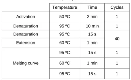
Oligonucleotides sequences used are shown in Table 5 and qPCR protocol in Table 6.
Table 4. Retrotranscription protocol.
Temperature Time
Reverse transcription 37 ºC 15 min
Inactivation of reverse transcriptase 85 ºC 5 s
Conservation 4 ºC pause
Table 5. Primer sequences used for qPCR.
Gene Primer Forward Primer Reverse
MCT1 5’-AATGCTGCCCTGTCCTCCTA-3’ 5’-CCCAGTACGTGTATTTGTAGTCTCCAT-3’
MCT2 5’-CAGCAACAGCGTGATAGAGCT-3’ 5’-TGGTTGCAGGTTGAATGCTAA-3’
MCT4 5’-CAGCTTTGCCATGTTCTTCA-3’ 5’-AGCCATGAGCACCTCAAACT-3’
P0 5’-AGCCCCAGCCCTATCCTGGC-3’ 5’- GCAGTGCAGGGTCACCTGGG-3’
Ubiq 5’-CAGCCACCAAGACTGACCAA-3’ 5’-CATTCACCAGTGCTATGAGGGA-3’
Table 6. qPCR protocol.
Temperature Time Cycles
Activation 50 ºC 2 min 1 Denaturation 95 ºC 10 min 1 Denaturation 95 ºC 15 s 40 Extension 60 ºC 1 min Melting curve 95 ºC 15 s 1 60 ºC 1 min 1 95 ºC 15 s 1
18 – Ana Temporão | Masters Dissertation
Immunohistochemistry
Adult animals were anesthetized intraperitoneally with a mixture of 10 µL/g of Ketanarkon 100 (1 mg/ml, Streuli) with 0.1% Rompun (Bayer) in PBS, sacrificed by cervical dislocation, and dissected to collect sciatic nerves, muscles (gastrocnemius, tibialis, soleus, extensor digitalis longus (EDL)) and hind paw skin. Tail was also collected for validation of the previously performed genotyping.
After fixations, tissues were cryoprotected (sucrose 20% overnight at 4ºC), embedded in optimal cutting temperature medium (OCT; Cell Path, KMA-0100-00A), and sectioned according to Table 7.
Tissues were immunostained following the procedures detailed below. Images were then acquired using Zeiss LSM 700 confocal microscope and processed with FIJI software (Schindelin et al., 2019).
Table 7. Settings for fixation and sectioning of different tissues.
Tissue Fixation Sections Thickness
Sciatic nerve 4% PFA; 1h Transversal 10 µm
Gastrocnemius 4% PFA; 15 min Longitudinal 25 µm Tibialis Soleus 4% PFA; 10 min EDL
Hind paw skin Zamboni fixative; 2h Transversal 50 µm
Teased fibers of sciatic nerve
PFA-fixed fixed mouse sciatic nerves were stripped of perineurium and small bundles of fibers separated using thin needles. Individual myelinated fibers were spread on a TESPA (Sigma-Aldrich, 101698432)-coated glass slide, allowed to dry and stored at -80ºC until used.
For immunostaining, samples were initially washed with PBS and permeabilized with 0.2% Triton X-100 for 15 min at RT. Afterwards, fibers were blocked with 1% BSA and 0.2% Triton X-100 for 1h at RT, and incubated with primary antibodies overnight at 4ºC or RT (Table 8). Next, samples were washed and fluorescently labeled with secondary antibodies for 2h at RT (Table 8). Lastly, nuclei were counterstained with DAPI and tissues were mounted with Vectashield.
Cross sections of sciatic nerve
10μm-thick cryosections of mouse sciatic nerve were washed with PBS and permeabilized as previously described. Secondly, samples were blocked and incubated with primary antibodies overnight at 4ºC or RT (Table 8). Next, sections were washed, incubated with secondary antibodies and counterstained with DAPI (Table 8) before mounting with Vectashield.
FEUP | ICBAS –
19
Intraepidermal nerve fibers
Hind paw skin was sectioned at 50 μm thickness, blocked with 1% NGS and 0.15% Triton X-100 for 3h at RT and incubated with primary antibodies overnight at 4ºC. Next, sections were fluorescently labeled with secondary antibodies for 2H at RT, stained with DAPI and mounted.
Z-stacks were obtained from four different frames of three or more sections per animal. The number of intraepidermal fibers was counted and normalized to the length of dermal-epidermal junction in the superimposed image.
Neuromuscular junctions
Muscle sections were washed with PBS, and blocked with 4% BSA and 0.5% Triton X-100 for 5h at RT. Then, samples were incubated with primary antibodies (Table 8) for 48h at 4ºC, washed and stained with secondary antibodies (Table 8) for 2h at RT. Finally, samples were counterstained with DAPI and mounted.
Table 8. List of primary antibodies, secondary antibodies and stains used for immunohistochemistry. Primary antibodies
Antigen Animal of origin Dilution Product reference
MCT1 Rabbit 1:200 Homemade
MCT4 Rabbit 1:200 Santa Cruz sc-50329
Neurofilament-145 Rabbit 1:200 Millipore AB1987
Neurofilament-200 Mouse 1:400 Sigma N0142 Clone N52
PGP9.5 Rabbit 1:400 Ultraclone RA95101
Secundary antibodies
Antigen Conjugated fluorophore Animal of origin Dilution Product reference
Mouse IgG Alexa Fluor® 488 Goat 1:200 Life Tech R37120
Mouse IgG Alexa Fluor® 594 Goat 1:200 Life Tech R37121
Rabbit IgG Alexa Fluor® 488 Goat 1:200 Life Tech A11034
Rabbit IgG Alexa Fluor® 594 Goat 1:200 Life Tech A11037
Stains
Conjugated fluorophore Dilution Product reference α-Bungarotoxin Alexa Fluor® 488 1:500 Thermo Scientific B13422
Phalloidin Alexa Fluor® 488 1:20 Thermo Scientific A12379 4',6-Diamidino-2-Phenylindole, Dihydrochloride
FEUP | ICBAS –
21
3. Results and Discussion
3.1. In vitro study of the intracellular response of SCs to neurotransmitters
Active axons can release ATP and glutamate (Glu) into the narrow periaxonal space, where they reach high local concentrations (Samara et al., 2013; Stys, 2011). Myelinating and non-myelinating SCs, as well as their common precursors, express various ion channels and G protein-coupled receptors at their surface that may act as activity sensors. Purinergic and glutamatergic receptors are among this set of proteins, so that SCs can detect ATP and Glu as extrasynaptic signals of axonal activity (Samara et al., 2013).
In the CNS, it was suggested that neuronal activity induces astrocytes and oligodendendrocytes to provide metabolic substrates and enzymes to neurons (Barros, 2013; Frühbeis et al., 2013). Peripheral glia may play a similar role by providing the metabolic support to axons.
In order to dissect whether (and how) the metabolic status of SCs is modulated by neuronal activity, we devised a strategy to assess the amplitude of lactate production and release by cultured SCs when stimulated by glutamate. If SCs promptly increase the generation of glycolysis products to support axons during periods of their activity, short-term exposure of SCs to neurotransmitters should be enough to trigger a feedback response.
With this in mind, we first evaluated whether rat SCs in our cell-culture settings were able to readily sense the presence of glutamate and initiate a feedback response through the activation of metabolism-related signaling pathways. Once we confirm that SCs in our in vitro system are responsive to glutamate and we establish a reliable protocol of glutamate-driven SC stimulation, we can use it coupled with a FRET system to screen potential fluctuations of the SC’s intracellular level of lactate arising from glutamate signaling. Hence, we worked in parallel on setting up a FRET imaging system for cultured cells expressing Laconic, a lactate-sensitive FRET probe (San Martín et al., 2013). As an initial attempt to reproduce the method using the available protocol for Laconic, we exposed HEK293T cells expressing Laconic to solutions that were reported to cause specific signal emission.
Of note, this in vitro approach is still ongoing and no conclusive data will be presented. A long series of optimization steps were needed to be carried out for the establishment of both systems.
3.1.1. Cell signaling cascades triggered in cultured SCs by glutamate exposure
During neuronal activity, glutamate extrasynaptically secreted by axons can interact with either ionotropic or metabotropic receptors in SCs (Verkhratsky & Kirchhoff, 2007). The former are ligand-gated ion channels subdivided into NMDA, AMPA and kainate receptors, which exhibit distinct functional properties (Verkhratsky & Kirchhoff, 2007). Metabotropic glutamate receptors (mGluR), in turn, are G-protein-coupled receptors that control intracellular second messenger signaling cascades. In comparison with ionotropic receptors, mGluR tend to be activated by a
22 – Ana Temporão | Masters Dissertation
stronger and/or longer stimulus (Verkhratsky & Kirchhoff, 2007). As such, following the abovementioned strategy to test if SCs promptly respond to axonal cues of activity, we expect to trigger ionotropic signaling in SCs shortly exposed to low concentrations of glutamate.
Recently, it was reported that ionotropic glutamate stimulation in primary rat SC cultures triggered the robust activation of PI3K/AKT/S6RP and ERK signaling pathways, in particular through interaction with NMDA receptors (Campana et al., 2017). These signaling pathways may be associated with glycolytic metabolism(Bhaskar & Hay, 2007; Marat & Haucke, 2016; Perkinton et al., 2002). The response was transient and bimodal, peaking upon a treatment of 80 µM glutamate for 20min and ceasing when glutamate concentrations exceeded 250 µM. This work matched perfectly the first part of our hypothesis. Thus, we followed the same protocol (Campana et al., 2017) as an attempt to replicate the outcome in our cell cultures, since more or less defined parameters in in vitro contexts can influence the type or magnitude of the effect.
The methodology involvedstarving subconfluent rat SCs for 1 hour in serum-free medium in order to reduce their metabolism to the basal levels. Then, different concentrations of Glu were applied for 10min, the proteins were extracted from treated and non-treated cells, and thelevels of total and phosphoryled AKT, S6RP and ERK were assessed by Western Blot (WB) (Table 9, step 1). Surprisingly, in our cell culture settings, no apparent effect of Glu was observed. The removal of fetal bovine serum (FBS), even when combined with the absence of pituitary extract, was not sufficient to reduce the cell metabolism to the baseline, which is required to reveal the specific response to Glu. Exploring the small deviations from the reported protocol, we found that loading half of the protein mass into the WB gel could be a potential cause for such outcome.
Moreover, whereas enhanced chemiluminescence (ECL) was the method adopted by Campana and colleagues for WB detection, we used Odyssey® Infrared Imaging System. We believe that this divergence is not a limiting factor. On the contrary, infrared imaging counteracts the dynamic light-producing enzymatic reactions on which ECL relies, suppressing this way the need for optimizing reaction times and imaging (Mathews et al., 2009). Furthermore, the highly-sensitive method that we used allows a broader detection range than ECL and the simultaneous imaging of different proteins on the same blot, which increases the detection efficiency.
In order to enhance the outcome of Glu-mediated stimulation of SCs, we used a multi-step optimization process (Table 9). We changed the culture medium to contain Nrg1 type III instead of bovine pituitary extract (BPE), as the latter possess undefined – and possibly variable – composition. We also performed the next assay on confluent populations of SCs as a way to collect more proteins from each culture plate (Table 9, step 2). Since we obtained a good outcome from the first optimized stimulation of confluent cells, we presumed that high cell density allowing us to have enough material would not be a parameter negatively affecting the outcome of our experiments. Additionally, we tried to strengthen SC starving step to lower their metabolic baseline (Table 9, steps 3-4).
In parallel, as a way to prevent operation errors, we minimized the number of conditions to test, doing also technical replicates for each condition. Since we were interested on evaluating the shortest axonal stimulation that triggers a glial response, we tested exposing cells for 5 min
FEUP | ICBAS –
23
to 40 µM or 80 µM glutamate, concentrations described to induce the stronger effect, in comparison with 20 min-long stimulation.The initial trials of cell starvation exhibited variable efficiency, even when the experiment was repeated using the same settings. An activation of AKT and/or ERK pathways following Glu exposure was visible when the starving was successful. A rise in ERK phosphorylation was observed specially for 5 min-long treatment with Glu. However, the results regarding the AKT signaling pathway were not linear. The phosphorylation of AKT was either robust or absent, while no clear effects on S6RP phosphorylation were observed.
We found in the literature that Glu at high concentration (2 mM) signals through metabotropic glutamate receptors in SCs to enhance Nrg1-induced phosphorylation of ERK, but not AKT (Saitoh et al., 2016). In that work, the metabolic baseline of primary cultured SCs was reached after starving cells during 6h in 1% FBS medium, and Glu stimulation was done for 30min in the presence of different picomolar concentrations of Nrg1 type III. Bearing this in mind, we questioned whether ionotropic glutamate signaling in SCs may similarly modulate ErbB receptor-mediated cellular signaling.
To test this possibility, we tried to combine the approaches described in the two referred papers (Table 9, steps 5-6). After the application of an optimized starving method, subconfluent cultured SCs were stimulated for 10 min with Nrg1 at concentrations ranging from tens of picomolar to few nanomolar, in the absence or presence of 80 μM glutamate. Cells efficiently reached the basal levels of activity, and usually the magnitude of activation of AKT/S6RP and ERK signaling increased in function of the concentration of Nrg1. Intriguingly, the addition of Glu generally decreased the effect induced by Nrg1, with exception of one assay where AKT phosphorylation was enhanced by Glu in the presence of at least 0.1 nM Nrg1. Furthermore, Glu alone induced variable types of response, either reducing or increasing the phosphorylation of the ERK, AKT and/or S6RP.
We supposed that the level of cell confluence may have largely influenced the outcome. Cultured SCs displayed ~85% confluence at the onset of the experiments as suggested by Campana and colleagues (2017). However, the method of starving described in the same article was substantially weaker compared to the treatment that we adopted. We observed that in standard culture conditions, our rat SCs would continue to replicate until the cytoplasm was compacted and little space would be visible between cells. Consequently, subconfluent cells may rely more on the presence of nutrients and growth factors, and may be more susceptible to starving-induced stress. As such, SCs in this set of tests may be more stressed upon starvation induced by removal of Fsk and Nrg1 from the culture medium for 24h plus FBS for 2h. The fact that AKT/S6RP signaling is also associated with survival responses and that stress activates ERK signaling (Yarden & Sliwkowski, 2001) can potentially explain the observed phenotype. We believe that exposing subconfluent SCs to a strong starvation regime induces more stress. In this context, residual levels of Nrg1 may trigger the activation of AKT/S6RP and ERK signaling in order to generate the survival response to stress. The additional presence of Glu seems to counteract the survival response of SCs to Nrg1. In line with our hypothesis, glutamate may signal
24 – Ana Temporão | Masters Dissertation
the metabolic demands of axons to SCs, leading to the glial mobilization of energy reserves, which are already depleted under these conditions. Thus, we believe that cell confluence is a critical factor to take into account in these experiments.
Having this in mind, we lastly followed the same procedure on a confluent culture of rat SCs, adding Nrg1 at concentrations in the order of hundreds of picomolar, alone or combined with 80 μM glutamate (Figure 7). Under these conditions, the presence of Glu appeared to enhance the Nrg1-induced phosphorylation of AKT, S6RP, and ERK. Moreover, the effect of Glu was visible even in the absence of Nrg1, and peaked when the lowest concentration of Nrg1 was added to the culture (Figure 8).
Firstly, these observations suggest that cell confluence is an important parameter to take into consideration in our experimental settings. Indeed, when cells in culture are establishing more and tighter contacts with each other, they form a more stable cell population. That means that confluent cells are less sensitive and can adapt much more efficiently to stress conditions. We further suppose that it can result in more consistent responses to external factors. Nevertheless, this experiment has to be repeated exactly with the same technical conditions to evaluate the reproducibility of the outcome.
Additionally, it seems that the number of times that SCs are passaged in culture influences the type of response triggered by stimulation. In Campana et al. (2017), it was mentioned that cells used were passaged no more than 7 times. We followed this indication without attention if a given protocol was repeated on cells at the same passage level. However, it may have compromised the replication of the results. Having a look on the assays number 2 and 3 where only Glu stimulation was tested (Table 9), there are clear differences on the type of response exhibited by confluent cells at the fourth passage (P4) versus at the sixth passage (P6) exposed to the same experimental settings. The metabolic baseline, as well as a robust Glu-induced increase of Erk and/or Akt phosphorylation, were reached in both tests for cells at P6; whereas a mild efficacy of the starving and a low phosphorylation of AKT and ERK were observed in cells at P4 in the best case scenario.
This difference may partially follow differences in physiological events in the developing versus mature PNS. In fact, since myelin-forming cells are highly energy demanding during lineage progression and particularly myelination (Rinholm et al., 2011; Sánchez-Abarca et al., 2001), it is plausible to assume that glia-to-axon metabolic support does not occur or is minimized at that period. In mature PNS, in turn, a growing body of evidence supports the idea that SCs provide axons with metabolites, as we intend to prove during neuronal activity. Thus, even though we used confluent cultured cells, the state of cell maturity (as reflected by the number of SC passages) may modulate their plasticity to develop physiological adjustments through subcellular signaling upon stimulation with neurotransmitters.
FEUP | ICBAS –
25
Table 9. Main steps of optimization for the glutamate stimulation of cultured rSCs and respective outcomes (continues on the next page).
Purpose Culture Starving Stimulation Observations n
1 Follow procedure described in Campana et al. (2017) ~85% cell confluence SCs at passage no. 4 FBS/Fsk/BPE in DMEM Fsk/BPE in DMEM; 1h 0, 20, 40, 80, 100, 250, 500, 2000 μM Glu Positive Ctr: Nrg1 5nM 10 min
Low mass of protein loaded Baseline not reached
Total AKT, ERK and S6RP variable No reproduction of phosphorylation levels
1
Fsk in DMEM;
1h 1
2
Load 40μg protein Use media with
more defined composition Reduce number of conditions-test ~98% cell confluence a) SCs at passage no. 6 b) SCs at passage no. 4 FBS/Fsk/Nrg1 in DMEM Fsk/Nrg1 in DMEM; 1h 0, 40, 80 μM Glu 5, 20 min a) No replicates b) 3/4 replicates a) Baseline reached
Robust phosphorylation of AKT and ERK (the longer/stronger exposure the less robust)
b) Baseline not reached
Low phosphorylation of AKT and ERK (peaking at 5min 80μM Glu)
2
3 Test a longer and stronger starving ~98% cell confluence a) SCs at passage no. 6 b) SCs at passage no. 4 FBS/Fsk/Nrg1 in DMEM Fsk in DMEM; 5-6h 0, 40, 80 μM Glu 5, 20 min Triplicates a) Baseline reached
Robust phosphorylation of ERK, particularly for 5min Glu exposure
AKT/S6RP signaling pathway seems to be unaffected by Glu exposure
b) Less efficient starving
Total AKT, ERK and S6RP variable
Robust phosphorylation of ERK only for 5min Glu exposure
Low phosphorylation of S6RP but not AKT
2 4 Test an even longer and stronger starving ~98% cell confluence SCs at passage no. 4 FBS/Fsk/Nrg1 in DMEM Fsk in DMEM, 5-6h FBS in DMEM, 24h + DMEM 2h 0, 80 μM Glu Positive Ctr: Nrg1 5nM 5 min Duplicates
The longer starving is even more efficient, and cells are still responsive to stimuli
Signal coming from the stimulated cells is weaker in the longer starving
