FACULDADE DE FARMÁCIA
Modulation of Human Apurinic/apyrimidinic
Endonuclease 1 (APE1) Functions for Breast
Cancer Therapy
Patrícia Isabel da Silva Guerreiro
Orientador: Professor Doutor Nuno Filipe Rocha Guerreiro de Oliveira
Co-orientadora: Professora Doutora Joana Paiva Gomes Miranda
Tese especialmente elaborada para a obtenção do grau de Doutor em Farmácia, especialidade de Toxicologia.
Júri
Presidente: Doutora Matilde da Luz dos Santos Duque da Fonseca e Castro, Professora Catedrática e Diretora da Faculdade de Farmácia da Universidade de Lisboa Vogais: Doutora Maria de Lourdes Pinho de Almeida Souteiro Bastos, Professora Catedrática
da Faculdade de Farmácia da Universidade do Porto
Doutor Fernando Manuel Gomes Remião, Professor Associado com Agregação da Faculdade de Farmácia da Universidade do Porto
Doutor António Sebastião Rodrigues, Professor Auxiliar da Faculdade de Ciências Médicas da Universidade Nova de Lisboa
Doutor Nuno Filipe Rocha Guerreiro de Oliveira, Professor Auxiliar com Agregação da Faculdade de Farmácia da Universidade de Lisboa, Orientador
Doutora Ana Paula Marreilha dos Santos, Professora Auxiliar da Faculdade de Farmácia da Universidade de Lisboa
Doutora Rita Alexandra do Nascimento Cardoso Guedes, Professora Auxiliar da Faculdade de Farmácia da Universidade de Lisboa
Trabalho financiado pela Fundação para a Ciência e a Tecnologia (FCT, Portugal) através da bolsa de doutoramento SFRH/BD/70293/2010 e do financiamento estratégico atribuído ao iMed.ULisboa
(Pest-OE/SAU/UI4013/2011 e UID/DTP/04138/2013).
Nothing in life is to be feared, it is only to be understood.
Now is the time to understand more, so that we may fear less.
Marie Curie | 1867-1934
Nobel Prize in Physics, 1903 Nobel Prize in Chemistry, 1911
ACKNOWLEDGEMENTS
First and foremost, I want to express my special gratitude to my supervisors.
To my supervisor Professor Nuno Oliveira my special thank for the guidance, tireless support, availability and critical thinking during numerous scientific discussions essential for the accomplishment of this thesis.
I also want to thank my co-supervisor Professor Joana Miranda for the support and helpful observations, especially in fundamental experimental issues and scientific writing.
A special word to Professor Matilde Castro for inviting me to apply to a doctoral grant and for introducing me to my supervisors. I also want to demonstrate my profound appreciation for your solicitude and availability despite your occupied schedule.
I also thank to Ana Sofia Fernandes for receiving me in the Chemical Biology and Toxicology group of the Research Institute for Medicines (iMed.ULisboa) and giving me the first insights in cell culture techniques. I appreciate the sharing of scientific knowledge as well as the collaboration in experimental work.
To Professor Fernando Antunes, a special thanks to the relentless support and availability regarding the biochemical activity assays.
To Sílvia Estácio and Professor Rita Guedes, thank you for showing me a piece of the emerging world of computational tools in drug discovery and for the important collaboration in the SBVS study based on docking analyses.
I also appreciate the collaboration of Nuno Saraiva in flow cytometry studies.
To all my former and current friends and colleagues of the Chemical Biology and Toxicology group, I would like to thank the friendship and bench sharing. A special thanks to Pedro Pinheiro and João Costa for the constant help and precious support. I also want to express my gratitude to Ana Matos for being a good listener and having always an encouraging word.
I also thank to my colleagues from Farmácia Holon Morais Soares.
To my dear friends Mara, Cláudia and Filipa, I am thankful for the support, friendship and sharing of important moments of your life.
To my family, especially to my parents, grandparents and little sister, I sincerely express my gratitude for the constant support, trust and unconditional love. I want to thank you for helping me to grow as a person and for believing in me. Importantly, I acknowledge your unmeasurable understanding for being less present, particularly in this last year. My little sister, my best friend, you have always something to say to encourage me.
To Sérgio, my bench colleague and my love, your attention, understanding, caring, encouragement and unconditional love were essential to accomplish this thesis. You appeared in my life in the hardest part of this work and, certainly, you are the best gift.
Finally, I want to acknowledge Faculty of Pharmacy, Universidade de Lisboa, and iMed.ULisboa for providing the infrastructures and conditions to perform this thesis. A special acknowledgement to Fundação para a Ciência e Tecnologia for funding this work through the PhD grant SFRH/BD/70293/2010 and strategic funding to iMed.ULisboa (Pest-OE/SAU/UI4013/2011 and UID/DTP/04138/2013).
ABSTRACT
DNA repair is required for the maintenance of genome stability. In the last years DNA repair pathways have emerged as important targets for cancer therapy. Since standard anticancer agents are mainly DNA-damaging drugs, its combination with DNA repair inhibitors may contribute to improve treatment outcomes. Among the multiple effectors involved in DNA repair, the multifunctional base excision repair (BER) protein apurinic/apyrimidinic endonuclease 1 (APE1) is one of the most attractive druggable targets in this field.
APE1 is the major endonuclease in BER participating in the repair of different DNA lesions including toxic abasic sites. In addition to the DNA repair activity, APE1 also acts independently as a reduction/oxidation signalling protein modulating the activation and DNA binding ability of several transcription factors implicated in cell survival and tumour promotion and progression. In this context, this thesis is focused on the combination of APE1 pharmacological inhibitors with conventional anticancer agents in the highly aggressive human breast cancer MDA-MB-231 cell line.
Endonuclease activity has been the most studied function of APE1 in cancer therapy. Methoxyamine (MX), a commercially available indirect inhibitor of APE1 DNA repair function, was evaluated in combination with doxorubicin (Dox) in MDA-MB-231 cells. The chemotherapeutic drug Dox is widely used in the treatment of advanced breast cancer and may act, in part, by inducing oxidative DNA damage. MX had little effects in viability and colony formation of MDA-MB-231 cells. However, a significant increase in the frequency of micronucleated cells and an alteration in the pattern of micronuclei distribution were observed suggesting an increase in Dox genotoxicity. The differential results obtained in terms of cytotoxicity and genotoxicity showed that a therapeutic strategy based on APE1 inhibition is likely to have no relevance for the improvement of outcomes of Dox treatment.
Although several putative inhibitors of APE1 endonuclease activity have been reported they still lack potential to be translated to the clinical setting. Therefore, in this thesis a structure-based virtual screening (SBVS) study based on molecular docking analysis of National Cancer Institute (NCI) database of compounds was performed to identify novel small-molecule inhibitors of APE1. The evaluation of SBVS study most promising compounds in a fluorescence-based APE1 endonuclease activity assay revealed three APE1 inhibitors. Compound 22 was a potent APE1 inhibitor showing inhibitory effects at nanomolar concentrations, while compounds 37 and 41 inhibited the enzyme in the
micromolar range. These novel scaffolds for the design of more potent APE1 inhibitors did not affect the viability of non-tumourigenic human breast epithelial MCF10A cell line highlighting their promising features.
The importance of APE1 modulation is beyond its functions in DNA repair. Therefore, E3330, a commercially available inhibitor of APE1 redox function, was also evaluated as single agent and in combination with the taxane drug docetaxel (DTX) in MDA-MB-231 cells. DTX has anti-migratory and anti-angiogenic effects and is frequently used in advanced breast cancer refractory to anthracycline-based regimens. Consequently, relevant endpoints of cell migration and invasion were studied in addition to cell viability, proliferation and cell cycle profile assessment. Minor effects were observed in cell proliferation. However, E3330 alone significantly reduced the collective cell migration evaluated by the wound-healing assay without affecting chemotaxis and chemoinvasion. The combination of E3330 with DTX significantly decreased invasion of MDA-MB-231 cells suggesting a potential therapeutic role in metastatic breast cancer.
The results described in this work emphasise the importance of preclinical studies of APE1 functions in cancer therapy and highlight the potential of novel drug combinations based on APE1 inhibitors reinforcing the role of targeting DNA repair in cancer treatment.
KEYWORDS
Apurinic/apyrimidinic endonuclease 1 (APE1), breast cancer therapy, DNA repair, novel APE1 inhibitors, redox activity.
RESUMO
As células de mamífero desenvolveram um conjunto de mecanismos que atuam de forma regulada e coordenada para proteger a integridade do genoma. Estes mecanismos incluem as vias de reparação de DNA responsáveis pela reparação de lesões resultantes da exposição constante do material genético a agentes químicos e físicos exógenos, assim como a produtos endógenos resultantes dos processos celulares como é o caso das espécies reativas de oxigénio (ROS). As várias vias de reparação de DNA podem atuar de forma coordenada ou independente de acordo com a complexidade das lesões.
Nos últimos anos, as proteínas das vias de reparação de DNA têm sido consideradas importantes alvos terapêuticos no tratamento do cancro. A atual terapêutica do cancro baseia-se na utilização de agentes indutores de lesões de DNA com o objetivo de induzir a acumulação de lesões citotóxicas que ultrapassem a capacidade de reparação das células tumorais culminando na morte celular. Para além disso, as células tumorais apresentam frequentemente mutações em genes que codificam proteínas envolvidas na reparação. Desta forma, a inibição de uma via de reparação, responsável por compensar a incapacidade de reparação do DNA da via que apresenta as mutações, poderá ser uma estratégia terapêutica que aumente a eficácia e diminua a toxicidade dos agentes antitumorais. Por outro lado, existe uma redundância de funções nos mecanismos de reparação de DNA que pode contribuir para a resistência aos fármacos antitumorais. Neste contexto, a modulação das vias de reparação de DNA constitui uma importante estratégia terapêutica no tratamento do cancro.
A via de reparação por excisão de bases (BER) é a principal responsável pela remoção e reparação de pequenas lesões induzidas por oxidação, alquilação e desaminação de bases do DNA. A BER também reconhece e remove o uracilo presente no DNA e está envolvida na reparação de locais abásicos citotóxicos espontaneamente induzidos durante o metabolismo celular ou formados como produtos intermédios durante a reparação de bases lesadas. A BER participa ainda na reparação de quebras de cadeias simples (SSBs).
De acordo com o tipo de lesão e o tamanho do fragmento de nucleótidos a remover após a excisão da base por DNA glicosilases, a reparação pode ocorrer através de duas sub-vias da BER designadas por short-patch (SP-BER) e long-patch (LP-BER).
A enzima endonuclease apurínica/apirimidínica 1 (APE1) é a principal endonuclease nas células de mamíferos responsável pela incisão do local abásico resultante da remoção das lesões não volumosas do DNA. Esta enzima multifuncional participa nas duas sub-vias da BER, apresentando-se como um importante alvo terapêutico para aumentar a sensibilidade a diferentes agentes antitumorais como demonstrado por vários estudos pré-clínicos e clínicos. Para além disso, alterações de expressão e da distribuição intracelular da APE1 têm sido correlacionadas com o prognóstico de diversos tipos de cancro, nomeadamente o cancro da mama.
Apesar de ser uma importante enzima de reparação, a APE1 também tem um papel independente como proteína de redução/oxidação em vias de sinalização celular, modulando a ativação e a capacidade de ligação ao DNA de vários fatores de transcrição envolvidos na sobrevivência celular e na progressão tumoral. A modulação da função redox da APE1 constitui também uma estratégia terapêutica relevante uma vez que está envolvida na regulação simultânea de vários processos necessários à metastização.
Neste âmbito, a presente tese visa a avaliação de uma estratégia terapêutica que combina inibidores da atividade endonucleásica e/ou da função redox da APE1, comercialmente disponíveis, com fármacos antitumorais atualmente utilizados na terapêutica do cancro da mama. Para além disso, perante a relevância da função de reparação de DNA da APE1 e a inexistência de inibidores da APE1 com potencial para serem utilizados na clínica, este trabalho pretende também identificar novos inibidores da atividade endonucleásica desta enzima.
Os tumores da mama constituem o tipo de cancro mais comum nas mulheres. A elevada mortalidade do cancro da mama deve-se, principalmente, ao desenvolvimento de metástases. Uma vez que a APE1 tem sido associada a prognósticos menos favoráveis no cancro da mama, a linha celular MDA-MB-231, representativa de adenocarcinoma da mama agressivo, com capacidades invasivas in vitro e tumorigénicas in vivo foi selecionada como modelo de cancro da mama em estadio avançado neste trabalho.
A antraciclina doxorrubicina (Dox) é um dos fármacos antitumorais mais utilizados no tratamento do cancro da mama em estadio avançado. Apesar da complexidade dos mecanismos de ação da Dox não se encontrar completamente elucidada, este fármaco pode induzir a formação de ROS que por sua vez podem provocar a oxidação de bases de DNA. A Dox também é um fármaco com propriedades genotóxicas reconhecidas, nomeadamente com características aneugénicas e clastogénicas, que
também pode induzir SSBs. Ambos os tipos de lesões podem ser alvos de reparação pela BER. Desta forma, a metoxiamina (MX), um inibidor indireto da função da APE1 comercialmente disponível e em estudos clínicos de fase I, foi estudada em combinação com a Dox em células MDA-MB-231. Várias concentrações dos compostos e diferentes protocolos de exposição foram adotados para mimetizar a administração dos fármacos na clínica. Diferentes métodos foram também utilizados para avaliar os efeitos da combinação do fármaco citotóxico Dox com a MX em termos de viabilidade celular (ensaio do MTT e do violeta de cristal), proliferação celular (ensaio de formação de colónias) e genotoxicidade. Enquanto que a utilização da MX apresentou um ligeiro efeito sensibilizador da viabilidade e proliferação das células tratadas com Dox, um efeito significativo foi observado na genotoxicidade. O ensaio do micronúcleo em células com a citocinese bloqueada (ensaio CBMN) revelou um aumento pronunciado na frequência das células micronucleadas e uma alteração no padrão de distribuição dos micronúcleos sugerindo um aumento da genotoxicidade da Dox na presença da MX. Contudo, estes resultados mostram que as células MDA-MB-231 têm uma capacidade para tolerar os efeitos genotóxicos da combinação do inibidor da APE1 com a Dox evidenciando uma ausência de relevância desta estratégia terapêutica para melhorar os resultados do tratamento do cancro da mama com Dox. Estes resultados podem estar associados aos diferentes mecanismos de ação descritos para a Dox e à redundância das vias de reparação.
Apesar de, atualmente, vários possíveis inibidores da actividade endonucleásica da APE1 se encontrarem descritos na literatura, estes não têm evidenciado potencial para a sua utilização na clínica. Desta forma, neste trabalho também foi proposta a identificação de novos inibidores diretos da APE1. Um estudo computacional de screening virtual baseado na estrutura (SBVS) e em análises de docking molecular foi realizado com os compostos químicos depositados no repositório do National Cancer Institute (NCI). Os compostos mais promissores identificados no estudo computacional foram avaliados num ensaio bioquímico de atividade endonucleásica da APE1 baseado na fluorescência para identificar potenciais inibidores desta enzima. Os compostos 22, 37 e 41 mostraram a capacidade para inibir a APE1. De facto, o composto 22 demonstrou ser um dos mais potentes inibidores da APE1 identificados até à data, inibindo a enzima quando utilizado em concentrações na ordem dos nanomolar. Por sua vez, concentrações na ordem dos micromolar dos compostos 37 e 41 inibiram a APE1. A inibição da APE1 foi também avaliada num ensaio mecanisticamente diferente para confirmar o carácter inibidor destes compostos. É de notar que os compostos 22, 37 e 41 não apresentaram efeitos na viabilidade celular
das células de mama não tumorais MCF10A. Para além disso, a presença de grupos sulfonato e isotiocianato no composto 22 evidencia a relevância destes grupos funcionais para o desenvolvimento de novos inibidores potentes da APE1.
A importância da modulação da APE1 não se deve apenas à sua função na reparação do DNA mas também à sua atividade como factor de sinalização redox. Neste contexto, devido ao papel emergente das espécies reativas e da regulação redox no desenvolvimento de agentes antitumorais, o inibidor redox comercialmente disponível da APE1, o E3330 foi estudado individualmente e em combinação com o taxano docetaxel (DTX) em células MDA-MB-231. O DTX é um fármaco antitumoral frequentemente utilizado no cancro da mama avançado refratário ao tratamento com antraciclinas. O seu mecanismo de ação baseia-se na indução de uma hiperestabilização dos microtúbulos. Para além disso, o DTX apresenta propriedades antimigratórias e antiangiogénicas. Consequentemente, para além dos efeitos na viabilidade, proliferação e ciclo celular, também foram avaliados os efeitos na migração e invasão celular. Enquanto que os efeitos observados na proliferação celular foram ligeiros, o E3330 diminuiu significativamente a migração celular coletiva sem afetar a quimiotaxia e a quimioinvasão. Por sua vez, a combinação do E3330 com o DTX reduziu significativamente a invasão das células MDA-MB-231 sugerindo um potencial terapêutico no cancro da mama metastático. É de notar, que até à data, este parece ser o primeiro estudo focado na avaliação da combinação terapêutica destes dois compostos.
De um modo geral, os resultados descritos nesta tese realçam a importância de se efetuarem estudos pré-clínicos sobre as diferentes funções da APE1 na terapêutica do cancro e evidenciam o potencial de novas combinações de fármacos e do desenvolvimento de novos inibidores da APE1, reforçando o seu papel no tratamento do cancro.
PALAVRAS
-
CHAVEEndonuclease apurínica/apirimidínica 1 (APE1), terapia do cancro da mama, reparação de DNA, novos inibidores da APE1, atividade redox
LIST OF PUBLICATIONS AND COMMUNICATIONS
From the results presented in this thesis, the following full papers were published in international peer-reviewed journals:
1. Guerreiro PS, Corvacho E, Costa JG, Saraiva N, Fernandes AS, Castro M,
Miranda JP, Oliveira NG. The APE1 redox inhibitor E3330 reduces collective cell migration of human breast cancer cells decreases chemoinvasion and colony formation when combined with docetaxel. Chem Biol Drug Des. (submitted).
2. Guerreiro PS, Estácio SG, Antunes F, Fernandes AS, Pinheiro PF, Costa JG,
Castro M, Miranda JP, Guedes RC, Oliveira NG. Structure-based virtual screening toward the discovery of novel inhibitors of the DNA repair activity of the human apurinic/apyrimidinic endonuclease 1. Chem. Biol. Drug Des. 2016; 88:915–925.
3. Guerreiro PS, Fernandes AS, Costa JG, Castro M, Miranda JP, Oliveira NG.
Differential effects of methoxyamine on doxorubicin cytotoxicity and genotoxicity in MDA-MB-231 human breast cancer cells. Mutat Res. 2013; 757:140–147.
Under the scope of this thesis, the following book chapter was also published:
1. Rodrigues AS, Gomes BC, Martins C, Gromicho M, Oliveira NG, Guerreiro PS, et al.. DNA repair and resistance to cancer therapy, in: C. Chen (Ed.), New Research Directions in DNA repair, InTech, Rijeka, 2013, pp. 489–528.
The work herein presented contains methods described in the following full-length papers published during this thesis:
1. Costa JC, Saraiva N, Guerreiro PS, Louro H, Silva MJ, Miranda JP, Castro M, Batinić-Haberle I, Fernandes AS, Oliveira NG. Ochratoxin A-induced cytotoxicity, genotoxicity and reactive oxygen species in kidney cell: an integrative approach of complementary endpoints. Food Chem Toxicol. 2016; 87:65–76.
2. Ribeiro IAC, Faustino CMC, Guerreiro PS, Frade RFM, Bronze MR, Castro MF, Ribeiro MHL. Development of novel sophorolipids with improved cytotoxic activity toward MDA-MB-231 breast cancer cells. J Mol Recognit. 2015; 28:155–165.
3. Bandarra S, Fernandes AS, Magro I, Guerreiro PS, Pingarilho M, Churchell MI, Gil OM, Batinić-Haberle I, Gonçalves S, Rueff J, Miranda JP, Marques MM, Beland FA, Castro M, Gaspar JF, Oliveira NG. Mechanistic insights into the cytotoxicity and genotoxicity induced by glycidamide in human mammary cells. Mutagenesis. 2013; 28:721–729.
4. Gonçalves S, Fernandes AS, Oliveira NG, Marques J, Costa J, Cabral MF, Miranda J, Guerreiro PS, Castro M. Cytotoxic effects of cadmium in mammary epithelial cells: protective role of the macrocycle [15]pyN5. Food Chem Toxicol. 2012; 50:2180–2187.
During this thesis, the following oral communications were presented in scientific meetings:
1. Guerreiro PS, Corvacho E, Miranda JP, Costa JG, Fernandes AS, Castro M,
Oliveira NG. Inhibition of APE1 redox function with E3330: effects on the proliferation and migration of MDA-MB-231 breast cancer cells. XLV Reunião Anual da Sociedade Portuguesa de Farmacologia, 2015, Lisbon, Portugal.
2. Guerreiro PS, Estácio S, Miranda JP, Antunes F, Costa JG, Fernandes AS,
Castro M, Guedes R, Oliveira NG. APE1 DNA repair activity: a druggable target for the development of chemotherapeutic sensitizers. 5th Post-Graduate iMed.UL Students Meeting, 2013, Lisbon, Portugal.
3. Guerreiro PS, Estácio S, Miranda JP, Antunes F, Fernandes AS, Castro M,
Guedes R, Oliveira NG. Development of APE1 inhibitors as potential sensitizing agents for chemotherapeutic drugs. XLIII Reunião Anual da Sociedade Portuguesa de Farmacologia, 2013, Porto, Portugal.
The following poster communications were also presented in scientific meetings:
1. Guerreiro PS, Corvacho E, Miranda JP, Costa JG, Fernandes AS, Castro M,
Oliveira NG. Targeting APE1 redox function with E3330: effects on the migration of MDA-MB-231 cells. 51st Congress of the European Societies of Toxicology (Eurotox 2015), 2015, Porto, Portugal.
2. Guerreiro PS, Corvacho E, Costa JG, Fernandes AS, Castro M, Saraiva N,
Miranda JP, Oliveira NG. Evaluation of an inhibitor of APE1 redox function in docetaxel-treated MDA-MB-231 cells. 7th iMed.ULisboa Postgraduate Students Meeting, 2015, Lisbon, Portugal.
3. Guerreiro PS, Corvacho E, Miranda JP, Costa JG, Fernandes AS, Castro M,
Oliveira NG. Targeting APE1 redox activity to modulate the migration of MDA-MB-231 breast cancer cells. 6th iMed.ULisboa Postgraduate Students Meeting, 2014, Lisbon, Portugal.
4. Guerreiro PS, Miranda JP, Estácio S, Antunes F, Costa JG, Fernandes AS,
Castro M, Guedes R, Oliveira NG. Small-molecule inhibitors of APE1 DNA repair activity as potential sensitizers of chemotherapy. The DNA damage response in cell physiology and disease - EMBO Conference, 2013, Cape Sounio, Grécia.
5. Guerreiro PS, Miranda JP, Fernandes AS, Costa JG, Castro M, Oliveira NG.
Effect of APE1 inhibitors on the cytotoxicity and genotoxicity of doxorubicin in MDA-MB-231 cells. 49th Congress of the European Societies of Toxicology (Eurotox 2013), 2013, Interlaken, Switzerland.
6. Guerreiro PS, Estácio S, Miranda JP, Fernandes AS, Castro M, Guedes R,
Oliveira NG. APE1 inhibitors as potential sensitizing agents for chemotherapeutic drugs. 4th Post-Graduate iMed.UL Students Meeting, 2012, Lisbon, Portugal.
7. Guerreiro P, Fernandes AS, Miranda JP, Castro M, Oliveira NG. Effect of the
indirect APE1 inhibitor methoxyamine on the cytotoxicity induced by doxorubicin in metastatic breast cancer cells. 3rd Post-Graduate iMed.UL Students Meeting, 2011, Lisbon, Portugal.
TABLE OF CONTENTS
Acknowledgements ... i
Abstract ... iii
Resumo ... v
List of publications and communications ... ix
Table of contents ... xiii
List of figures ... xvii
List of tables... xix
Abbreviations ... xx
Chapter 1 – General introduction ... 1
1.1. DNA repair pathways and cancer therapy – A brief overview ... 2
1.2. The base excision repair (BER) pathway... 7
1.3. Overview of human apurinic/apyrimidinic endonuclease 1 (APE1) functions .... 12
1.4. DNA repair activity of APE1 as a therapeutic target in cancer ... 18
1.4.1. Structural insights into the DNA repair active site of APE1 in the context of drug discovery ... 19
1.4.2. Currently available small-molecule inhibitors of APE1 DNA repair activity . 22 1.5. Redox function of APE1 as a therapeutic target in cancer ... 27
1.5.1. APE1 in redox signalling ... 28
1.5.2. Currently available small-molecule inhibitors of APE1 redox function ... 30
1.6. APE1 in cancer ... 33
1.7. APE1 in breast cancer: role and therapeutic opportunities ... 35
1.8. References... 38
Chapter 2 – Aim ... 57
Chapter 3 – Differential effects of methoxyamine on doxorubicin cytotoxicity and genotoxicity in MDA-MB-231 human breast cancer cells ... 61
3.2. Introduction ... 63 3.3. Materials and methods ... 65 3.3.1. Chemicals ... 65 3.3.2. Cell culture ... 65 3.3.3. MTT reduction assay ... 65 3.3.4. Crystal violet (CV) staining assay ... 66 3.3.5. Colony formation assay ... 66 3.3.6. DHE fluorimetric assay ... 66 3.3.7. Cytokinesis-block micronucleus (CBMN) assay ... 67 3.3.7.1. Experimental protocol ... 67 3.3.7.2. Micronucleus (MN) scoring ... 67 3.3.7.3. Cell proliferation assessment ... 67 3.3.8. Statistical analysis ... 68 3.4. Results ... 69 3.4.1. Cytotoxicity profile of Dox in MDA-MB-231 cells ... 69 3.4.2. Effect of MX on Dox cytotoxicity ... 69 3.4.3. Dox-induced ROS in MDA-MB-231 cells ... 72 3.4.4. Assessment of the effect of MX in the genotoxicity of Dox ... 73 3.5. Discussion... 75 3.6. References... 79
Chapter 4 – Structure-based virtual screening toward the discovery of novel inhibitors of the DNA repair activity of the human apurinic/apyrimidinic endonuclease 1 ... 85
4.1. Abstract ... 86 4.2. Introduction ... 87 4.3. Materials and methods ... 90 4.3.1. Chemicals ... 90
4.3.2. Virtual screening library ... 90 4.3.3. Structure-based virtual screening (SBVS) and molecular docking studies . 90 4.3.4. Fluorescence-based APE1 endonuclease activity assay ... 92 4.3.5. Fluorescence quenching assay ... 93 4.3.6. Plasmid DNA nicking assay ... 93 4.3.7. Cytotoxicity evaluation of active compounds ... 94 4.4. Results and discussion... 95 4.4.1. SBVS and docking studies ... 95 4.4.2. Inhibition of APE1 endonuclease activity ... 97 4.4.3. Ligand docking binding modes ... 100 4.4.4. Evaluation of the cytotoxicity of potential APE1 inhibitors in MCF10A cells…. ... 102 4.5. Conclusions ... 104 4.6. References... 105
Chapter 5 – The APE1 redox inhibitor E3330 reduces collective cell migration of human breast cancer cells decreases chemoinvasion and colony formation when combined with docetaxel ... 111
5.1. Abstract ... 112 5.2. Introduction ... 113 5.3. Materials and methods ... 116 5.3.1. Chemicals ... 116 5.3.2. Cell culture ... 116 5.3.3. Fluorescence-based APE1 endonuclease activity assay ... 116 5.3.4. Crystal violet (CV) staining assay ... 117 5.3.5. MTS reduction assay ... 117 5.3.6. Colony formation assay ... 117 5.3.7. Cell DNA content analysis ... 118 5.3.8. In vitro wound healing assay ... 118
5.3.9. Chemotaxis and chemoinvasion assays ... 119 5.3.10. Statistical analysis ... 119 5.4. Results ... 120 5.4.1. APE1 endonuclease activity in the presence of E3330 ... 120 5.4.2. Effect of E3330 alone or in combination with DTX on the viability and colony formation of MDA-MB-231 cells ... 120 5.4.3. Cell cycle distribution of MDA-MB-231 cells treated with E3330 alone or in combination with DTX ... 122 5.4.4. Migration of MDA-MB-231 cells treated with E3330 alone or in combination with DTX ... 124 5.4.5. E3330 reduces invasion of DTX-treated MDA-MB-231 cells ... 124 5.5. Discussion... 126 5.6. Conclusions ... 131 5.7. References... 132
Chapter 6 – Concluding remarks and future perspectives ... 137
References ... 147
LIST OF FIGURES
Figure 1.1 Sources of DNA damage and general overview of DNA repair pathways and
biological responses to DNA lesions.. ... 3
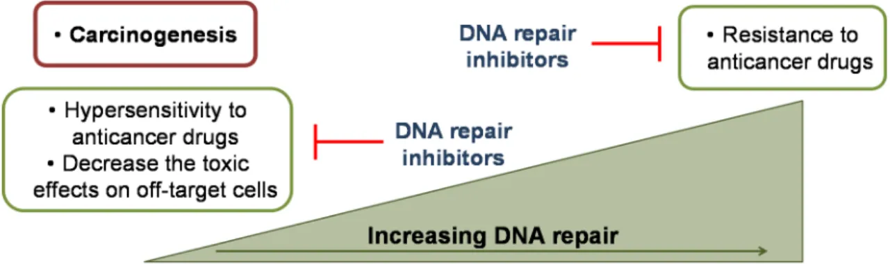
Figure 1.2 Rationale for the use of DNA repair inhibitors in cancer therapy.. ... 7 Figure 1.3 Chemical structures of some base lesions and apurinic/apyrimidinic (AP)
sites commonly recognized and repaired by base excision repair pathway.. ... 8
Figure 1.4 Overview of base excision repair (BER) sub-pathways: short-patch BER
and long-patch BER. ... 10
Figure 1.5 APE1 is a DNA repair protein with an independent redox function. APE1
participates in base excision repair pathway. ... 13
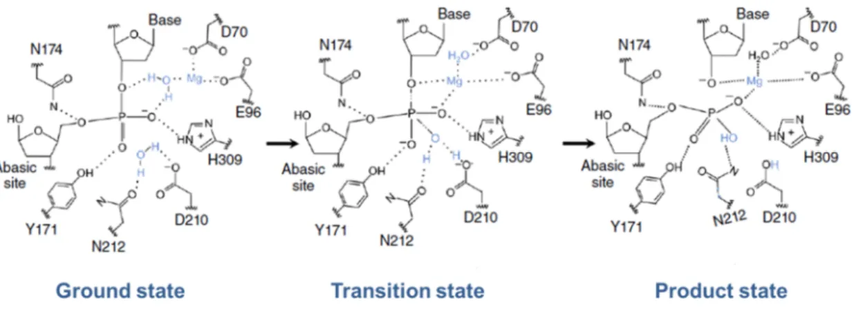
Figure 1.6 Schematic representation of the primary structure of human APE1. ... 14 Figure 1.7 Scheme of the suggested catalytic mechanism of APE1 endonuclease
activity. ... 22
Figure 1.8 Structures of several reported inhibitors of APE1 DNA repair activity. ... 24 Figure 1.9 Scheme of the hypothesised mechanism of activation of transcription
factors by APE1 redox function. ... 30
Figure 1.10 Structures of APE1 redox inhibitors. ... 31 Figure 1.11 Rationale for the combination of standard chemotherapeutic drugs used in
the advanced breast cancer treatment with APE1 inhibitors. ... 37
Figure 3.1 Cytotoxic effects of doxorubicin (Dox) in MDA-MB-231 cells. ... 69 Figure 3.2 Doxorubicin-induced cytotoxicity in the presence of methoxyamine (MX) in
MDA-MB-231 cells. ... 70
Figure 3.3 Colony formation assay of MDA-MB-231 cells treated with methoxyamine
(MX) and doxorubicin (Dox)... 71
Figure 3.4 Effect of the simultaneous inhibition of the DNA repair activity and redox
domain of APE1 in the viability of MDA-MB-231 cells treated with doxorubicin (Dox) as assessed by the MTT assay. ... 72
Figure 3.5 Effect of doxorubicin (Dox) in the intracellular levels of superoxide anion in
MDA-MB-231 cells evaluated by the dihydroethidium (DHE) probe. ... 72
Figure 3.6 Effect of methoxyamine (MX) in the proliferative indices of Dox-treated cells
Figure 3.7 Effect of methoxyamine (MX) in Dox-induced micronuclei in MDA-MB-231
cells as evaluated by cytokinesis-block micronucleus (CBMN) assay. ... 74
Figure 4.1 Virtual screening protocol and experimental methodologies to identify novel
chemical entities from the NCI database showing a high affinity for the APE1 enzyme. ... 95
Figure 4.2 APE1 X-ray structures. ... 96 Figure 4.3 Effect of the identified compounds in the APE1 endonuclease activity. ... 98 Figure 4.4 Best poses of active compounds inside the APE1 binding pocket. ... 102 Figure 4.5 Effect of the compounds in the viability of MCF10A cells evaluated by the
MTS assay. ... 103
Figure 5.1 Chemical structure of the APE1 redox inhibitor E3330. ... 114 Figure 5.2 APE1 endonuclease activity in the presence of the redox inhibitor
E3330. ... 120
Figure 5.3 Cytotoxic effects of docetaxel (DTX) in MDA-MB-231 cells. ... 121 Figure 5.4 Effect of E3330 on viability and colony formation of MDA-MB-231 cells
treated with docetaxel (DTX). ... 122
Figure 5.5 Cell cycle distribution of MDA-MB-231 cells treated with E3330 and/ or
docetaxel (DTX). ... 123
Figure 5.6 Effect of the simultaneous exposure to E3330 (30 µM) and docetaxel (DTX;
0.5 nM) on the migration of MDA-MB-231 cells after a 24 h period of incubation with compounds. ... 124
Figure 5.7 Effect of the simultaneous exposure to E3330 (30 µM) and docetaxel (DTX;
0.5 nM) on the invasion of MDA-MB-231 cells. ... 125
Figure S1 Mass spectra (ESI-) of compounds 22, 37 and 41. ... 162 Figure S2 Determination of the mode of enzymatic inhibition of compound 22. ... 163
LIST OF TABLES
Table 1.1 Anticancer agents and DNA repair pathways involved in the repair of DNA
damage-induced by cancer treatment. ... 6
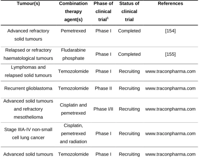
Table 1.2 Clinical trials completed or planned with methoxyamine (MX) in cancer... 25 Table 4.1 Structure, lipophilicity (LogP), GoldScore, and IC50 values of the identified
APE1 inhibitors... 99
Table S1 Structure of the 30 compounds identified in the in silico study and selected to
be tested in the first screen using the fluorescence-based APE1 endonuclease activity assay. ... 156
ABBREVIATIONS
3-AB 3-aminobenzamide
3-D Three-dimensional
4-AN 4-amino-1,8-naphthalimide AAG/MPG N-methylpurine-DNA glycosylase
AP Apurinic/apyrimidinic
AP-1 Activator protein 1
APE1 Human apurinic/apyrimidinic endonuclease 1
ATP Adenosine triphosphate
B-Raf B-type Raf kinase
BCNU 1,3-bis(2-chloroethyl)-1-nitrosourea
BER Base excision repair
BHQ-2 Black hole quencher-2
BN Binucleated
BRCA1/2 Breast cancer 1/2, early onset gene CREB cAMP response element-binding protein CRT0044876 7-nitro-1H-indole-2-carboxylic acid
CV Crystal violet
DDR DNA damage response
DHE Dihydroethidium Dox Doxorubicin dRP 5’-deoxyribose phosphate DSB Double-strand break DMSO Dimethylsulfoxide E3330 (2E)-2-[(4,5-dimethoxy-2-methyl-3,6-dioxo-1,4-cyclohexadien-1-yl)methylene] undecanoic acid
Egr-1 Early growth response protein 1 EMSA Electrophoretic mobility shift assay
5-FU 5-Fluorouracil
FA Fanconi anemia
FapyG 2,6-diamino-4-hydroxy-5-formamidopyrimidine
FEN1 Flap endonuclease 1
GA Genetic algorithm
GG-NER Global-genome nucleotide excision repair GOLD 5.1.0 Genetic Optimization for Ligand Docking H2O2 Hydrogen peroxide
HER2/neu Human epidermal growth factor receptor 2 HIF-α Hypoxia-inducible factor 1 alpha
1H NMR Proton nuclear magnetic resonance
HPLC-DAD High-performance liquid chromatography with a diode array detector
HR Homologous recombination
HR-ESI-MS High-resolution electrospray ionization mass spectrometry Hsp70 Heat shock protein 70
HTS High-throughput screening ICL Interstrand cross-link IdUrd 5-iodo-2’-deoxyuridine
IR Ionising radiation
LP-BER Long-patch BER
MBC Metastatic breast cancer
MBD4 Methyl-CpG binding domain 4 DNA glycosylase
MDM2 Mouse double minute 2
MDR1 Multidrug resistance gene MMP Matrix metalloproteinase
MMR Mismatch repair
MMS Methyl methanesulfonate
MNBN Micronucleated binucleated MOE Molecular Operating Environment
mRNA Messenger RNA
MTS Mitochondrial targeting sequence MTT Thiazolyl blue tetrazolium bromide MUTYH MutY homolog DNA glycosylase
MX Methoxyamine hydrochloride
nCaRE Negative calcium responsive elements
NCI/DTP National Cancer Institute/Developmental Therapeutics Program NDI Nuclear division index
NEIL1/2/3 NEI endonuclease VIII-like 1/2/3 NER Nucleotide excision repair NF-кB Nuclear factor-kappa B NHEJ Non-homologous end-joining NIR Nucleotide incision repair NSCLC Non-small cell lung cancer
NTHL1 E. coli NTH endonuclease III-like DNA glycosylase NLS Nuclear localization sequence
NPM1 Nucleophosmin 1 protein
O2•- Superoxide anion
OGG1 8-Oxoguanine-DNA glycosylase
5’-P 5’-Phosphate
3’-PG 3’-Phosphoglycolate
3’-PUA 3’-Phospho-α,β-unsaturated aldehyde PARP Poly(ADP-ribose)polymerase
PCNA Proliferating cell nuclear antigen
PDB Protein Data Bank
PNKP Polynucleotide kinase/phosphatase Polβ/δ/ε DNA polymerase β/δ/ε
PTEN Phosphatase and tensin homolog
PTH Parathyroid hormone
QSAR Quantitative structure-activity relationship Ref-1 Redox effector factor 1
RFC Replication factor C
RMSD Root mean square deviation
RNase H Ribonuclease H
ROS Reactive oxygen species
rRNA Ribosomal RNA
SBVS Structure-based virtual screening
SCCHN Squamous cell carcinoma of the head and neck
shRNA Short-hairpin RNA
siRNA Small-interfering RNA
SMUG1 Single-strand-selective monofunctional uracil-DNA glycosylase 1
SP-BER Short-patch BER
SSB Single-strand break
STAT3 Signal transducer and activator of transcription 3 TAMRA Carboxytetramethylrhodamine
TBHP Tert-butylhydroperoxide
TC-NER Transcription-coupled nucleotide excision repair
TDG Thymine-DNA glycosylase
THF Tetrahydrofuran
TLS Translesion synthesis
TMZ Temozolomide
TNBC Triple-negative breast cancer
UNG Uracil-DNA glycosylase
XRCC1 X-ray cross-complementing factor 1 YB-1 Y-box-binding protein 1
C
HAPTER
1
GENERAL
INTRODUCTION
This Chapter contains information published in:
Rodrigues AS, Gomes BC, Martins C, Gromicho M, Oliveira NG, Guerreiro PS, et al.. DNA repair and resistance to cancer therapy, in: C. Chen (Ed.), New Research Directions in DNA repair, InTech, Rijeka, 2013, pp. 489-528.
1.1. DNA REPAIR PATHWAYS AND CANCER THERAPY
–
A BRIEF OVERVIEWDNA is continuously exposed to numerous sources of damage which include exogenous chemical and physical agents and endogenous genotoxic insults related to physiological and cellular processes namely the reactive species generated by metabolism and the replication errors [1,2]. Cells have evolved a set of tightly regulated, coordinated and redundant surveillance mechanisms to protect the genome stability. These complex molecular pathways are collectively named DNA damage response (DDR) and they detect, signal, control cell cycle progression, promote the DNA repair and activate cell death machineries to counteract the thousands of lesions that threat the genome integrity (Fig. 1.1) [1–6].
In general, a DNA lesion is recognized by several sensor proteins of DDR. These DNA damage sensors initiate signalling pathways that can slow down or transiently arrest the cell cycle progression increasing the available time for DNA repair [2,4]. Cells have multiple DNA repair pathways to prevent the accumulation of a wide diversity of DNA lesions. These DNA repair mechanisms target different types of DNA damage and they can act independently or interact according to the complexity of DNA lesions [1,2].
The major DNA repair pathways include the a) direct reversal repair, b) mismatch repair (MMR) c) base excision repair (BER), d) nucleotide excision repair (NER), e) translesion synthesis (TLS), f) homologous recombination (HR), g) non-homologous end-joining (NHEJ) and h) the Fanconi anemia (FA) pathways [1,2]. The present work is focused on human apurinic/apyrimidinic endonuclease 1 (APE1), a key BER enzyme considered a promising target for cancer therapy, including breast cancer. In this context, a detailed description of the other mentioned DNA repair pathways is beyond the scope of this thesis. Several comprehensive reviews on this topic have been published in the last years [1,2,7–13].
Although the majority of the DNA repair systems require the coordinated action of several proteins, a small group of DNA lesions can be directly repaired by a single protein mechanism without the incision of the DNA backbone or the base excision. The
direct reversal repair is a simple and error-free DNA repair process with high substrate specificity [14,15]. In addition, the BER pathway identifies and excises bases damaged by alkylation, oxidation, deamination, depurination/depyrimidination, removes the uracil from DNA and also repairs DNA single-strand breaks (SSBs) [2,16]. While BER repairs small adducts, NER ensures the removal of bulky helix-distorting base lesions which
segment containing the damaged base, usually with 24–32 oligonucleotides of length, has to be excised and replaced to complete the repair. NER may proceed through two sub-pathways depending on the substrate: the global-genome NER (GG-NER) targets lesions in the entire genome and the transcription-coupled NER (TC-NER) is involved in the removal of transcription-blocking damage to allow an accurate gene expression [13,16,17]. The MMR eliminates mispaired bases and small insertion-deletion loops generated during DNA replication [2,16,18]. Moreover, mammalian cells use the TLS to
Figure 1.1 Sources of DNA damage and general overview of DNA repair pathways and biological responses to DNA lesions [2,3,6]. Abbreviations: ROS - reactive oxygen species; RNS - reactive
nitrogen species; DR - direct reversal repair; MMR - mismatch repair; BER - base excision repair; NER - nucleotide excision repair; TLS - translesion synthesis; HR - homologous recombination; NHEJ - non-homologous end-joining. DNA damage Exogenous sources - UV light - Ionizing radiation - Environmental chemicals - Dietary genotoxicants - Chemotherapeutic agents Endogenous sources - Oxidative stress (ROS/RNS)
- DNA replication errors - Replication fork arrest
DNA Damage Response
DNA Repair DR MMR BER NER HR TLS NHEJ Cell cycle checkpoints Senescence Apoptosis Transcription/ Replication
Efficient Repair Repair Failure
Genome Integrity Cell Viability Mutations/ Chromosomal aberrations Blocked transcription/ Blocked replication Senescence Apoptosis Cancer Ageing ↑ risk
bypass the lesions that could be incompletely repaired and often remain in the DNA during replication jeopardizing the progress and the fidelity of replicative polymerases [8,19].
For DNA double-strand breaks (DSBs), two major DNA repair pathways are available: the NHEJ and the HR. NHEJ repairs the DSBs by promoting the direct ligation of broken DNA ends. Conversely, HR requires an homologous undamaged DNA template to carry out the repair of DSBs, interstrand cross-links (ICLs) and to restart stalled replication forks. Thus, NHEJ is considered an error-prone mechanism which can be activated in all phases of the cell cycle whereas HR is generally classified as an error-free repair strategy primarily restricted to late S and G2 phase. In fact, HR is more prominent after the DNA replication because an intact sister chromatid can be used as template for repair [2,16,20,21]. For ICLs repair, which cannot be completely removed by MMR and/or NER, HR cooperates with the FA pathway. In general, the repair of these lesions can occur only in the S-phase during the DNA replication [2,22].
The outcomes of DNA lesions are unpredictable despite cell efforts for the genome maintenance. They depend not only on the nature of the damage but also on the number and location of DNA lesions, cell type, cell cycle phase and differentiation stage [6]. An inaccurate repair and/or the overwhelming of the DNA repair mechanisms yield the accumulation of DNA lesions (Fig. 1.1). The blockage of transcription or replication is a common consequence inducing senescence or cell death, particularly in non-proliferating cells, and contributing for ageing [6,23]. On the other hand, in proliferating cells permanent mutations appear during the replication cycles of damaged DNA. Similarly, structural chromosomal aberrations may be generated during the DSBs repair and dysregulate gene expression. The transmission of these alterations to descendant cells can also result in mutations. The inactivation of certain tumour suppressor genes, the activation of oncogenes and an enhanced genomic instability are potential effects of these harmful mutations. Consequently, if these alterations constitute a selective advantage for cells they may contribute to an uncontrolled and sustained cellular proliferation leading to the development of precancerous lesions and ultimately to cancer progression [2,6,23,24]. Mutations can also affect genes required for DNA repair such as breast cancer 1/2, early onset genes (BRCA1 and BRCA2). Both are tumour suppressor genes which encode proteins involved in DDR and HR and inherited germline mutations in BRCA1 and BRCA2 have been correlated with a higher susceptibility to develop hereditary breast and ovarian cancer among others [25–27]. The epigenetic alterations which involve the chromatin remodelling, histones modification and the DNA methylation also display a
carcinogenic role through the inactivation of the DDR and DNA repair effectors and enabling the malignant transformation of cells and tumour development [2,6,23,24].
Another important aspect regarding DNA repair and cancer is the resistance to anticancer agents. Cancer therapy is challenging and generally it involves the use of different strategies to target specific molecular and cellular features of cancer cells in order to cure, or at least hamper the tumour progression.
The most fundamental hallmark of cancer cells is their ability to proliferate at higher rates than non-tumour cells which renders the cell cycle an attractive target in cancer therapy [28]. In addition to the inhibitors of the mitotic spindle and the targeting of growth signalling pathways, cell cycle is frequently disrupted by the DNA-damaging agents. Actually, many chemotherapeutic regimens are based on DNA-damaging drugs often used in complementary combinations [2,8]. Their effectiveness depends on their ability to directly or indirectly damage DNA with the generation of cytotoxic lesions that overwhelm the DNA repair capacity and ultimately induce cell death of rapid proliferating cancer cells (Table 1.1). In view of this, the rationale for their use in cancer therapy is, as abovementioned, the genomic instability and replicative stress of tumour cells as well as the presence of mutator phenotypes in DNA repair genes which can impair or even inactivate DNA repair pathways increasing the cell death induced by DNA-damaging drugs [2,8,29].
However, the efficacy of DNA-damaging drugs is frequently precluded by toxicity issues and cancer resistance to therapy. Although non-tumour cells have a lower replication rate and are usually proficient in the DDR being less susceptible to the effects of ionising radiation (IR) and chemotherapeutic drugs, their DNA can also be damaged with the subsequent development of side effects [8]. In respect to the resistance to DNA-damage agents, there is a redundancy of DNA repair mechanisms that may lead to the activation of another DNA repair pathway upon failure of the first attempt to repair the lesions. Moreover, cancer cells may also present an increased activity of the DNA-damage signalling and DNA repair pathways preventing the accumulation of toxic lesions and promoting their survival [2,8,16,30,31].
Table 1.1 Anticancer agents and DNA repair pathways involved in the repair of DNA damage-induced by cancer treatment. The major DNA repair pathways contributing to the repair of lesions are represented in bold. Adapted from [2,8,29,32].
In this context, the modulation of DNA repair is a targeted therapeutic approach to improve the outcomes of cancer treatments (Fig. 1.2). DNA repair inhibitors may not only overcome the resistance to anticancer agents but also increase their sensitivity
and specificity decreasing the toxic side effects. This is particularly important in tumours with somatic mutations that lead to the inactivation of a certain DNA repair pathway. In these circumstances cancer cells will be more dependent on the remaining
Anticancer agents Types of DNA lesions DNA repair
pathways Radiotherapy and radiomimetics
Ionizing radiation; Bleomycin DSBs; SSBs; Base damage
NHEJ; BER; HR Monofunctional alkylators
Alkylsulphonates (e.g. busulfan); Nitrosoureas (e.g. BCNU); Temozolomide (TMZ)
Base damage; Replication lesions; Bulky DNA
adducts
DR; BER; MMR; HR; NER; TLS; FA Bifunctional alkylators
Nitrogen mustards (e.g. cyclophosphamide); Mytomicin C; Cisplatin
DSBs; DNA crosslinks; Replication lesions; Bulky
DNA adducts
NER; HR; FA; TLS; MMR
Antimetabolites
5-Fluorouracil (5-FU); Thiopurines (e.g. 6-mercaptopurine); Folate analogues (e.g. methotrexate); Gemcitabine/Troxacitabine
Base damage; Replication lesions
BER; MMR
Topoisomerase inhibitors
Camptothecin (Topo I); Etoposide (Topo II); Anthracyclines (Topo II)
DSBs; SSBs; Replication lesions; Oxidative base
damage
HR; NHEJ; FA; BER
Taxanes
Docetaxel; Paclitaxel Taxanes-induced ROS [32,33] -oxidative base
damage (?)
BER (?)
Replication inhibitors
DNA repair pathways which could be targeted with DNA repair inhibitors to induce a hypersensitivity to anticancer drugs and decrease the toxic side effects [2,8,16]. The concept of synthetic lethality has also emerged as a therapeutic approach in tumours carrying genetic defects [8,29,33]. The underlying principle of synthetic lethality is the cells ability to tolerate and maintain viability upon inactivation of only one pathway due
to mutations of genes that code relevant proteins [8,29,33]. The use of chemical inhibitors to abrogate a second and redundant DNA repair pathway promotes the accumulation of DNA damage and induces cell death [8,29,33]. Synthetic lethality has been notably relevant in tumour cells harbouring mutations in proteins associated with
defects in HR which have shown to be sensitive to inhibitors of proteins involved in BER, namely poly(ADP-ribose)polymerase 1 (PARP1) [29,34,35]. These are clinical opportunities to personalised therapy since it requires the knowledge of tumour genetic alterations to improve the specificity of the treatment, the overall survival and to the use of DNA repair inhibitors in monotherapy limiting the number of drugs administered and
consequently the toxicity of therapeutic agents.
Figure 1.2 Rationale for the use of DNA repair inhibitors in cancer therapy. A jeopardised DNA repair increases the sensitivity to anticancer drugs and decreases the toxic side effects. An increased DNA repair activity is related with the resistance to anticancer agents. Targeting DNA repair is an opportunity to improve the cancer therapy outcomes [2,8,32].
1.2. THE BASE EXCISION REPAIR
(
BER)
PATHWAYThe basic steps of the base excision repair pathway were first described in the 70s decade by Tomas Lindahl1 after the identification of the Escherichia coli uracil-DNA glycosylase [9,36,37]. BER is known to play a critical role in the removal and replacement of several small non-bulky DNA lesions generated by oxidation, alkylation or deamination and the uracil from DNA. It is also the major DNA repair pathway responsible for repairing the apurinic/apyrimidinic (AP) sites spontaneously produced during the cellular metabolism or as intermediates of this pathway during the repair of
1
Awarded with the Nobel Prize in Chemistry 2015 along with Paul Modrich and Aziz Sancar for mechanistic studies in the DNA repair field.
base modifications inflicted by endogenous or exogenous genotoxicants (Fig. 1.3). The SSBs can occur directly by the attack of ROS to the deoxyribose or indirectly upon repair being also a substrate of BER [9,10,38]. In this context, it is essential to highlight the pivotal role of the human APE1, a topic that will be thoroughly described in this and in the next sections of the present thesis.
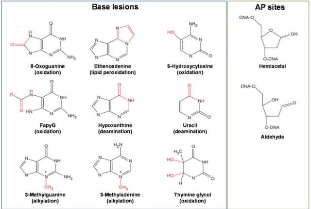
Figure 1.3 Chemical structures of some base lesions and apurinic/apyrimidinic (AP) sites commonly recognized and repaired by base excision repair pathway. Adapted from [9]. Abbreviation:
FapyG - 2,6-diamino-4-hydroxy-5-formamidopyrimidine.
In general, BER comprises five coordinated enzymatic steps (Fig. 1.4) which include the recognition and excision of the damaged base by a DNA glycosylase, the incision of the AP site by APE1 or the AP lyase function of a bifunctional DNA glycosylase, the removal of the 5’- or 3’- terminal blocking groups, the DNA synthesis by a DNA polymerase for filling the resulting gap and, finally, the nick sealing carried out by a DNA ligase [9,10,39,40].
Firstly, a lesion-specific DNA glycosylase recognizes and removes the damaged or inappropriate base. The hydrolytic cleavage of the N-glycosidic bond linking the incorrect base and the DNA sugar-phosphate backbone carried out by DNA glycosylases produces an AP site [41,42]. The intact AP sites generated by the monofunctional DNA glycosylases (UNG, TDG, SMUG1, MUTYH, MBD4 and AAG/MPG) are further processed by APE1 which catalyses the hydrolysis of the
phosphodiester backbone 5’ to the AP site producing a SSB with a deoxyribose phosphate (dRP) group at the 5’ end and a 3’-OH termini. In the case of bifunctional DNA glycosylases (such as OGG1, NTHL1, NEIL1, NEIL2 and NEIL3) which also have an intrinsic AP lyase activity, the AP site incision is performed by the DNA glycosylase itself 3’ to the AP site. The resulting SSB presents a 3’-non-ligatable group which should be removed to provide the proper substrate to be channeled to the following step of BER [10,39–41]. NTH1 and OGG1, primarily responsible for the excision of the oxidative lesions 8-oxo-7,8-dihydroguanine (8-oxoG) and 2,6-diamino-4-hydroxy-5-formamidopyrimidine (FapyG), have a weak lyase activity. They catalyse the AP site processing via a β-elimination reaction which creates an intermediate with a 5’-phosphate (5’-P) and a phospho-α,β-unsaturated aldehyde (PUA) moiety. The 3’-blocking group is removed by APE1 intrinsic 3’-phosphodiesterase activity [10,39,40]. Regarding the NEIL glycosylase family responsible for the removal of oxidised pyrimidines, formamidopyrimidines, 5’-hydroxyuracil and urea from the DNA, the AP site incision is accomplished through a β, δ-elimination reaction with the formation of a SSB carrying a 3’-phospate (3’-P) group. Since the 3’-P moiety is not a suitable substrate for APE1, it can be removed by the phosphatase activity of polynucleotide kinase/phosphatase (PNKP) in an APE1-independent BER pathway [10,39,40,43,44]. However, the APE1-independent repair of AP sites is inefficient in the APE1 replacement during repair of the most commonly generated AP lesions.
The next steps of BER can proceed through two sub-pathways: the short-patch BER (SP-BER) or the long-patch BER (LP-BER) [10,39,40]. The aforementioned base excision and the cleaning of the terminal blocking groups are common to both sub-pathways. However, they diverge in the length of the DNA fragment to be replaced and
in the subsets of enzymes involved in the DNA synthesis and nick sealing. The SP-BER seems to be the predominant SP-BER sub-pathway and only one nucleotide is displaced to continue the repair. Then the DNA polymerase β (Polβ) lyase activity removes the 5’-dRP moiety and promotes the DNA synthesis from the 3’-OH group through its DNA polymerase activity to fill the existing gap. To complete the repair, the
nick sealing is mediated by the integrated action of DNA ligase IIIα and the scaffold protein X-ray cross-complementing factor 1 (XRCC1) [10,39,40]. When 5’ end cleaning
lyase activity of Polβ is incapable to remove the 5’-blocking groups present in oxidised AP sites BER occurs through LP-BER [10,39,40]. For example, the C1’-oxidised
2’-deoxyribonolactone is repaired by LP-BER to avoid the formation of a DNA-protein cross-link with the Polβ [39,45]. LP-BER requires the strand displacement synthesis of a fragment usually with 2-12 nucleotides [10]. The DNA synthesis is mostly assigned to
the replicative DNA polymerases δ and ε (Polδ/ε) although Polβ may also participate in the initiation of LP-BER [46,47]. The replication factor C (RFC) and the proliferating cell
nuclear antigen (PCNA) support the Polδ/ε and the flap endonuclease 1 (FEN1).
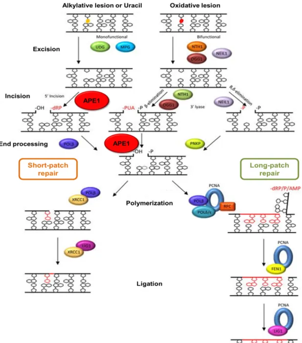
Figure 1.4 Overview of base excision repair (BER) sub-pathways: short-patch BER and long-patch BER. Both sub-pathways require the recognition and excision of the damaged base by a DNA glycosylase. After the strand incision at the abasic sites, 5’- and 3’- ends are processed by different enzymes according to the blocking groups. The repair may proceed through SP-BER or LP-BER to accomplish the DNA synthesis and ligation. Adapted from [40].
During the synthesis of the displaced oligonucleotide containing the 5’-sugar phosphate, PCNA functions as an accessory sliding clamp for Polδ/ε that is loaded into the DNA by RFC [10,39,48]. A flap intermediate is generated and further excised by the
structure-specific FEN1 [10,39,49]. Consequently, the substrate for DNA ligase I is created and double-stranded DNA is restored by nick sealing [10,39,40].
The selection of SP-BER or LP-BER remains to be fully clarified. The nature of the DNA damage, the intracellular levels of ATP, the protein-protein interactions, the cell cycle phase and the differentiation stage of cells have been reported to influence the sub-pathway choice.
The type of the DNA lesion determines the BER initiating DNA glycosylase and the 5’- and 3’-blocking moieties, which will define the DNA polymerase to be recruited and thus the BER sub-pathway [10,47,50–52]. Moreover, nick sealing reactions depend on the intracellular ATP availability to generate a covalent phosphodiester bond linking the 5’- and 3’-DNA strand ends. In the presence of high ATP levels the catalytic activity of the complex DNA ligase IIIα/XRCC1 is favored being the SP-BER the predominant mechanism. Under conditions of ATP depletion the DNA repair is shifted from SP-BER to LP-BER due to the promotion of strand displacement DNA synthesis by Polβ and allowing the DNA ligation by DNA ligase I [53,54].
In BER the intermediates are transferred to the next enzyme in a sequential and tightly orchestrated stepwise process to prevent the exposure of the cellular components to potentially harmful molecules [52]. Among the protein-protein interactions regulating the switch between SP-BER and LP-BER, XRCC1 and SSBs sensor proteins PARPs have a critical role in BER coordination [39,52]. XRCC1 is regarded as a scaffold protein interacting with several BER proteins to stimulate their recruitment for the SSBs, to promote their stabilisation and modulating their activities. In addition to DNA ligase IIIα [55], XRCC1 has been described to interact with proteins implied in different stages of both sub-pathways [56] such as the PCNA [57], APE1 [58], PNK [59,60], Polβ [61–63], NEILs [43,44,64], PARP1 and 2 [63,65,66]. The enrollment of PARP enzymes, especially PARP1, to the DNA-strand break is an early event of BER and probably occurs after the strand incision carried out by APE1 or a bifunctional DNA glycosylase. The activated proteins catalyse a nicotinamide adenine dinucleotide (NAD+)-dependent synthesis of poly(ADP-ribose) chains which are a signal to the recruitment of multiple enzymes of BER for the damaged DNA site. PARPs functionally and physically interact with APE1, XRCC1, Polβ, PCNA and DNA ligase IIIα to boost the DNA repair. In the presence of an overwhelming DNA damage, PARPs can be overactivated leading to NAD+ and ATP shortage and, consequently, triggering cell death by apoptosis or necrosis [67,68].
The cell cycle stage may also govern the SP-BER or LP-BER selection. The LP-BER shares proteins with the replicative machinery (e.g. FEN1, Polβ/δ/ε, DNA ligase I) suggesting its involvement in the repair of replication-associated DNA lesions. This hypothesis is supported by the detection at replication foci of protein complexes containing the aforementioned proteins. Thus the LP-BER can be more dominant at S/G2 cell cycle phases while a faster BER pathway such as SP-BER will be required in G1 phase (reviewed in [52]).
1.3. OVERVIEW OF HUMAN APURINIC
/
APYRIMIDINIC ENDONUCLEASE 1(
APE1)
FUNCTIONSAPE1 is undoubtedly a key DNA repair enzyme that plays a crucial role in genome integrity as the major endonuclease in BER. Among the several physiological functions of APE1, its endonuclease activity and consequently its role as an upstream key player in SP-BER and LP-BER are of utmost importance as it was mentioned in the previous section. However, the therapeutic opportunities of targeting APE1 should not be restricted to the modulation of the DNA repair activity. APE1 is considered an ubiquitous multifunctional protein essential for the regulation of cellular response to oxidative stress and vital for the maintenance of genome stability and cell integrity [69– 71]. Importantly, APE1, also designated redox effector factor 1 (Ref-1), has another major function since it independently acts as a reduction/oxidation signalling protein modulating the activation and DNA binding ability of several transcription factors which promote the expression of genes implicated in cell survival and in tumour promotion and progression (Fig. 1.5) [71–73]. Moreover, the understanding of the regulation as well as the role of distinct APE1 activities in cellular homeostasis is required to efficiently develop inhibitors of the different APE1 functions.
APE1 is a member of the Xth family of class II AP endonucleases due to its structural homology with the exonuclease III (Xth) enzyme of Escherichia coli [73–76]. In mammalian cells APE1 contributes for approximately 95% of the endonuclease activity in eukaryotic organisms [73,74]. APE1 is a monomeric α/β globular protein with 318 amino acid in length and a molecular weight of approximately 35 kDa [71,77]. Despite lacking the initial N-terminal fragment of 35 residues, the first X-ray crystal structure of APE1 revealed two main domains displaying similar topologies [77]. Both N-terminal and C-terminal domains have a six-stranded β-sheet encircled by α-helices folding together to form the four layered α/β sandwich structural core and resembling the motifs observed in the E. coli homologue exonuclease III and DNase I-like proteins
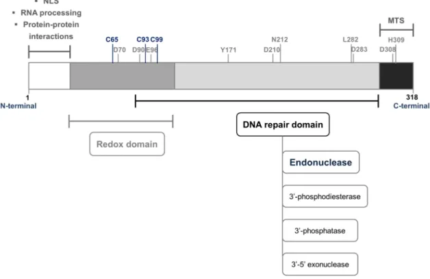
[71,77]. The first 40 N-terminal amino acids of APE1 structure have a disordered folding [78,79]. Notably, the C-terminal region is highly conserved among organisms from different kingdoms while the N-terminal fraction is almost restricted to mammals [80–82]. Noteworthy, the protein structural organization enables both DNA repair and redox activity to be physically and functionally independent since they are encoded by non-overlapping domains of APE1 [71,72,83]. While the catalytic site responsible for its endonuclease activity is located within the C-terminal domain, the redox regulatory function of APE1 is assigned to N-terminal portion which also harbours a complex bipartite nuclear localization signal (NLS) (Fig. 1.6) [71,81,83–85].
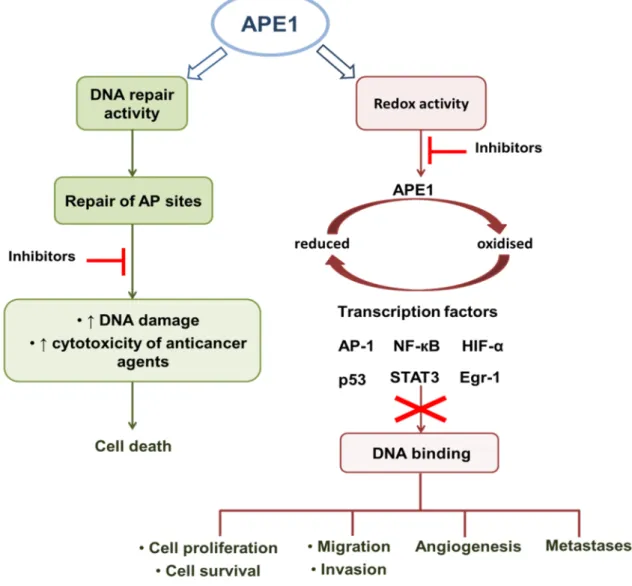
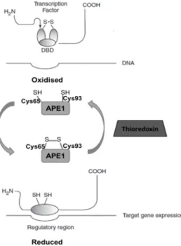
Figure 1.5 APE1 is a DNA repair protein with an independent redox function. APE1 participates in base excision repair pathway. The inhibition of APE1 endonuclease activity increases the DNA damage and might improve the cytotoxic effects of anticancer agents causing the tumour cells death. The inhibition of the reduction/oxidation signalling function of APE1 reduces the activation of several transcription factors modulating the expression of genes involved in cancer promotion and progression [71,72]. Abbreviations:
AP – apurinic/apyrimidinic; AP-1 – activator protein 1; Egr-1 – early growth response protein 1; HIF-α – hypoxia inducible factor 1 alpha; NF-кB – nuclear factor-kappa B; STAT3 – signal transducer and activator of transcription 3.
The DNA repair function of APE1, particularly its endonuclease activity, is essential for the repair of toxic AP sites spontaneously generated, chemically induced or resulting as intermediate products in the enzymatic hydrolysis of DNA bases damaged by alkylating and oxidising agents (e.g. ROS) carried out during BER [86]. In the intracellular milieu, AP sites are an equilibrium mixture of four species corresponding 99% to a mixture of equal parts of the two hemiacetal enantiomers α- and β-2-deoxy-D-ribofuranose and approximately 1% to the ring-opened aldehyde and hydrated aldehyde forms [87]. The ring-opened forms are highly reactive providing a site for DNA cleavage by a β-elimination reaction [87,88]. If left unrepaired, non-coding AP sites can be cytotoxic and mutagenic threatening the integrity of cell function and survival [87,89]. The cytotoxicity of AP sites may result from the ability to induce replication forks stalling with the generation of SSBs which can be converted into DSBs after replication [87,89,90]. The ring-opened AP sites can also react with nuclear proteins yielding DNA-protein complexes which can hamper the DNA replication [87,89]. Another cytotoxic mechanism of AP sites may be related with the interference with topoisomerases DNA cleavage activity or the irreversible trapping of topoisomerase-DNA covalent complexes [87,89,91].
Figure 1.6 Schematic representation of the primary structure of human APE1. The active domains and their major functions are displayed. The essential residues for APE1 redox function are highlighted in blue and the amino acids involved in the endonuclease activity are depicted in grey. Adapted from [81,85,89]. Abbreviations: NLS – nuclear localization signal; MTS – mitochondrial targeting sequence.
![Figure 1.1 Sources of DNA damage and general overview of DNA repair pathways and biological responses to DNA lesions [2,3,6]](https://thumb-eu.123doks.com/thumbv2/123dok_br/15134642.1011221/37.892.130.768.291.1036/figure-sources-general-overview-pathways-biological-responses-lesions.webp)
![Figure 1.10 Structures of APE1 redox inhibitors. These compounds were described in [200–205]](https://thumb-eu.123doks.com/thumbv2/123dok_br/15134642.1011221/65.892.132.767.345.790/figure-structures-ape-redox-inhibitors-compounds-described.webp)
