Development of
New Peptide-Drug
Conjugates for
Cancer Therapy
Abigail Filipe Ferreira
Biochemistry Master’s Dissertation presented to Faculty of Sciences, University of Porto
New Peptide-Drug
Conjugates for
Cancer Therapy
Abigail Filipe Ferreira
Master Degree in Biochemistry Department of Chemistry and Biochemistry 2015
Supervisor
Dr. Nuno Filipe de Sousa Vale,
FCT Researcher, UCIBIO/REQUIMTE – Faculty of Sciences of the University of Porto
Co-Supervisor
Dr. Paula Alexandra de Carvalho Gomes,
Associated Professor, Faculty of Sciences of the University of Porto
AGRADECIMENTOS (ACKNOWLEDGEMENTS)
Em primeiro lugar, tenho que agradecer ao Dr. Nuno Vale, por ser um verdadeiro orientador, em todos os sentidos da palavra, e também um professor. Obrigada pelo incansável acompanhamento diário, pelo esforço que dedicou a este projeto, pela enorme capacidade de trabalho, pela paciência, ajuda, otimismo e palavras de incentivo nas alturas que mais precisei. Não há palavras suficientes para agradecer tudo o que fez por mim durante este projeto.
À Professora Paula Gomes, por ter coorientado este projeto, mostrando sempre preocupação em acompanhar o trabalho que estava a ser desenvolvido e por toda a força e confiança que sempre me transmitiu.
À Iva Fernandes, agradeço a oportunidade de realizar os ensaios biológicos. Obrigada por tudo o que me ensinaste, pela tua paciência e por estares sempre disponível para me ajudar.
Agradeço também à Sílvia Maia, por realizar as análises de espetrometria de massa e pela ajuda na sua interpretação.
A todos os que passaram pelo laboratório 2.28 durante a realização deste trabalho tenho que agradecer por me proporcionarem o melhor ambiente de trabalho que podia pedir. Obrigada ao Stephane Azevedo, que na sua curta passagem por este laboratório, teve sempre a capacidade de me pôr um sorriso na cara. Ao Lorenzo Cianni, por proporcionar uns jantares inesquecíveis. E ao Rafael Almeida pela simpatia e boa disposição.
Mas tenho que deixar um agradecimento (muito) especial às meninas “da casa”, Ana, Joana, Luísa, Mariana e Rita. Obrigada Ana por seres a melhor química (e não só!) de sempre e pela tua ajuda e disponibilidade absolutamente incansável. Obrigada Luísa e Mariana por todas as gargalhadas que demos em conjunto (algumas das quais não irei esquecer). Joana, obrigada por seres única e não teres medo de o mostrar. E Rita, porque entendes uma outra faceta de mim melhor que ninguém, obrigada por tudo!
Aos meus amigos bioquímicos que de alguma forma também me apoiaram nesta difícil jornada (Margarida, Ricardo, Mariana, …), obrigada.
À minha família e especialmente aos meus pais, obrigada por acreditarem em mim e me puxarem para cima, sempre. Vocês sabem o quanto vos amo.
ABSTRACT
In developed countries, cancer is one of the major causes of death, along with cardiovascular diseases. One of the main reasons that can be attributed to this high mortality is the failure of current treatment options. The hard distinction of normal and cancer cells, but also the phenomenon of resistance and the inefficacy to treat metastases, are the main obstacles when treating cancer patients.
Gemcitabine (2•,2•-difluorodeoxycytidine), commercially available as Gemzar® by Eli Lilly and Company, is a nucleoside analogue which has been proven efficient against a wide range of solid tumors. The use of gemcitabine hydrochloride was approved in 1996 by the Food and Drug Administration (FDA) as first-line treatment for patients with locally advanced (non-resectable Stage II or Stage III) or metastatic (Stage IV) pancreatic adenocarcinoma previously treated with fluorouracil (5-FU). Gemcitabine is activated in vivo via phosphorylation of its 5’-hydroxyl group to gemcitabine monophosphate by deoxycytidine kinase, and is subsequently phosphorylated by intracellular kinases to the triphosphate form. However, gemcitabine may be deaminated to its inactive uridine metabolite, 2’,2’-difluorodeoxyuridine, by cytidine deaminase (CDA), which is present at high levels in both human plasma and liver.
This project aims at the chemical modification of gemcitabine and subsequent conjugation to Cell-Penetrating Peptides (CPPs), in an effort to both prevent (or retard) deamination of that drug and facilitate its delivery into cancer cells, taking advantage of the fact that all CPPs are able to efficiently pass through cell membranes while being non-cytotoxic and carrying a wide variety of cargos inside cells.
Two CPP-drug conjugates have been successfully synthesized, purified and characterized by HPLC and LC-MS. The time-dependent kinetics of gemcitabine release from hydrolysis of these new conjugates was studied in phosphate-buffered saline (PBS) at physiological pH and temperature. Furthermore, the biological activity of these two new conjugates was evaluated against three different human tumoral cell lines: MKN-28 (human gastric cancer), Caco-2 (heterogeneous human epithelial colorectal adenocarcinoma) and HT-29 (human colon adenocarcinoma).
RESUMO
Nos países desenvolvidos, o cancro é uma das principais causas de morte, juntamente com as doenças cardiovasculares. Uma das razões fundamentais que pode ser atribuída à elevada mortalidade desta doença é a baixa taxa de sucesso das opções terapêuticas atualmente disponíveis. O tratamento de doenças oncológicas enfrenta alguns obstáculos, como a difícil distinção entre células saudáveis e células tumorais (que conduz a sérios efeitos secundários associados à terapia), mas também o aparecimento de fenómenos de resistência a quimioterapêuticos e a ineficácia do tratamento de metástases.
A Gemcitabina (2•,2•- difluorodesoxicitidina) é um análogo de nucleósido comercializado pela farmacêutica Eli Lilly and Company como Gemzar® cuja atividade contra uma grande variedade de tumores sólidos foi já extensivamente comprovada. O uso de cloridrato de gencitabina foi aprovado em 1996 pelo órgão governamental dos Estados Unidos da América responsável pelo controlo dos alimentos e medicamentos (Food and Drug Administration, USA) como opção terapêutica de primeira linha para doentes com adenocarcinoma pancreático localmente avançado (inoperável, fase II ou II) ou metastático (fase IV) previamente tratados com 5-fluorouracilo (5-FU, um outro análogo de nucleósido). A gencitabina é ativada in vivo por fosforilação na posição 5’- a monofosfato de gencitabina pela enzima desoxicitidina cinase, e é subsequentemente fosforilada por outras cinases intracelulares à forma trifosfatada, que tem capacidade de inibir a síntese de ADN. No entanto, a gencitabina também pode ser inativada, sendo convertida no metabolito uridínico pela enzima citidina desaminase, presente em elevados níveis no plasma e fígado humano.
Com este projeto pretendeu-se modificar quimicamente a gencitabina e subsequentemente conjugar o seu derivado a Péptidos Penetradores Celulares (CPPs), esperando que estes novos pro-fármacos consigam aumentar a entrada deste agente nas células cancerígenas, tirando partido da propriedade intrínseca dos CPPs, péptidos capazes de atravessar eficazmente membranas celulares, podendo transportar para o interior de diferentes tipos de células inúmeras moléculas, sem comprometer a integridade da membrana.
Foram sintetizados dois conjugados Fármaco–Péptido, que foram purificados e caracterizados por Cromatografia Líquida de Alta Pressão e por Espetrometria de Massa acoplada a Cromatografia Líquida de Baixa Pressão. A cinética de libertação de gencitabina na sua forma livre foi estudada em condições fisiológicas e foi ainda avaliada a atividade biológica destes novos conjugados contra três linhas tumorais humanas: MKN-28 (cancro gástrico), Caco-2 e HT-29 (provenientes do cólon).
INDEX
AGRADECIMENTOS (ACKNOWLEDGEMENTS) ... i
ABSTRACT ... ii
RESUMO ... iii
INDEX ... iv
LIST OF FIGURES ... viii
LIST OF TABLES ... xiii
LIST OF ABBREVIATIONS ... xiv
OBJECTIVES ... xvii
1. INTRODUCTION ... 1
1.1. Cancer Management ... 1
1.2. Gemcitabine ... 2
1.2.1. Biodistribution and Mechanism of Action ... 3
1.2.2. Resistance to Gemcitabine ... 5
1.2.3. Modifications of Gemcitabine ... 6
1.2.3.1. Modifications at the 4-(N)-site of gemcitabine ... 7
1.2.3.1.1. PEG – Gemcitabine ... 7
1.2.3.1.2. Folate-PEG – Gemcitabine ... 8
1.2.3.1.3. Valproic acid – Gemcitabine (LY2334737) ... 9
1.2.3.1.4. Squalenoyl – Gemcitabine ... 10
1.2.3.1.5. Lipophilic Prodrugs ... 12
1.2.3.1.6. H–Gemcitabine: Hoechst conjugated to gemcitabine ... 13
1.2.3.2. Modifications at the 5’-site of gemcitabine ... 14
1.2.3.2.1. CP–4126 ... 14
1.2.3.2.2. NEO6002 ... 15
1.2.3.2.4. Gemcitabine prodrugs incorporating D-enantiomer amino acids ... 17
1.3. Cell-Penetrating Peptides... 19
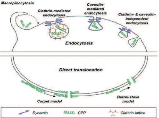
1.3.1. Cellular Uptake Mechanisms of CPPs ... 21
1.3.2. Applications of CPPs ... 22
1.3.3. Solid Phase Peptide Synthesis (SPPS) ... 23
2. EXPERIMENTAL ... 28
2.1. Reagents, Solvents and Equipments ... 28
2.2. Peptide Synthesis by SPPS ... 29
2.2.1. Manual Synthesis ... 29
2.2.1.1. Experimental setup ... 29
2.2.1.2. Preparation of the resin... 29
2.2.1.3. Kaiser Test... 30
2.2.1.4. Coupling of Amino Acids and Deprotection Cycles ... 31
2.2.2. SPPS Assisted by Microwave Energy ... 32
2.3. Cleavage: separating the peptide from the resin and removing the side chain protecting groups 33 2.4. Structural modification of Gemcitabine ... 34
2.4.1. Synthesis of N-[3-(S-trityl)sulfanyl]propanoylgemcitabine (3) ... 35
2.4.2. Synthesis of N-(3-sulfanyl)propanoylgemcitabine (4) ... 35
2.5. First approach to the synthesis of the conjugate Gemcitabine –Linker–Penetratin: Strategy A .... 35
2.6. Synthesis of the Gemcitabine–Linker–CPP conjugates: Strategy B ... 36
2.7. Purification of the conjugates ... 36
2.8. Stability studies ... 37
2.8.1. Calibration curve... 37
2.8.2. Study of conjugates’ stability at physiological pH and temperature ... 37
2.9. In vitro growth inhibition assays ... 38
2.9.1. Cell cultures ... 38
2.9.1.2. Cell lines ... 38
2.9.1.3. Culture medium ... 38
2.9.1.4. Trypsinization of the cell lines ... 38
2.9.1.5. Evaluation of cell viability ... 39
2.9.1.6. Maintaining cells in exponentially growth phase ... 39
2.9.2. Sulforhodamine B (SRB) assay: evaluation of the antiproliferative activity of the compounds . 39 3. RESULTS AND DISCUSSION ... 40
3.1. Peptide Synthesis ... 40
3.1.1. Manual Synthesis of Cys-Penetratin ... 40
3.1.2. Manual Synthesis of Cys-pVEC ... 42
3.1.3. Automated MW-assisted Synthesis of Cys-Penetratin ... 43
3.1.4. Automatic MW-assisted Synthesis of Cys-pVEC ... 44
3.2. Structural modification of Gemcitabine ... 46
3.2.1. Synthesis of N-[3-(S-trityl)sulfanyl]propanoylgemcitabine (3) ... 46
3.2.2. Synthesis of N-(3-sulfanyl)propanoylgemcitabine (4) ... 47
3.2.3. Synthesis of N-{3-[S-(2-sulfanyl)pyridyl]sulfanyl}propanoyl-gemcitabine (7) ... 48
3.3. Synthesis of the Drug-CPP Conjugates ... 50
3.3.1. Strategy A – direct conjugation of compound (4) with Cys-Penetratin ... 50
3.3.2. Strategy B – conjugation of compound (7) with Cys-Penetratin ... 53
3.3.3. Strategy B – conjugation of compound (7) with Cys-pVEC ... 56
3.4. Studies on the stability of Drug-CPP conjugates ... 59
3.4.1. Calibration curve... 59
3.4.2. Time-dependent kinetics of the conjugates’ hydrolysis ... 60
3.4.3. Time-dependent kinetics of the Gemcitabine–Linker–Penetratin conjugate (over 22 days) ... 62
3.5. Antiproliferative activity of the conjugates – SRB assay ... 63
4. CONCLUSIONS AND FUTURE PERSPECTIVES ... 66
6. SUPPLEMENTARY INFORMATION ... 77
6.1. Stability studies – periodic HPLC analysis over 6 days ... 77
6.1.1. Gemcitabine–Linker–Penetratin Conjugate ... 77
6.1.2. Gemcitabine–Linker–pVEC Conjugate ... 80
6.2. Stability of the Gemcitabine–Linker–Penetratin conjugate – periodic HPLC analysis over 22 days 83 6.3. LC-MS chromatograms of the CPPs and the conjugates ... 85
LIST OF FIGURES
Figure 1: Chemical structure of Gemcitabine (2!,2!-difluorodeoxycytidine), where 4-(N)- and 5’- sites are
highlighted. ... 3
Figure 2: Mechanism of intracellular activation of gemcitabine. Adapted from [17]. ... 4
Figure 3: Chemical structures of polyethylene glycol (PEG) and of a PEG – gemcitabine conjugate. ... 7
Figure 4: Chemical structure of a Folate-PEG – gemcitabine conjugate. ... 9
Figure 5: Chemical structure of LY2334737, a prodrug of gemcitabine tested in Phase I clinical trials. ... 10
Figure 6: Chemical structure of squalenoyl – gemcitabine (SQdFdC). ... 10
Figure 7: General structure of lipophilic 4-(N)-acyl – gemcitabine conjugates. ... 12
Figure 8: Chemical structure of Hoechst – Gemcitabine. ... 13
Figure 9: Chemical structure of gemcitabine derivative CP–4126. ... 14
Figure 10: Chemical structure of NEO6002. ... 15
Figure 11: Chemical structures of gemcitabine derivatives Gem-1, Gem-2, Gem-3 and Gem-4. ... 16
Figure 12: Gemcitabine prodrugs with D- and L-amino acids. ... 17
Figure 13: CPPs as delivery vectors – intracellular delivery of CPP-cargo complexes. Reproduced from [84] 20 Figure 14: Different mechanisms of cellular uptake CPPs can use to penetrate into cells. Reproduced from [98]... 21
Figure 15: General procedures in SPPS, following the Fmoc/tBu orthogonal protection scheme. ... 25
Figure 16: Symphony X Multiplex Peptide Synthesizer ©, available in our laboratory. ... 26
Figure 17: CEM Liberty1 Microwave Peptide Synthesizer™, used in this project. ... 26
Figure 18: Experimental setup for manual SPPS. ... 29
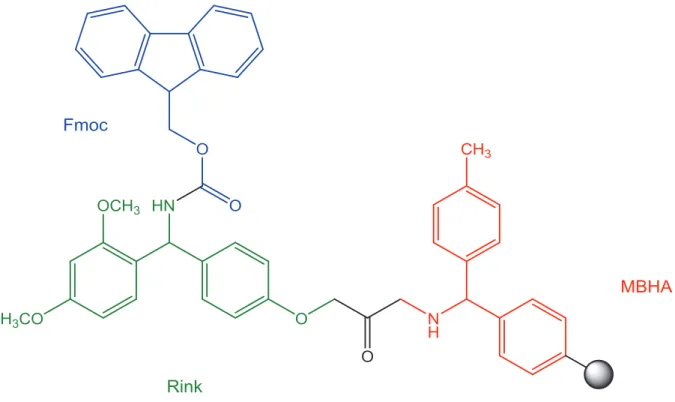
Figure 19: Chemical structure of the Fmoc-Rink Amide MBHA resin (polystyrene base polymer represented by the grey sphere). ... 30
Figure 20: Ninhydrin general reaction with primary amines, resulting in the formation of a chromophore. 31 Figure 21: Synthesis of new CPP-Gemcitabine conjugates (5). a) 3-(S-trityl)sulfanylpropanoic acid (2), TBTU, DIEA, DMF, 0 ºC - rt, 24h, (45%); b) DCM/TFA 1:1, Et3SiH 0 ºC, 1h (95%); c) 2,2-disulfanyldipyridine (6), MeOH, AcOH, rt, 24h (60%); d) DMF, rt, 24 h (95% for Pen; 60% for pVEC); e) 5% DMSO in H2O/ACN (3:1), pH 8 (dil. NH4OH), 24h. ... 34
Figure 22: Synthesis of Gemcitabine-dithiopyridine (7). ... 36
Figure 23: Chromatogram of the crude product obtained in the manual synthesis of Cys-Penetratin, acquired with a HPLC-DAD system, with a C18 column, using ACN in water with 0.05% TFA as eluent, in gradient mode (0 – 100%), for 30 minutes, at a flow rate of 1 mL/min and detection at " = 220 nm. The arrow indicates the chromatographic peak that was identified as the target peptide by LC-ESI/Orbitrap MS (mass spectrum on Figure 24). ... 41
Figure 24: Mass spectrum (LC-ESI/Orbitrap MS, positive mode) of the Cys-Penetratin peptide (manual synthesis). ... 41
Figure 25: Chromatogram of the product of the manual synthesis of the peptide Cys-pVEC, acquired with a HPLC-DAD system, with a C18 column, using ACN in water with 0.05% TFA as eluent, in gradient mode (0 – 100%), for 30 minutes, at a flow of 1 mL/min and detection at " = 220 nm. ... 42 Figure 26: Chromatogram of the product of the synthesis of the peptide Cys-Penetratin in the CEM Liberty1 MW-assisted synthesizer, acquired with a HPLC-DAD system, with a C18 column, using ACN in water with 0.05% TFA as eluent, in gradient mode (0 – 100%), for 30 minutes, at a flow of 1 mL/min and detection at " = 220 nm. ... 43 Figure 27: Mass spectrum (LC-ESI/MS Orbitrap, positive mode) of the Cys-Penetratin peptide (MW-assisted synthesis). ... 43 Figure 28: Chromatogram of the product of the synthesis of the peptide Cys-pVEC in the CEM Liberty1 MW-assisted synthesizer, acquired with a HPLC-DAD system, with a C18 column, using ACN in water with 0.05% TFA as eluent, in gradient mode (0 – 100%), for 30 minutes, at a flow of 1 mL/min and detection at " = 220 nm. ... 44 Figure 29: Mass spectrum (LC-ESI/MS Orbitrap, positive mode) of the Cys-pVEC peptide (MW-assisted synthesis). ... 45 Figure 30: Mass spectrum (LC-ESI/MS Orbitrap, positive mode) of compound (3). ... 46 Figure 31: Chromatogram of compound (4), acquired with a HPLC-DAD system, with a C18 column, using ACN in water with 0.05% TFA as eluent, in gradient mode (0 – 100%), for 30 minutes, at a flow of 1 mL/min and detection at " = 270 nm. ... 47 Figure 32: Mass spectrum (LC-ESI/MS Orbitrap, positive mode) of compound (4). ... 48 Figure 33: Chromatogram of compound (7), acquired with a HPLC-DAD system, with a C18 column, using ACN in water with 0.05% TFA as eluent, in gradient mode (0 – 100%), for 30 minutes, at a flow of 1 mL/min and detection at " = 220 nm. ... 49 Figure 34: Mass spectrum (ESI-IT MS Orbitrap, positive mode) of compound (7). ... 49 Figure 35: Chromatogram of the Cys-Penetratin peptide, acquired with a HPLC-DAD system, with a C18 column, using ACN in water with 0.05% TFA as eluent, in gradient mode (15 – 40%), for 30 minutes, at a flow of 1 mL/min and detection at " = 220 nm. ... 50 Figure 36: Chromatogram of the reaction mixture obtained in the attempt of producing the target Gemcitabine–Linker–Penetratin conjugate, following strategy A; data acquired with a HPLC-DAD system, with a C18 column, using ACN in water with 0.05% TFA as eluent, in gradient mode (15 – 40%), for 30 minutes, at a flow of 1 mL/min and detection at " = 220 nm. ... 51 Figure 37: Chromatogram of the reaction mixture obtained in the attempt of producing the target Gemcitabine–Linker–Penetratin conjugate, following strategy A; data acquired with a HPLC-DAD system, with a C18 column, using ACN in water with 0.05% TFA as eluent, in gradient mode (15 – 40%), for 30 minutes, at a flow of 1 mL/min and detection at " = 270 nm. ... 51 Figure 38: Mass spectrum (LC-ESI/MS Orbitrap, positive mode) relative to the most intense peak of the HPLC. ... 52 Figure 39: Chromatogram of the Cys-Penetratin peptide, acquired with a HPLC-DAD system, with a C18 column, using ACN in water with 0.05% TFA as eluent, in gradient mode (0 – 100%), for 30 minutes, at a flow of 1 mL/min and detection at " = 220 nm. ... 53 Figure 40: Chromatogram of the crude Gemcitabine–Linker–Penetratin conjugate, acquired with a HPLC-DAD system, with a C18 column, using ACN in water with 0.05% TFA as eluent, in gradient mode (0 – 100%), for 30 minutes, at a flow of 1 mL/min and detection at " = 220 nm. ... 53 Figure 41: Mass spectrum (LC-ESI/MS Orbitrap, positive mode) of the Gemcitabine–Linker–Penetratin conjugate. ... 54
Figure 42: Chromatogram of the Gemcitabine–Linker–Penetratin conjugate after purification, acquired with a HPLC-DAD system, with a C18 column, using ACN in water with 0.05% TFA as eluent, in gradient mode (0 – 100%), for 30 minutes, at a flow of 1 mL/min and detection at " = 220 nm. ... 55 Figure 43: Chemical structure of the Gemcitabine – Linker – Penetratin conjugate (exact mass 2696.3691 Da). ... 55 Figure 44: Chromatogram of the Cys-pVEC peptide, acquired with a HPLC-DAD system, with a C18 column, using ACN in water with 0.05% TFA as eluent, in gradient mode (0 – 100%), for 30 minutes, at a flow of 1 mL/min and detection at " = 220 nm. ... 56 Figure 45: Chromatogram of the Gemcitabine–Linker–pVEC conjugate, acquired with a HPLC-DAD system, with a C18 column, using ACN in water with 0.05% TFA as eluent, in gradient mode (0 – 100%), for 30 minutes, at a flow of 1 mL/min and detection at " = 220 nm. ... 56 Figure 46: Mass spectrum (LC-ESI/MS Orbitrap, positive mode) of the Gemcitabine–Linker–pVEC conjugate. ... 57 Figure 47: Chemical structure of the Gemcitabine – Linker – pVEC conjugate. ... 57 Figure 48: Chromatogram of the Gemcitabine–Linker–pVEC conjugate after purification, acquired with a HPLC-DAD system, with a C18 column, using ACN in water with 0.05% TFA as eluent, in gradient mode (0 – 100%), for 30 minutes, at a flow of 1 mL/min and detection at " = 220 nm. ... 58 Figure 49: Schematic representation of the Peptide-Drug conjugates, highlighting the hydrolysable bonds. ... 59 Figure 50: Calibration curve by HPLC analysis of gemcitabine solutions with different concentrations, with detection at " = 270 nm. ... 59 Figure 51: Graphical representation of the time-dependent release kinetics of free gemcitabine from the hydrolysis of Gemcitabine–Linker–Penetratin and Gemcitabine–Linker–pVEC conjugates in PBS (pH 7.4) at 37 ºC (" = 270 nm). ... 60 Figure 52: Graphical representation of the time-dependent kinetics of Gemcitabine–Linker–Penetratin and Gemcitabine–Linker–pVEC conjugates’ hydrolysis in PBS (pH 7.4) at 37 ºC (" = 220 nm). ... 61 Figure 53: Graphical representation of the 22 days study on the time-dependent release kinetics of free gemcitabine from the hydrolysis of Gemcitabine–Linker–Penetratin conjugate in PBS (pH 7.4) at 37 ºC (" = 270 nm). ... 62 Figure 54: Graphical representation of the 22 days study on the time-dependent kinetics of Gemcitabine– Linker–Penetratin conjugate’s hydrolysis in PBS (pH 7.4) at 37 ºC (" = 220 nm). ... 62 Figure 55: Effect of Gemcitabine–Linker–Penetratin and Gemcitabine–Linker–pVEC conjugates and free gemcitabine on the growth of different human tumoral cell lines evaluated by Sulforhodamine B assay. Cells were treated with a broad concentration range (6.3 – 100.0 µM) of each compound for 48 h. Each value represents the mean ± SEM (n = 3 – 6). **p <0.001, ***p <0.0001 (significant decrease vs control). . 64 Figure 56: Stability study of the Gemcitabine–Linker–Penetratin conjugate: HPLC analysis performed in the beginning of the study (0 h). Chromatogram acquired with a HPLC-DAD system, with a C18 column, using ACN in water with 0.05% TFA as eluent, in gradient mode (0 – 100%), for 30 minutes, at a flow of 1 mL/min and detection at " = 220 nm. ... 77 Figure 57: Stability study of the Gemcitabine–Linker–Penetratin conjugate: HPLC analysis performed after 24 h. Chromatogram acquired with a HPLC-DAD system, with a C18 column, using ACN in water with 0.05% TFA as eluent, in gradient mode (0 – 100%), for 30 minutes, at a flow of 1 mL/min and detection at " = 220 nm. ... 77 Figure 58: Stability study of the Gemcitabine–Linker–Penetratin conjugate: HPLC analysis performed after 48 h. Chromatogram acquired with a HPLC-DAD system, with a C18 column, using ACN in water with 0.05%
TFA as eluent, in gradient mode (0 – 100%), for 30 minutes, at a flow of 1 mL/min and detection at " = 220 nm. ... 78 Figure 59: Stability study of the Gemcitabine–Linker–Penetratin conjugate: HPLC analysis performed after 72 h. Chromatogram acquired with a HPLC-DAD system, with a C18 column, using ACN in water with 0.05% TFA as eluent, in gradient mode (0 – 100%), for 30 minutes, at a flow of 1 mL/min and detection at " = 220 nm. ... 78 Figure 60: Stability study of the Gemcitabine–Linker–Penetratin conjugate: HPLC analysis performed after 144 h. Chromatogram acquired with a HPLC-DAD system, with a C18 column, using ACN in water with 0.05% TFA as eluent, in gradient mode (0 – 100%), for 30 minutes, at a flow of 1 mL/min and detection at " = 220 nm. ... 79 Figure 61: Stability study of the Gemcitabine–Linker–pVEC conjugate: HPLC analysis performed in the beginning of the study (0 h). Chromatogram acquired with a HPLC-DAD system, with a C18 column, using ACN in water with 0.05% TFA as eluent, in gradient mode (0 – 100%), for 30 minutes, at a flow of 1 mL/min and detection at " = 220 nm. ... 80 Figure 62: Stability study of the Gemcitabine–Linker–pVEC conjugate: HPLC analysis performed after 24 h. Chromatogram acquired with a HPLC-DAD system, with a C18 column, using ACN in water with 0.05% TFA as eluent, in gradient mode (0 – 100%), for 30 minutes, at a flow of 1 mL/min and detection at " = 220 nm. ... 80 Figure 63: Stability study of the Gemcitabine–Linker–pVEC conjugate: HPLC analysis performed after 48 h. Chromatogram acquired with a HPLC-DAD system, with a C18 column, using ACN in water with 0.05% TFA as eluent, in gradient mode (0 – 100%), for 30 minutes, at a flow of 1 mL/min and detection at " = 220 nm. ... 81 Figure 64: Stability study of the Gemcitabine–Linker–pVEC conjugate: HPLC analysis performed after 72 h. Chromatogram acquired with a HPLC-DAD system, with a C18 column, using ACN in water with 0.05% TFA as eluent, in gradient mode (0 – 100%), for 30 minutes, at a flow of 1 mL/min and detection at " = 220 nm. ... 81 Figure 65: Stability study of the Gemcitabine–Linker–pVEC conjugate: HPLC analysis performed after 144 h. Chromatogram acquired with a HPLC-DAD system, with a C18 column, using ACN in water with 0.05% TFA as eluent, in gradient mode (0 – 100%), for 30 minutes, at a flow of 1 mL/min and detection at " = 220 nm. ... 82 Figure 66: Stability study of the Gemcitabine–Linker–Penetratin conjugate: HPLC analysis performed in the beginning of the study (0 h). Chromatogram acquired with a HPLC-DAD system, with a C18 column, using ACN in water with 0.05% TFA as eluent, in gradient mode (0 – 100%), for 30 minutes, at a flow of 1 mL/min and detection at " = 220 nm. ... 83 Figure 67: Stability study of the Gemcitabine–Linker–Penetratin conjugate: HPLC analysis performed after 7 days. Chromatogram acquired with a HPLC-DAD system, with a C18 column, using ACN in water with 0.05% TFA as eluent, in gradient mode (0 – 100%), for 30 minutes, at a flow of 1 mL/min and detection at " = 220 nm. ... 83 Figure 68: Stability study of the Gemcitabine–Linker–Penetratin conjugate: HPLC analysis performed after 14 days. Chromatogram acquired with a HPLC-DAD system, with a C18 column, using ACN in water with 0.05% TFA as eluent, in gradient mode (0 – 100%), for 30 minutes, at a flow of 1 mL/min and detection at " = 220 nm. ... 84 Figure 69: Stability study of the Gemcitabine–Linker–Penetratin conjugate: HPLC analysis performed after 22 days. Chromatogram acquired with a HPLC-DAD system, with a C18 column, using ACN in water with 0.05% TFA as eluent, in gradient mode (0 – 100%), for 30 minutes, at a flow of 1 mL/min and detection at " = 220 nm. ... 84
Figure 70: LC-MS chromatogram of the Cys-Penetratin peptide, synthesized in the CEM Liberty1 MW assisted peptide synthesizer. ... 85 Figure 71: LC-MS chromatogram of the Cys-pVEC peptide, synthesized in the CEM Liberty1 MW assisted peptide synthesizer. ... 85 Figure 72: LC-MS chromatogram of the Gemcitabine–Linker–Penetratin conjugate. ... 86 Figure 73: LC-MS chromatogram of the Gemcitabine–Linker–pVEC conjugate. ... 86
LIST OF TABLES
Table 1: General steps of Fmoc/tBu SPPS. ... 32 Table 2: Experimental conditions for SPPS on the CEM Libery1 Peptide Synthesizer. ... 33 Table 3: Sequence and exact mass of the Cys-Penetratin peptide and m/z ratios of its adducts with H+ detected by ESI-IT MS. ... 40 Table 4. Sequence and exact mass of the Cys-pVEC peptide and m/z ratios of its adducts with H+ detected by ESI-IT MS. ... 45 Table 5: Exact mass of the Gemcitabine–Linker–Penetratin conjugate and m/z ratios of its adducts with H+. ... 52 Table 6: Exact mass of the Gemcitabine–Linker–pVEC conjugate and m/z ratios of its adducts with H+. ... 58 Table 7: Summary of the results of the antiproliferative activity of the compounds, with indication of the IC50 (µM). ... 64
LIST OF ABBREVIATIONS
5’-NT: 5’-nucleotidase AA: amino acid ACN: acetonitrile
AMP: Antimicrobial Peptide Boc: tert-butoxycarbonyl Bzl: benzyl
CARPA: complement activation-related pseudoallergy CDA: cytidine deaminase
CMP: cytidine monophosphate CPP: Cell-Penetrating Peptide Ct: C-terminus
Da: Dalton (unified atomic mass unit) dCK: deoxycytidine kinase
DCM: dichloromethane
DCTD: deoxycytidylate deaminase
dFdC: 2!,2!-difluorodeoxycytidine (gemcitabine)
dFdCMP / dFdCDP / dFdCTP: gemcitabine monophosphate / diphosphate / triphosphate dFdU: 2’,2’-difluorodeoxyuridine
dFdUMP / dFdUDP / dFdUTP: 2’,2’-difluorodeoxyuridine monophosphate / diphosphate / triphosphate
DMSO: dimethylsulfoxide
DIPEA: N-ethyl-N,N-diisopropylamine DMF: N,N-dimethylformamide
DNA: deoxyribonucleic acid EE: encapsulation efficiency
FBS: fetal bovine serum
FDA: Food and Drug Administration (USA) Fmoc: 9-fluorenylmethoxycarbonyl
Fmoc-AA-OH: amino acid residue N"-protected with an Fmoc group GAGs: glycosaminoglycans
HBTU: O-(benzotriazol-1-yl)-N,N,N•,N!-tetramethyluronium hexafluorophosphate hCNT: human concentrative nucleoside transporter
hENT: human equilibrative nucleoside transporter HOBt·H2O: 1-hydroxybenzotriazole (mono hydrated)
HPLC-DAD: High-Performance Liquid Chromatography with Diode-Array Detection HSR: hypersensitivity reactions
LC-MS: Liquid Chromatography hyphenated to Mass Spectrometry m/z: mass-to-charge ratio
MAP: Membrane Active Peptide
MAPS: Microwave-Assisted Peptide Synthesis
MBHA: 4-methylbenzhydrylamine-functionalized resin MS: Mass Spectrometry
MW: microwave radiation
MW-SPPS: Microwave-Assisted Solid-Phase Peptide Synthesis NMP: N-methyl-2-pyrrolidone
Nt: N-terminus
Pbf: pentamethyl-2,3-dihydrobenzofuran-5-sulfonyl PBS: phosphate-buffered saline
PEG: polyethylene glycol
Rink: (R,S)-2-{4-[amino(2,4-dimethoxyphenyl)methyl]phenoxy}acetic acid RP-MPLC: Reversed Phase Medium-Pressure Liquid Chromatography rt: retention time
SPPS: Solid Phase Peptide Synthesis
TBTU: O-(Benzotriazol-1-yl)-N,N,N’,N’-tetramethyluronium tetrafluoro borate t
Bu: tert-butyl
TCA: trichloroacetic acid TFA: trifluoroacetic acid TIS: triisopropylsilane Trt: trityl
OBJECTIVES
The main goal of the work developed in this thesis was the development of new gemcitabine prodrugs, obtained through chemical modification of that anticancer agent, followed by conjugation of the modified derivative with Cell-Penetrating Peptides Penetratin and pVEC, activated with an N-terminal cystein residue.
Cys-Penetratin: C-RQIKIWFQNRRMKWKK Cys-pVEC: C-LLIILRRRIRKQAHAHSK
Furthermore, the work also aimed at the study of the time-dependent kinetics of gemcitabine release from hydrolysis of the new conjugates under physiological conditions.
Gemcitabine – Linker – Penetratin Gemcitabine – Linker – pVEC
1. INTRODUCTION
1.1. Cancer Management
In developed countries, cancer is one of the major causes of death, along with cardiovascular diseases. This condition involves the abnormal growth of any type of cell with the potential to invade or spread to other parts of the body. One of the main reasons that can be attributed to the high mortality of this group of diseases is the failure of current treatment options. The hard distinction between normal and cancer cells and difficulty in targeting only the altered cells, the phenomenon of resistance, and the inefficacy to treat metastases are the main obstacles when treating cancer patients.
Cancer management and treatment plans depend on several factors, such as the type, location, and grade of the cancer as well as the patient’s health and personal choices. Treatments may be curative or aimed at improving life quality. There are many treatment options for cancer, including surgery, chemotherapy, radiotherapy, hormonal therapy and palliative care.
Although surgery is the primary method used in most isolated solid cancers and is often an important part of determining the definitive diagnosis, chemotherapy is a major treatment choice. This type of therapy is recurrently used in combination with hormonal therapy (pharmacotherapy) and with other cancer treatments, such as radiotherapy, surgery, and/or hyperthermia therapy. Chemotherapy consists on the administration of chemical substances, especially one or more anti-cancer drugs (chemotherapeutic agents), which are traditionally cytotoxic agents, killing cells that divide rapidly, one of the main properties of most cancer cells. However, these substances will also reach other cells in our body that divide rapidly under normal circumstances, as cells in the bone marrow, digestive tract, and hair follicles. This can lead to various severe side effects, frequently associated with chemotherapy. These adverse reactions in addition to the limited effectiveness of oncologic treatments make the development of new therapeutic options that are able to be more selective and effective an imperative need.
Alternative targeted therapies have been put forward, aiming at efficiently treating specific types of cancer while decreasing the severe side effects associated with these treatments. According to the National Cancer Institute of the United States of America (NCI, USA), targeted therapies are deliberately chosen or designed to interact with and act on specific molecular targets that are associated with cancer, whereas most standard chemotherapies act on all rapidly dividing normal and cancerous cells (many standard chemotherapies were identified because they kill cells) [1]. These targeted therapies are often cytostatic (that is, they block tumor cell proliferation), whereas standard chemotherapy agents are cytotoxic (that is, they kill tumor cells).
Still, there are some limitations to targeted therapeutic approaches: cancer cells can become resistant to them and, for this reason, targeted therapies may work best in combination; another current limitation of targeted therapies is that drugs for some identified targets are difficult to develop because of the target’s structure and/or the way its function is regulated in the cell. Moreover, although targeted therapies are designed to reduce side effects, some patients receiving this type of therapy suffer from substantial adverse effects, the most common including diarrhea and liver problems, such as hepatitis and elevated liver enzymes. Nevertheless, several experimental targeted therapies for specific types of cancer are currently being studied in clinical trials and some have already been approved by the Food and Drug Administration (FDA).
1.2. Gemcitabine
The current treatment of cancer using chemotherapy is largely based on the use of nucleoside analogues and gemcitabine, or 2!,2!-difluorodeoxycytidine (1, Figure 1), is one of such analogues. Despite the similarities with other nucleoside analogues, gemcitabine has many distinctive properties, including its spectrum of activity.
Gemcitabine is commercially available as Gemzar® and was developed by the pharmaceutical Eli Lilly and Company in the 1980s. Although gemcitabine was originally investigated for its antiviral effects [2], this drug has been proven efficient against a wide range of solid tumors and has since been developed as an active agent for cancer therapy. The FDA has also approved the use of gemcitabine (a) in combination with carboplatin for the treatment of advanced ovarian cancer that has relapsed at least 6 months after completion of platinum-based therapy [3]; (b) in combination with paclitaxel, for first-line treatment of metastatic breast cancer after failure of prior anthracycline-containing adjuvant chemotherapy, unless anthracyclines were clinically contraindicated [4]; (c) in combination with cisplatin for the treatment of non-small cell lung cancer (NSCLC) [5] and (d) as a single agent for the treatment of pancreatic cancer [6]. Gemcitabine is particularly effective against pancreatic cancer, and the use of its hydrochloride salt was approved by the FDA in 1996 as first-line treatment for patients with locally advanced (non-resectable Stage II or Stage III) or metastatic (Stage IV) pancreatic adenocarcinoma previously treated with fluorouracil (5-FU) [7].
However, treatment with gemcitabine leads to some severe adverse reactions, which are dependent on dosage and duration of the treatment, clinical indication and cancer stage, gender, age and patients’ clinical history. The most common include myelosuppression, hepatic and renal toxicity, cardiovascular alterations (such as hypotension, capillary leak syndrome,…) nausea/vomiting, fever, thrombocytopenia and dyspnea [8].
Figure 1: Chemical structure of Gemcitabine (2!,2!-difluorodeoxycytidine), where 4-(N)- and 5’- sites are highlighted.
1.2.1. Biodistribution and Mechanism of Action
Gemcitabine is a polar drug with low membrane permeability, and is primarily administered by intravenous infusions. It has a very short plasma circulation time and the elimination half-life depends upon the infusion time, age and gender of the patient, ranging from 42 to 94 min for short infusions and from 4 to 10 h for infusions of 70 min [9]. An increased toxicity is associated with longer infusions and, at higher doses, major toxicity can be observed, such as neutropenia, reversible hepatic transaminase increase and high levels of hepatoxicity, renal toxicity, myelosuppression, thrombocytopaenia, anemia, proteinuria, nausea and vomiting, mild flulike syndrome, and mild skin rash. Also, gemcitabine is rapidly cleared from the body upon its enzymatic conversion in the blood, liver, kidney, and various tumor tissues [10, 11].
Upon administration, gemcitabine is transported in the plasma and crosses the plasma membrane into cells via nucleoside transporters. These transporters can be sodium-dependent (human concentrative nucleoside transporter, hCNTs) or sodium-independent (human equilibrative nucleoside transporter, hENTs) [12]. There are two equilibrative nucleoside transporters (hENT1 and hENT2) and three concentrative nucleoside transporters (hCNT1, hCNT2, and hCNT3). Kinetic studies have shown that gemcitabine intracellular uptake is preferentially directed by hENT1 and, to a lesser extent, by hCNT1 and hCNT3 [13, 14]. Also, several studies have shown the importance of the presence of the hENT1 transporter for an optimal response to gemcitabine [15].
Once inside the cells, gemcitabine needs to be activated, and undergoes a series of phosphorylation steps catalyzed by intracellular enzymes (Figure 2). First, deoxycytidine kinase (dCK) phosphorylates gemcitabine to a monophosphate derivative, dFdCMP. Then, nucleoside monophosphate kinase (UMP/CMP) phosphorylates dFdCMP to gemcitabine diphosphate (dFdCDP) and finally diphosphate kinase yields gemcitabine triphosphate (dFdCTP), the active form of this drug [16].
4-(N)-site
Figure 2: Mechanism of intracellular activation of gemcitabine. Adapted from [17].
However, gemcitabine can also be inactivated inside cells. This inactivation is due to the conversion of gemcitabine into its inactive uridine metabolite, 2’,2’-difluorodeoxyuridine (dFdU), catalyzed by cytidine deaminase (CDA), as well as to the deamination of gemcitabine monophosphate into dFdUMP, which is catalyzed by deoxycytidylate deaminase (DCTD). Furthermore, the phosphorylated metabolites of gemcitabine are inactivated via reduction by cellular 5’-nucleotidase (5’-NT). CDA is present at high levels in both human plasma and liver, and the enzymatic conversion of gemcitabine rapidly clears it from the body [16].
In its triphosphate form, dFdCTP, gemcitabine acts as an antimetabolic drug, being a competitive substrate of deoxycytidine triphosphate to DNA polymerases, and causes cell death by apoptosis. This action is possible because of the analogy between dFdCTP and deoxycytidine triphosphate: gemcitabine triphosphate is incorporated into DNA during replication (making it an S-phase-specific drug) thus inhibiting chain elongation of DNA because DNA polymerases are unable to proceed after one deoxynucleotide is added [18]. This action is called “masked chain termination”, and it appears to lock the drug into DNA because proof-reading exonucleases are unable to remove gemcitabine nucleotide from this penultimate position [19]. The inhibitory action of gemcitabine is further improved by its nondetection in the DNA chain.
Additionally, because the diphosphate form of gemcitabine (dFdCDP) inhibits ribonucleoside diphosphate reductase (RNR), an enzyme of DNA synthesis which permits the formation of nucleoside triphosphates, there is a significant decrease in the concentration of cellular deoxycytidine triphosphate (dCTP) and a change in the ratio of dCTP/dFdCTP in favor of dFdCTP.
The accumulation of gemcitabine triphosphate and the intracellular reduction of dCTP results in the inhibition of dFdCMP inactivation by DCTD, which requires sufficient concentrations of dCTP to be active [20]. Active metabolites of gemcitabine can also inhibit ribonucleotide reductase (RR), dCMP deaminase, and CTP synthetase, thus enhancing gemcitabine activation, due to a unique property called self-potentiation.
1.2.2. Resistance to Gemcitabine
The rise of drug resistance is a major obstacle in the treatment of many forms of cancer, and this phenomenon usually appears very rapidly after the beginning of the treatment, when cells are typically sensitive to the drug. It is critical to have a better understating oh this phenomenon so that drugs can be replaced by more efficient substituents, combination therapies can be developed or the targeting of the drugs can be improved.
There are many factors contributing to the development of resistance to gemcitabine, from transporters expression to abnormal levels of the enzymes involved in the activation, metabolism and mechanism of action of gemcitabine. One of the major causes of resistance to gemcitabine can be attributed to alterations in transporters. hENT1 is a preferential transporter and studies have found a strong correlation between gemcitabine resistance and a deficiency of hENT1 expression in human breast and pancreatic cancer cells [21, 22]. Additionally, studies have shown that the determination of hENT1 but also hCNT3 expression can be used as a prognostic marker for patients treated with gemcitabine [23, 24].
There are many enzymes involved in the metabolism of gemcitabine, and alterations on the expression levels of any of these enzymes can make cells resistant to this drug or, on the other hand, make it more cytotoxic. One of the key enzymes is deoxycytidine kinase (dCK), responsible for the first phosphorylation of gemcitabine. This is the rate-limiting enzyme involved in the metabolism of gemcitabine and its expression has been postulated to correlate with gemcitabine resistance [25]. This can be caused by mutations of the dCK gene [26] or by low levels of dCK enzyme activity [27]. Also, high levels of the catabolic enzymes cytidine deaminase (CDA), dCMP deaminase and 5!-nucleotidase (5!-NT) are associated with resistance to gemcitabine [28].
Another factor in gemcitabine resistance is the overexpression of ribonucleotide reductase (RR) [29]. This enzyme is mainly responsible for the conversion of ribonucleosides to deoxyribonucleoside triphosphates (dNTPs), which are essential for DNA polymerization and repair. Both regulatory subunit RRM1 and catalytic subunit RRM2 influence the cytotoxic action of gemcitabine. RRM1 overexpression through transfection of a lung cancer cell line resulted in gemcitabine resistance and the reduction of RRM1 expression through RNA interference
abrogated the induced gemcitabine resistance [30]. As for the RRM2 subunit, its overexpression is associated with resistance to gemcitabine and down regulation of RRM2 by siRNA enhanced gemcitabine cytotoxicity, both in vitro and in vivo in pancreatic adenocarcinoma [31].
The aberrant expression of genes associated with cellular survival and apoptosis upon treatment with gemcitabine is also implicated in resistance to this drug [32, 33]. Once gemcitabine is incorporated into DNA, the cell enters a process of cell death, mainly through apoptosis, in response to cytotoxic drug treatment [34, 35]. One of the main proteins involved in the cell death mechanism and apoptosis is p53 [36]. The expression of p53 plays an important role in apoptosis pathways, inducing cell cycle arrest and in the higher concentration ranges, p53 induces apoptosis. In sensitive malignant cells, a variety of anticancer drugs have been shown to produce extensive apoptosis, but it has been suggested that the inability of some cells to undergo apoptosis is similar to the mechanism of gemcitabine resistance. Human lung cancer expressing the mutation of the p53 gene does not undergo apoptosis after gemcitabine treatment [37].
1.2.3. Modifications of Gemcitabine
From the previous sections it can be inferred that, although gemcitabine is an effective treatment to patients with different types of cancer and mainly to treat pancreatic adenocarcinoma, it has low membrane permeability and is extensively degraded by CDA into an inactive metabolite in the liver. Moreover, the increasing levels of resistance also reduce gemcitabine cytotoxicity. Thus, a frequent administration schedule at high drug doses is required, and this leads to serious side effects. To date, various innovative approaches have been developed to overcome these disadvantages, including combination therapy to determine the utility of adding a second cytotoxic drug or radiotherapy to this treatment regimen, but clinical trials have shown modest improvements in disease-free survival, and did not result in statistically significant improvements in survival with reported survival rates reaching 20% to 40% at 1 year [38, 39]. The FDA has also approved the use of gemcitabine in combination with carboplatin, paclitaxel and cisplatin for the treatment of patients with ovarian, breast and non-small cell lung cancer (NSCLC), respectively.
A different strategy consists on chemically modifying gemcitabine, in order to enhance transport and cytotoxicity. These modifications can provide (i) protection against deamination, (ii) better storage and (iii) prolonged release in the cell, (iv) a possible use in the case of deoxycytidine kinase deficiency, and (v) transporter deficiency. These new gemcitabine-based systems have the potential to improve the clinical outcome of a chemotherapy strategy and make these new molecules into prodrugs (a biologically inactive derivative of a parent drug molecule that usually requires an enzymatic or chemical transformation within the body to release the active drug and has improved delivery properties in comparison to the parent molecule [40]).
Most modifications on the gemcitabine structure are performed at the 4-(N)- and 5’-positions, as these two functional groups have significant roles in the action of this drug: the inactivation of gemcitabine results from deamination on the 4-(N)-site, whereas the necessary phosphorylation to make it active occurs in the hydroxyl group at the 5’-position. Some of the new molecules resulting from the modification of gemcitabine and their most relevant properties are described below.
1.2.3.1. Modifications at the 4-(N)-site of gemcitabine
1.2.3.1.1. PEG – Gemcitabine
Polyethylene glycol (PEG, Figure 3) refers to a family of differently-sized polymers with high solubility in water and excellent biocompatibility that have been conjugated to several molecules, from antitumor agents, proteins, peptides or low molecular-weight drugs, due to a relevant number of advantages resulting from the PEGylation process [41]. This technique has become the leading approach to (a) increase plasma half-life of the PEGylated products, (b) provide enzyme protection and (c) accumulate the PEGylated product in tumor zones according to the “enhanced permeability and retention” effect (EPR) [42-44]. However, PEG can activate the complement system, which has been recently proposed as a major underlying or contributing cause of infusion reactions, referred to as complement activation-related pseudoallergy (CARPA), an anaphylactic-type reaction within the Type I category of hypersensitivity reactions (HSRs) [45]. Clinical trials of several derivatives of PEG coupled to anticancer drugs are already under way or have been completed [46].
Figure 3: Chemical structures of polyethylene glycol (PEG) and of a PEG – gemcitabine conjugate.
Because of encouraging results from studies with other anticancer agents, and given that the anticancer effects of gemcitabine are limited by its rapid metabolization and consequently short half-life, as well as its low tumor uptake, the coupling of PEG to gemcitabine has been tested by
several research teams. In a recent paper, the synthesis of a PEG – gemcitabine construct, by conjugating the amino group at the 4-(N)-position of gemcitabine to an N-hydroxysuccinimide derivative of PEG, has been carried out (Figure 3) [47]. It is known that the cellular uptake of PEG-drug conjugates occurs through endocytosis and that conjugates are retained in transport vesicles which traffic along the endolysosomal scaffold, which are acidic in nature [48, 49]. The endolysosomal transport vesicles allow cleavage of the amide bonds between PEG and gemcitabine, thanks to the acidic nature of these vesicles, and thereby allow its prolonged release. Vandana’s team was able to demonstrate a colocalization of PEG-gemcitabine in lysosome and endosome after 24 h incubation, and an enhanced retention in cancer cells after 3 days of incubation in comparison to parent gemcitabine, using confocal analysis. Moreover, pharmacokinetic studies have shown consistently higher (by 21-fold) bioavailability of PEG-gemcitabine over unmodified PEG-gemcitabine, after 1 h of intravenous administration in mice and, in MiaPaCA 2 and PANC-1 cells, PEG – gemcitabine was more effective in comparison to native gemcitabine.
1.2.3.1.2. Folate-PEG – Gemcitabine
Folate, or folic acid, has been used as a targeting agent in cancer nanotherapeutics, because its receptor is overexpressed in several types of cancers (lung, breast, kidney, and ovarian) [50-52], while in normal human tissues its receptors may have limited distribution. To improve the effect of PEG – gemcitabine, folic acid has been inserted into PEG – gemcitabine conjugates, where the drug was coupled through its 4-(N)-amino group to the carboxylic end of an aminocarboxylated PEG (NH2-PEG-COOH), while folic acid was linked through its carboxylic
function to the amino group of the polymer (
Figure 4). The active targeting and cytotoxic superiority of Folate-PEG – Gemcitabine, compared to the nontargeted PEG – gemcitabine, were investigated [53]. All conjugates were able to release the drug in a pH-dependent manner with no role played by enzymes, which confirms previous studies showing that the protection of the amino group of gemcitabine prevents enzymatic degradation [54]. Polymer conjugation of gemcitabine increased drug plasma half-life by reducing its kidney clearance, which was dependent on the polymer’s molecular weight. Folic acid–PEG derivatives were also found as less toxic than PEG derivatives, which was explained by a slower entry into the cell by endocytosis and a cytotoxic activity only after the release of gemcitabine. Moreover, derivatives targeting folate receptors need a receptor-mediated endocytosis mechanism for cell penetration in cells that do not overexpress the folate receptor. In contrast, in KB-3-1 cells, which overexpress folate receptors, derivatives with folate are certainly less cytotoxic than parent gemcitabine but are more cytotoxic than gemcitabine only coupled to PEG [53].
Figure 4: Chemical structure of a Folate-PEG – gemcitabine conjugate.
1.2.3.1.3. Valproic acid – Gemcitabine (LY2334737)
An orally available prodrug of gemcitabine has been developed, consisting of gemcitabine linked to valproic acid via an amide bond at the 4-(N)-position. This molecule is called LY2334737 (Figure 5), and is able to circumvent the rapid deamination of gemcitabine into dFdU, and thus bypass hydrolysis in enterocytes and portal circulation, avoiding the extensive first pass metabolism that occurs with unmodified gemcitabine, underlying its poor oral bioavailability due to an extensive deamination by CDA.
The stability of the prodrug was tested on a pH range from 1 to 8, to check the possibility of delivering an intact prodrug into systemic circulation after passing through the gastrointestinal tract following oral administration. LY2334737 degradation is pH-dependent, with about 21% degradation at pH 1 and no degradation between pH 6 to 8, after 4 h of incubation at 40 °C [55]. Circulating levels of LY2334737 are detectable several hours after oral administration. In addition, a gradual release of gemcitabine following cleavage of the amide bond should enhance efficacy, since more cancer cells should be exposed to a continuous effective cytotoxic level of gemcitabine. The researchers have found that, in mice bearing HCT-116 human colon tumor xenografts, a much smaller dose via oral administration has the same efficacy that a high dose administered via intraperitoneal injection. Phase I trials of LY2334737 either as monotherapy or in combination with other agents were carried out to determine the maximum tolerated dose and dose limiting toxicities of daily administration [56].
Figure 5: Chemical structure of LY2334737, a prodrug of gemcitabine tested in Phase I clinical trials.
1.2.3.1.4. Squalenoyl – Gemcitabine
Squalene is a triterpene that is an intermediate in the cholesterol biosynthesis pathway. Both in vitro and in vivo model experiments suggest a tumor-inhibiting role for squalene [57] and, therefore, it has been linked to various nucleoside analogues [58].
Couvreur’s team was interested in the pharmacological activity of gemcitabine covalently coupled at 4-(N)-position with 1,1’,2-tris-nor-squalenoic acid to obtain Sq-gemcitabine [4-(N)-tris-nor-squalenoyl-gemcitabine, SQdFdC] (Figure 6).
Those authors investigated the anticancer activity of Sq-gemcitabine in vitro on resistant murine and human leukemia cells (L1210 10K and CEM/ARAC8C, respectively). L1210 10K cells were characterized by a lower expression of cytoplasmic dCK and CEM/ARAC8C by a deficiency in hENT1 transporters [59, 60], hence respectively representing two major resistance factors to gemcitabine. After 72 h of incubation at different concentrations, Sq-gemcitabine demonstrated 3.26 and 3.22-fold higher cytotoxicity compared to gemcitabine with L1210 10K and CEM/ARAC8C, respectively [60]. After intravenous injection of Sq-gemcitabine in aggressive leukemia-bearing mice, a significant increase in survival time compared to gemcitabine was obtained. This increase was attributed to the high degree of localization of Sq-gemcitabine in the liver and spleen, which are the major metastatic organs. Sq-gemcitabine was found to be more efficient than gemcitabine, suggesting the considerable potential of this treatment for leukemia.
Studies on human pancreatic adenocarcinoma models were also performed. Sq-gemcitabine showed higher antiproliferative and cytotoxic effects in vitro compared to parent gemcitabine, on chemoresistant tumor cells (Panc-1) and sensitive cell lines (Capan-1 and BxPc-3), which were associated with significant DNA synthesis inhibition, S-phase arrest, and higher induction of apoptosis (caspase 3 activation) [61].
While the mechanism of entry and metabolization of gemcitabine into cells is known, the cellular uptake mechanism of Sq-gemcitabine, its subcellular localization, and its metabolization pathway have been studied only recently. An in vitro passive entry in cancer cells (MCF-7: human breast adenocarcinoma) and a preferential accumulation in endoplasmic reticulum, thanks to the high lipophilic level of squalene, were observed [62]. This passive input may explain the efficacy of Sq-gemcitabine on cell lines deficient in active transporters [63].
Finally, two important elements have contributed to the efficiency of this new drug: (a) the storage of gemcitabine in the endoplasmic reticulum allowed it to be protected from deamination by the presence of squalene in the 4-(N)-position, and (b) the progressive cleavage to its parent form allowed the metabolization of the cancer compound.
1.2.3.1.5. Lipophilic Prodrugs
To protect against the deamination of gemcitabine, it was proposed to covalently link the amino group in the 4-(N)-position with saturated and monounsaturated fatty acids, namely, with those bearing long C18 and C20 chains. In 1998, Eli Lilly patented the synthesis of lipophilic gemcitabine derivatives (Figure 7), which showed better cytotoxicity than unmodified gemcitabine.
Figure 7: General structure of lipophilic 4-(N)-acyl – gemcitabine conjugates.
A few years later, the synthesis of a series of 4-(N)-acyl derivative prodrugs of gemcitabine was carried out, first to prevent diffusion through liposome bilayers and later to be encapsulated into other particles [64]. Liposomal formulations containing these lipophilic prodrugs of gemcitabine increased the drug entrapment efficacy with respect to conventional liposomes, but their encapsulation efficiency (EE) closely depended on the length of the 4-(N)-acyl chain, the phospholipids, and the presence of cholesterol. Better results were obtained by incorporating 4-(N)-lauroyl-gemcitabine (GemC12) and 4-(N)-stearoyl-gemcitabine (GemC18) in liposomes composed by DSPC/DSPG 9:1. Furthermore, while parent gemcitabine is rapidly converted into inactive metabolite by CDA which is widely distributed in plasma, C12 and C18 derivatives were both stable in plasma. After 24 h, more than 60% of unmodified GemC12 and GemC18 prodrugs were still present, whereas only 40% of unmodified gemcitabine was present after 8 h of incubation of the drug in plasma. The prodrugs were also stable the pH range 4!9, which confirms the stability of the acyl-drug amide linkage. In vitro studies have shown that cytotoxicity of free or encapsulated GemC12 and GemC18 derivatives were 2- and 7-fold (in KB and HT-29 cells line, respectively) greater than that of parent gemcitabine. Encapsulation of the C18 derivatives into liposomes produced an increase of plasma availability: the AUC was 50 times higher than for
unmodified gemcitabine, resulting in the increased accumulation in tumor cells and a high level of antitumoral efficacy in mice grafted with HT-29 and KB 396p cells [65].
The increase of anticancer activity observed with these lipophilic derivatives compared with parent gemcitabine was obtained at the expense of their solubility in aqueous media. Indeed, with their highly lipophilic properties, these compounds proved difficult to reconcile with intravenous administration, and hence encapsulation is necessary. The modification of lipophilic behavior and encapsulation could be considered a good and versatile antitumoral approach against several tumors which become less sensitive to the parent drug.
1.2.3.1.6. H–Gemcitabine: Hoechst conjugated to gemcitabine
Dasari and his team developed a new conjugate, composed of the DNA binding agent Hoechst conjugated to gemcitabine [66, 67]. H–Gemcitabine (
Figure 8) is designed to reduce the systemic toxicity of gemcitabine by targeting it to the necrotic core of tumors, and also protect against deamination of its 4-(N) amine via protection with an amide bond. H–Gemcitabine targets tumors via its Hoechst moiety, which binds extracellular DNA (E-DNA) present in the necrotic core of tumors. The authors showed that H–Gemcitabine is significantly more effective than free gemcitabine at treating xenograft human colon tumors in nude mice, and also has a maximum tolerated dose equivalent to that of gemcitabine [67].
1.2.3.2. Modifications at the 5’-site of gemcitabine
1.2.3.2.1. CP–4126
In an attempt to enhance cellular uptake, prolong cell retention, and increase the half-life of gemcitabine with a less hydrophilic moiety, conjugation with a fatty acid side chain at the drug’s 5’-site has also been carried out [68, 69]. In this connection, CP–4126 has been developed through esterification of the 5’-position of the nucleoside sugar moiety with elaidic acid, a monounsaturated fatty acid with a trans-double bond at carbon 9 (Figure 9).
Figure 9: Chemical structure of gemcitabine derivative CP–4126.
Due to its different molecular design, CP–4126 is absorbed by cancer cells independent of hENT1 levels, which improves its efficacy in tumors with low or no hENT1 expression [70]. CP– 4126 needs to be converted into gemcitabine by nonidentified esterases, releasing the fatty acid and allowing the free drug to be phosphorylated [71]. In vitro tests have shown that IC50 of gemcitabine increased up to 200-fold in deficient nucleoside transport cell lines, but there was no difference with CP–4126, suggesting a nucleoside transporter-independent transport in the cell of the fatty acid derivative [72]. Inside the cell, CP–4126 was localized in the membrane and the cytosolic fraction, leading to long retention after removal of the cell culture medium. This accumulation caused a slower and prolonged release of the gemcitabine from the lipophilic derivative. CP–4126 is active in cells with deficient nucleoside membrane transport [73], but on the other hand, activity of parent gemcitabine and CP–4126 was comparable in the cell lines without resistance, while in dCK deficient cells, both compounds were inactive, because CP–4126 depends, like free gemcitabine, on activation by dCK.
In contrast to parent gemcitabine, which was highly toxic via the oral route, CP–4126 was administered orally with various schedules and an efficient antitumor activity [74]. CP–4126 is currently in a phase II clinical trial with patients with solid tumors [71], to investigate the use of CP– 4126 as a second-line treatment for advanced, metastatic pancreatic cancer in patients refractory
to first line gemcitabine treatment, where the resistance mechanism is likely due to impaired drug entry into tumor cells.
1.2.3.2.2. NEO6002
NeoPharm has synthesized a novel gemcitabine-cardiolipin conjugate via a succinate linker to enhance the uptake and efficacy by a prolonged release of gemcitabine in cancer cells [75]. Cardiolipin (CL) is a major membrane phospholipid specifically localized in mitochondria. At the cellular level, CL has been shown to have a role in the triggering of apoptosis [76]. The conjugate is called NEO6002 (Figure 10) and in vitro studies showed that this conjugate can improve cytotoxic efficacy and potentially overcome NT-deficient (nucleoside transporters) gemcitabine-resistant tumors, indicating a different internalization route of NEO6002 [77]. NeoPharm’s studies on NEO6002 showed evidence of cytotoxicity against various cancer cell lines, including A549 (human lung), BxPC-3 (human pancreas), MX-1 (human breast), HT-29 (human colon), and P388 (murine leukemia) [78]. In vivo studies showed that mice treated with gemcitabine for six daily 27 "mol/kg-doses were all moribund, whereas no mouse treated with NEO6002 died. This suggested that NEO6002 was less toxic at equimolar dosage when compared to gemcitabine.
1.2.3.2.3. Phosphoramidate Gemcitabine
Because the obligatory phosphorylation is often the rate-limiting step in the activation process of many anticancer drugs and is therefore still one of the limiting factors for the therapeutic use of nucleoside analogues, the use of modified nucleotide prodrugs incorporating a phosphate protecting group, has led to the selective release of the monophosphorylated nucleoside analogue [79]. This modification was tested on gemcitabine to get (a) resistance to chemical degradation, (b) passive diffusion across cell membranes, and (c) release of the monophosphorylated metabolite, independent of kinase expression. These prodrugs were designed to undergo intracellular activation to generate an unstable phosphoramidate intermediate anion, followed by a spontaneous cyclization and PѸN bond cleavage by water to liberate the nucleoside monophosphate [80].
In 2009, Perigaud et al. synthesized four derivatives of gemcitabine, N-(n-butylamino)-O-(S-pivaloyl-2-thioethyl)-O-5’-gemcitabine phosphoramidate diester (Gem-1), N-(isopropylamino)-O-(S-pivaloyl-2-thioethyl)-O-5’-gemcitabine phosphoramidate diester (Gem-2), (S-pivaloyl-2-thioethyl)-O-5’-gemcitabine phosphoramidate diester (Gem-3), and N-(benzylamino)-O-(S-(2,2-dimethyl-3-hydroxypropionyl)-2-thioethyl)-O-5’-gemcitabine phosphoramidate diester (4) (Figure 11). A better cytotoxicity level was obtained in preliminary in vitro tests with Gem-1, Gem-2, and Gem-3 compared to the parent gemcitabine in L1210 10K cell line (23.7 ± 1.2 "M, 18.3 ± 1.5 "M, 9.7 ± 9.0 "M, 36.7 ± 11.6 "M, respectively) [81].
The purpose of these modifications was to overcome resistance in tumors deficient in dCK by intracellular delivery of gemcitabine 5#-monophosphate after cell-membrane crossing by passive diffusion [82]. In vitro tests on many cancer cell lines have shown that the prodrugs are less active than unmodified gemcitabine in wildtype cell lines but more active than the parent drug in dCK deficient cell lines (AG6000 and CEM-dCK). However, after blocking the equilibrative nucleoside transport, the inhibition of tumor growth was no longer observed with the prodrugs, indicating that their antitumor activity was mediated by cell entry implying equilibrative nucleoside transport.
1.2.3.2.4. Gemcitabine prodrugs incorporating D-enantiomer amino acids
Gemcitabine prodrugs with D- and L-amino acids (Figure 12) were synthesized by Tsume’s team. Their chemical stability in buffers, resistance to glycosidic bond metabolism, enzymatic activation, permeability in Caco-2 cells and mouse intestinal membrane and antiproliferative activity in cancer cells were determined and compared to that of parent gemcitabine [83].
Figure 12: Gemcitabine prodrugs with D- and L-amino acids.
As expected, prodrugs containing D-amino acids were enzymatically more stable than their L-configuration counterparts. The activation of all gemcitabine prodrugs was 1.3 – 17.6-fold faster in cancer cell homogenate than their hydrolysis in buffer, suggesting enzymatic action. The enzymatic activation of amino acid monoester prodrugs containing D-amino acids in cell homogenates was 2.2-10.9-fold slower than for amino acid monoester prodrugs with L-amino acids. All prodrugs exhibited enhanced resistance to glycosidic bond metabolism by thymidine phosphorylase compared to parent gemcitabine. Also, these gemcitabine prodrugs showed
![Figure 2: Mechanism of intracellular activation of gemcitabine. Adapted from [17].](https://thumb-eu.123doks.com/thumbv2/123dok_br/15723129.1070700/34.892.171.725.132.501/figure-mechanism-intracellular-activation-gemcitabine-adapted.webp)


![Figure 13: CPPs as delivery vectors – intracellular delivery of CPP-cargo complexes. Reproduced from [84]](https://thumb-eu.123doks.com/thumbv2/123dok_br/15723129.1070700/50.892.87.808.129.477/figure-cpps-delivery-vectors-intracellular-delivery-complexes-reproduced.webp)