CLINICAL SCIENCE
1. Department of Pathology, School of Medicine, University of São Paulo, São Paulo, Brazil.
2. Pediatric Institute of Clinical Hospital, School of Medicine, University of São Paulo, São Paulo, Brazil.
3. Department of Pathology of Institute Adolfo Lutz, São Paulo, São Paulo, Brazil.
E-mail: [email protected] Received for publication on July 11, 2007 Accepted for publication on September 09, 2007
MORPHOMETRIC EVALUATION OF NB84,
SYNAPTOPHYSIN AND AGNOR IS USEFUL FOR THE
HISTOLOGICAL DIAGNOSIS AND PROGNOSIS IN
PERIPHERAL NEUROBLASTIC TUMORS (PNTS)
Aparecida de Cássia Carvalho1, Edwin Roger Parra1, Maria Cláudia Zerbini2,
Venâncio Avancini Ferreira Alves3, Vera Luiza Capelozzi1, Leila Antonangelo1
Carvalho AC, Parra ER, Zerbini MC, Alves VAF, Capelozzi VL, Antonangelo L. Morphometric evaluation of NB84, synaptophysin and AgNOR is useful for the histological diagnosis and prognosis in peripheral neuroblastic tumors (pNTs). Clinics. 2007;62(6):731-40.
OBJECTIVE: To study the importance of NB84, synaptophysin and AgNOR and explore the quantitative association of these factors with diagnosis and outcome as well as the association between NB84 and AgNOR and other tumor and stromal factors in twenty-eight peripheral neuroblastic tumors.
METHODS: We assessed AgNORs, NB84, synaptophysin and several other markers in tumor tissues from 28 patients with primary neuroblastic tumors. The treatment included: surgery for stage 1, chemotherapy and bone marrow transplantation for most of stages 3 and 4. Histochemistry, immunohistochemistry and morphometry were used to evaluate the amount of tumor staining for AgNOR, NB84 and synaptophysin; the outcome for our study was survival time until death due to recurrent neuroblastic tumors.
RESULTS: Only stage (p<0.01), AgNOR (p<0.01), NB84 (p<0.01) and synaptophysin (p=0.01) reached statistical significance as prognostic indicators.
CONCLUSIONS: Determination of NB84 and synaptophysin are useful tools for the diagnosis of peripheral neuroblastic tumors The association of the evaluation of AgNOR expression by the tumor cells may provide an important contribution to the prognostic evaluation and management approach of the patients.
KEYWORDS: Neuroblastoma. AgNOR. DNA ploidy. NB84. Synaptophysin
INTRODUCTION
Peripheral neuroblastic tumors (pNTs) are a heteroge-neous group of tumors that do not have a simple and re-producible morphological diagnosis and of which progno-sis is variable, mainly due to the following features: a) pNTs
can arise in the adrenal medulla, sympathetic ganglia, and sympathetic paraganglia; b) these tumors exhibit complex histological findings; c) they do not have uniform termi-nology and criteria for the designation of types and subtypes and d) they are often diagnosed on the basis of a biopsy sample from the primary tumor or from a metastatic site, in which only a small portion of the tumor is avail-able for the histological examination. Furthermore, to ex-plain the biological relevance of the morphologic classifi-cation, pathologists now have to associate prognostic mark-ers to the histological examination to better identify pa-tients that will be assigned to different therapeutic protocols. Classical prognostic indicators such as age,1,-4
Pathology Committee5,6 Prognostic Group (INPC) are
usu-ally applied to large series of patients. Additionusu-ally, the cri-teria proposed by the INPC5,6 are only applicable when the
entire or at least the major portion of the primary tumor is excised. However, sometimes the pNTs have already metastasized at the clinical presentation or the tumor can-not be surgically excised. In these cases, one may decide to perform only a biopsy from the primary tumor or from the metastasis (lymph node, liver, skin, bone marrow, or rarely, other sites) to attain the diagnosis. If the biopsy specimen is inadequate for the classification of the tumor type or subtype, tumor markers must be associated to the histological evaluation. At the histological examination, pNTs basically consist of small round cells, scattered around the loose stroma, the neuropil. The small round cell component frequently requires a differential diagnosis with lymphoma, Ewing’s sarcoma and embryonal rhabdomyosa-rcoma, among others. Therefore, there is great interest in finding ways to identify which tumors are likely to recur and shorten survival, mainly in small series of patients.
In this regard, many authors have studied biological and diagnostic markers in tumors to discover which of these markers might be related to tumor diagnosis, recurrence and shortened survival. Because the monoclonal antibody NB84 directed to an unknown molecule of 57 kDa can provide considerable assistance in the recognition of the neuroblastic differentiation, NB84 has been thought to be important as an adjuvant to the diagnosis in the routinely processed specimens.7-9 In addition, as the nuclear and stromal
activ-ity of tumor cells have been shown to influence tumor in-vasion and metastases, specific markers such as AgNOR,10-12
DNA ploidy, 13-15 Ki-67, 16,17 p53, 15,16,18 collagen and neuropil
matrix components have also been targeted as potential use-ful tumor markers.16,19-22
To validate the importance of NB84, synaptophysin and AgNOR and to explore the quantitative relationship be-tween these factors and histological diagnosis and progno-sis, as well as the relationship between NB84, synapto-physin and AgNOR and other tumor and stromal factors, we studied these markers in peripheral neuroblastic tumors and herein report our results.
MATERIAL AND METHODS
Peripheral Neuroblastic Tumors (pNTs)
Out of 300 NTs diagnosed from 1992 to 2002, a total of 28 cases from the Surgical Pathology Archives were in-cluded in this study (Table 1). The inclusion criteria com-prised the following conditions: 1) specimen obtained by biopsy or surgical resection from the primary tumor; 2) no
chemotherapy or radiotherapy treatment prior to the diag-nosis; 3) sufficient amount of tissue in the paraffin blocks to perform serial sections for histochemical and immuno-histochemical preparations and more than 10 medium-power (100·) fields of adequate tissue for microscopic evalu-ation; and 4) medical records with adequate information. Small incisional biopsies from the primary or a metastatic site, possibly not representative of the tumor, were excluded from the study. Clinical staging was performed according to the revised International Neuroblastoma Staging System (INSS) criteria.23,24 Treatment included surgery for stage 1
(except for patient 13, who also received chemotherapy), chemotherapy and bone marrow transplantation for most of stages 3, 4; patient 4S received chemotherapy as well as undergoing surgery. All cases were reviewed and the clas-sification according to the INPC system was applied.5,6 The
following histological categories were defined: a) undiffer-entiated neuroblastoma (Figure 1A), undifferundiffer-entiated pleo-morphic neuroblastoma, poorly differentiated neuroblas-toma (Figure 1B) and differentiated neuroblasneuroblas-toma (Fig-ure 1C); b) atypical nodular ganglioneuroblastoma, inter-mixed ganglioneuroblastoma (Figure 1D) and classical nodular ganglioneuroblastoma and c) maturing ganglioneu-roma and mature ganglioneuganglioneu-roma. The histopathological classification and the staging data are shown in Table 1. The clinical records were reviewed for data concerning the relapse-free intervals and overall survival (Table 1).
Morphological Study
Immunohistochemistry
After surgical resection or biopsy, the tissue was forma-lin-fixed in 10% neutral-buffered formalin and processed for routine histological analysis. The expression of p53, Ki-67, NB84 and synaptophysin was evaluated by immunohis-tochemistry, using the avidin-biotin immunoperoxidase technique. The antibodies used were: NCL-NB84 monoclonal mouse (clone NB84a; dilution of 1:400; Novocastra Laboratories – Newcastle, UK); monoclonal mouse antihuman directed to p53 protein (dilution of 1:100; Dako A/S, Glostrup-Denmark); monoclonal antibody di-rected to Ki-67 antigen (MIB-1; dilution of 1:100; Immunotech, Marseille – France) and monoclonal antibody to synaptophysin (363 M; dilution of 1:400; BioGenex, CA-USA). Brownish nuclear, membrane or cytoplasmic stain-ing was considered to be evidence of the antigen expres-sion by the cells, according to Ki-67 (Figure 1E, 1F), p53 (Figure 1G), NB84 (Figure 1H) and synaptophysin expres-sion (Figure 1I), respectively. The positive index for each antibody was systematically determined in the “hot spot”
Ki-67, p53 and NB84 at a magnification of X1000 in noncoincident microscopic fields, counting a total of 1,000 tumor cells. The results were expressed as the index of posi-tive cells. Synaptophysin was quantified in an image analyzer system. The positive areas occupied by this struc-ture within a known tumor area were expressed in square
micrometers (mm2).
AgNOR. Slides for AgNOR evaluation were stained with the modified one-step silver colloid method26 in 3-mm
thick sections. A total of 100 cells per case were analyzed at a 1000x magnification and the results were expressed
in terms of mean area (mm2) occupied by the dark dots
in-side the nucleus of the tumor cells (Figure 1J).
Extracellular Matrix. Collagen fibers were stained with a 0.2% Sirius red solution (Direct Red 80, CI 35780, Aldrich, Milwaukee, WI) dissolved in aqueous saturated picric acid solution. This staining method has been used since 1964 for collagen staining in histological specimens. The enhancement of collagen birefringence promoted by the picrosirius polarization method is specific for
collagen-ous structures consisting of aggregates of orientated mol-ecules.27-29 Five different fields were randomly selected and
the density of collagen fibers was quantified at a magnifi-cation of x200, using an image analysis system (software Bioscan-Optimas 5.1, Bioscan Inc; Edmonds, Wash). The threshold for collagen was selected for individual cases, af-ter enhancing the contrast up to a point at which the fibers were easily identified. The amount of collagen fibers was expressed in square micrometers (Figure 1K, 1L).
DNA.Tissue sections were treated with 5N
hydrochlo-ric acid for 60 min at room temperature and then stained with the DNA Feulgen staining (Tripath Imaging Inc, Feul-gen Biological Stain Kit, Auto Cyte – TI, Inc Burlington, NC, USA), Slides containing pellets of diploid and tetra-ploid hepatocytes (Cell Analysis Systems) were used as con-trols. Following dehydration with ethyl alcohol and clear-ing in xylene, the slides were coverslipped with Entellan
(MERCK®). The evaluation of the nuclear DNA was
per-formed in a digital image analyzer, using the CAS – “Quan-titative DNA analysis” software (version 3.0). Control cells with a known DNA content (tetraploid rat hepatocytes)
Table 1 - Clinicopathologic data
Abbreviations: Recur (recurrence, 1 yes, 2 no), death (1 yes, 2 no), surv (survival); NR=never remitted; Histol. Categ.= histological categories: 1= neuroblastoma and 2=ganglioneuroblastoma. INPC PG = International Neuroblastoma Pathology Classification Prognostic Groups: FH = Favorable histology; UH= Unfavorable histology and UC= Unclassifiable; mo = months; synapt = synaptophysin
Case 1 2 3 4 5 6 7 8 9 10 11 12 13 14 15 16 17 18 19 20 21 22 23 24 25 26 27 28 Age (mo) 52 20 50 48 73 32 8 12 5 1 16 51 9 11 41 48 72 4 60 78 13 8 15 14 29 14 69 36 Stage 1 1 4 4 4 1 3 4 4S 3 1 3 1 3 4 4 4 4 4 4 1 1 3 1 3 3 4 1 Recur No No Yes NR NR No No No No No No Yes Yes No Yes NR Yes Yes Yes NR No No Yes No No Yes No No Death No No No Yes Yes No No Yes No No No No No No Yes Yes No No No Yes No No No No No Yes No No Overall Surv (mo) 108 104 97 11 16 74 58 16 52 49 47 50 39 37 29 10 33 29 44 14 23 20 16 13 118 92 87 80 Histol. Categ. 2 1 1 1 1 1 1 1 1 1 1 1 1 1 1 1 1 1 2 1 1 1 1 1 1 2 1 2 INPC PG FH UH UH UH UH FH UH FH FH FH UH UC FH FH UC UH UC UH FH UH FH FH FH FH UH FH UH FH AgNOR (mm2)
6.87 5.68 6.71 6.53 5.67 10.53 5.22 6.05 4.66 9.60 4.41 4.79 5.03 5.57 7.68 4.91 2.78 -3.97 4.23 4.32 4.30 5.03 5.25 4.39 5.03 3.49 6.81 Ki-67 n/ 1000 7 130 0 23 122 103 403 127 35 353 211 28 210 260 5 40 255 254 4 225 339 347 214 73 297 248 89 5 p53 n/ 1000 0 0 0 0 0 0 0 0 0 10 0 0 0 1 0 0 0 6 28 0 0 2 3 4 0 0 0 0 NB84 n/ 1000 180 35 3 0 383 272 0 0 268 420 220 18 603 313 468 9 229 205 165 147 0 324 313 386 369 492 0 9 Synapt (mm2)
39815.29 765.75 404.13 2095.73 6070.45 12943.75 2050.51 839.21 1056.62 9024.27 7815.42 386.07 208.99 736.82 16654.60 11377.19 605.37 2124.92 18863.18 2063.27 31.23 4881.43 15482.28 848.45 2981.25 6841.63 1940.79 26602.42 Collagen (mm2)
were analyzed to determine the optical density of each slide, allowing the system calibration (external control). DNA from endothelial cells present in all specimens was measured separately and was used as the internal control. The DNA content of 200 tumor cells from every slide was
analyzed. A frequency histogram using cell count vs. DNA
content was generated. The results were expressed as DNA index (ID), and the tumor cells were classified as: diploid (ID: 0.8 – 1.2); tetraploid (ID: 1.8 – 2.2) and aneuploid for the other indexes.16
Statistical Analysis
The association between age, tumor stage, histological categories, INPC Prognostic Group, AgNOR, Ki-67, p53, NB84, DNA, collagen and synaptophysin was verified in subgroups of patients stratified by tumor recurrence using Fisher’s exact test. For this analysis, AgNOR, Ki-67, p53, NB84, collagen and synaptophysin were dichotomized at the median. When applying the INPC pattern for age dis-tribution, three groups were obtained: <18 months, 18 to 60 and > 60 months. Survival was illustrated by Kaplan-Meier curves. Cox regression analysis was used to exam-ine the relationship between pathological variables and mortality, controlling for other potential confounders includ-ing patients’ age, stage, histological categories and INPC Prognostic Group. The impact of AgNOR was verified re-garding survival time and recurrence, added as covariable dichotomized at the median (low and high) and as a con-tinuous covariable. All statistical analyses were performed using the SPSS software program (SPSS Inc., Chicago, IL, version 9.0). The statistical significance was set at p< 0.05.
RESULTS
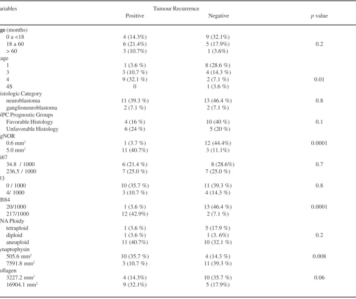
The patients’ ages at diagnosis ranged from 1 to 78 months (Table 1); thirteen (13) patients presented age < 18 months (46.4%), 11 (39.2%) patients presented age from 18 to 60 months and 4 (14.2%) patients presented age > 60 months. No statistical difference was found between age and tumor recurrence (p=0.2) (Table 2; Figure 2A). For clinical stage 9 patients were at stage I; 7 patients at stage 3; 11 patients at stage 4 and only one patient at stage 4S (Table 1). Of the patients at stages 1, 3 and 4S disease (17 pts, 7%) only 4 patients (14.3%) presented disease recur-rence. However, of the 11 patients (39.2%) at advanced stage (4), 9 (32.1%) presented recurrence, which showed statistical significance (p=0.01) when compared to the stage 1, 3 and 4S patients. Histological categories included 24 cases of neuroblastomas and 4 cases of ganglioneu-roblastomas. INPC Prognostic Groups included 14 cases of Figure 1 (Panels A to L). (A) Undifferentiating neuroblastoma: note
favorable histology, 11 cases of unfavorable histology and 3 cases were unclassifiable (Table 1). In this study, histo-logical categories and INPC prognostic groups classifica-tion did no present statistical associaclassifica-tion with tumor recur-rence. Eleven cases (40.7%) presented high expression of AgNOR (5.0 mm2) associated to tumor recurrence, whereas 12 cases (44.4%) presented low expression of this marker (0.6 mm2) without tumor recurrence, with a statistically
sig-nificant difference (p<0.01; Table 2, Figure 2B). Equally significant was the higher expression of NB84 (217/1000) in 12 (42.9%) cases with disease recurrence compared to low expression (20/1000) in 13 (46.4 %) cases without re-currence (p<0.01; Table 2, Figure 2C). A significant
dif-ference (p= 0.008) was found between the low amount
(505.6 mm2) of synaptophysin found in 10 cases(35.7%)
with tumor recurrence and the high expression (7591.8
mm2) of this neuroendocrine marker detected in 11 cases
(39.3%) without tumor recurrence (Table 2, Figure 2D). Difference between the high amount of collagen (16904.1
mm2) foundin 9 (32.1%) cases with tumor recurrence and
the low amount (3227.2 mm2) present in10 (35.7 %) cases
without tumor recurrence attained marginal significance (p=0.06, Table 2). No statistical difference was found for the low and high expressions of Ki67, p53, DNA Ploidy and presence or absence of tumor recurrence.
Table 2 - Differences in the frequency of clinical and of morphological variables (threshold: low and high) in subgroups of patients stratified by tumour recurrence.
Abbreviations: INPC – International Neuroblastoma Pathology Classification Variables
Age (months) 0 a <18 18 a 60 > 60 Stage
1 3 4 4S
Histologic Category neuroblastoma ganglioneuroblastoma INPC Prognostic Groups
Favorable Histology Unfavorable Histology AgNOR
0.6 mm2
5.0 mm2
Ki67
34.8 / 1000 236.5 / 1000 p53
0 / 1000 4/ 1000 NB84
20/1000 217/1000 DNA Ploidy
tetraploid diploid aneuploid Synaptophysin 505.6 mm2
7591.8 mm2
Collagen 3227.2 mm2
16904.1 mm2
Positive
4 (14.3%) 6 (21.4%) 3 (10.7%) 1 (3.6 %) 3 (10.7 %) 9 (32.1 %)
0 11 (39.3 %)
2 (7.1 %) 4 (16 %) 6 (24 %) 1 (3.7 %) 11 (40.7%) 6 (21.4 %) 7 (25.0 %) 10 (35.7 %)
3 (10.7 %) 1 (3.6 %) 12 (42.9%)
1 (3.6 %) 1 (3.6 %) 11 (40.7%) 10 (35.7 %) 3 (10.7 %)
4 (14.3%) 9 (32.1%)
Negative
9 (32.1%) 5 (17.9%) 1 (3.6%) 8 (28.6 %) 4 (14.3 %) 2 (7.1 %) 1 (3.6 %) 13 (46.4 %)
2 (7.1 %) 10 (40 %) 5 (20 %) 12 (44.4%)
3 (11.1%) 8 (28.6%)
7 (25.0 %) 11 (39.3 %)
4 (14.3 %) 13 (46.4 %)
2 (7.1 %) 5 (17.9 %)
1 (3. 6%) 10 (32.1 %)
4 (14.3 %) 11 (39.3 %) 10 (35.7 %) 5 (17.9%)
p value
0.2
0.01
0.8
0.1
0.0001
0.7
0.8
0.0001
0.2
0.008
Survival Time Analysis
The preliminary examination of the Kaplan-Meier
sur-vival curves (data not shown) demonstrated that, in this study, only patients with pathological stages 3 and 4 had different hazards for survival, with a median time of 92 and 29 months for each group, respectively (Figure 2E). The results of the Cox model are shown in Table 3. Two
vari-ables were significantly associated with survival time: pathological stage and AgNOR expression. We observed that stage was significantly related to survival time and re-currence, even when the AgNOR expression was not con-sidered (p = 0.004 and 0.003, respectively). However, when AgNOR was added as a covariable, its association with sur-vival and tumor recurrence was much stronger. The over-all likelihood ratio (survival vs recurrence) using only tumor stage was 29.71 and 60.0, respectively. When the AgNOR expression was associated (cutoff of 5.0 m2) to the stage,
the likelihood ratio for survival and recurrence changed to 38.13 and 65.9, respectively. We also observe that the prog-nostic information provided by the AgNOR estimation was maximized when this parameter was used as a continuous variable, that is, without using a cutoff level (41.65 and 65.58, respectively). The association between survival time and AgNOR expression is illustrated by the plots in
Fig-ure 2. The group with AgNOR < 5 mm2 appears in the
up-per curve. Their median survival time was not reached dur-ing the follow-up period of observation, but it was quite long, at 100.33 months. On the other hand, the cases that
presented AgNOR> 5 mm2 (lower curve) had a median
sur-vival time of only 92 months (p=0.05 by log rank test). Fig-ures 2G and 2H we present the cumulative hazard curves for recurrence, according to stage (log rank test 11.8; p = 0.008) and AgNOR as a continuous variable (log rank test 67.2; p < 0.001).
DISCUSSION
The majority of the small blue round-cell tumors can be diagnosed by the conventional histological analysis. However, diagnostic difficulties are often observed with the less differentiated ones, which show no neural, rhabdoid or epithelial differentiation at light microscopy. Immunohis-tochemistry has proved to be a useful adjunctive method for the differential diagnosis of these tumors.7,8,23 This is
particularly true for the identification of lymphomas and leukemia, which are the most frequent members of this group of tumors. However, the diagnosis of neuroblastomas is more complex. There are only a few available monoclonal
Table 3 - Cox Proportional Hazard Model Analysis of Survival Timei ando Recurrence
Variable Coefficient SE Likelihood ratio P value Stage -2.47 – 11.59 1.08 – 0.60 29.71 – 60.0 0.004 – 0.003
AgNOR 1.07 – 2.38 1.08 – 1.04 38.13 – 65.9 0.07 – 0.004
Stage AgNOR -2.26 - -0.94 1.15 – 0.63 32.48 0.008
0.76 – 1.94 1.14 – 1.08 53.60 0.003
AgNOR 41.65 – 65.58 0.00001 –
0.00001
antibodies with reactivity against these tumors cells and these antibodies present variable and limited specificity and sensitivity for the routinely processed material.8
In this study, we tested the monoclonal antibody NB84, which showed immunoreactivity in approximately 90% of the tumors. Similar results were obtained by Thomas and
coleagues8, who observed reliable staining of 17/19 (90%)
neuroblastomas, which was negative for all other tumors except for Ewing’s sarcomas, of which 9/14 (64%) showed mainly focal positivity. This finding suggests that NB84 provides considerable assistance in the recognition of neu-roblastomas and thus it is important to allow the most ap-propriate therapy to be established. Furthermore, we found a higher number of NB84 positive cells in the group of pa-tients that presented tumor recurrence, the majority of them undifferentiated or poorly differentiated neuroblastomas, once more showing that NB84 can be used to predict the behavior of these tumors. Compared to NB84, we found that synaptophysin, a neuroendocrine marker of well differen-tiated cells, was positive in 100% of the cases and highly expressed by tumors without potential for recurrence, mak-ing synaptophysin a potential marker to predict tumor behavior. Similar findings were also observed by Miettinen7
and Bozzi.30
For clinical and pathological use, it would be desirable that the aforementioned adjuvant methods could also pro-vide information related to tumor prognosis, in addition to being easy to perform as well as rapidly assessable and re-producible.9 Considering the large variety of tumors and the
diversity of their biological behaviors, there is no univer-sal marker that fulfills all these conditions. A variety of new markers has been proposed to aid in the diagnosis of some tumor types and to better evaluate the prognosis of these tumors. Recently, the quantitation of interphasic NORs has been associated with growth rate in neuroblastoma cell lines.11 A decreasing number of nucleolar organizer regions
was found to be associated with histopathological grades, high nerve growth factor receptor (TRKA) expression and a better survival.12 Addditionally, stage IVS patients, lymph
nodes and distant metastasis exhibited reduced nucleolar
organizing region counts when compared with stage IV.10
Therefore, for all these reasons, we should not be surprised at the fact that that staining for AgNOR provides impor-tant prognostic information about pNTs and our results have now confirmed the prognostic importance of AgNOR in pNTs. Whereas only three prior studies were able to show a significant association between staining for AgNOR in the tumor and survival10-12 our results suggest that staining
for AgNOR, used as a continuous variable, provides more prognostic information than the routine pathological
stag-ing.Although we were unable to identify a binary cutpoint
in AgNOR, a natural dividing point was the median of 5.0
mm2 of nucleoli area expressing AgNOR and this point
pro-vided a practical means to separate them into two groups: patients with an expected short survival versus patients with an expected longer survival. Thus, staining of the pNTs for AgNOR offers us the potential to offer the use of adjuvant chemotherapy in patients likely to recur after the surgical excision of pNTs. In summary, to reach this conclusion will require a larger study in a randomized and prospective trial and we also believe it is important to validate our quanti-tative assessment of AgNOR in additional pNTs.
Among the microenvironmental features, the extracel-lular matrix components (collagen density) seems to be an important prognostic parameter, mainly in some special cancer types16,19. Although several studies of extracellular
matrix proliferation in cancer have been carried out 20,21 the
association of these parameters with nuclear changes and oncogenic characteristics in pNTs, as well as the impact of these factors on prognosis have not been previously con-sidered. Although with marginal significance, our data showed that tumors with higher collagen density were at an upper risk for recurrence. The extracellular matrix is an important, although poorly understood, collagenous host response to tumor invasion22. Shimosato et al19 reported one
of the first studies about the impact of the extracellular ma-trix proliferation on cancer prognosis. They showed that tumors with a rich extracellular matrix tend to present higher blood vessel invasion and a greater incidence of lymph node metastasis, carrying a poor prognosis when compared to cases with low or no extracellular matrix pro-liferation. Regardless of the mechanism, the quantification of collagen can also provide prognostic information in pNTs. Other classic markers investigated in our study were Ki-67,17 p5315,18 and DNA ploidy,13,14,23 which did not show
association with NTs prognosis.
Finally, we should explain the negative results of classi-cal prognostic indicators such as age,1-4 histological category
or INPC Prognostic Group5,6 in our series. Based on the
lit-erature and the recent published article by Sano and col-leagues,31 the International neuroblastoma pathology
classi-fication adds independent prognostic information beyond the prognostic contribution of age. Age has been used as a prog-nostic factor for patients with peripheral neuroblastic tumors (pNTs). According to these authors, the analysis disclosed a cutoff of around 18 months for the optimal prognostic dis-tinction. The International Neuroblastoma Pathology Clas-sification (INPC) differentiates favorable from unfavorable histology based on the age-appropriate evaluation of histo-logical indicators (grade of neuroblastic differentiation, mi-tosis-karyorrhexis index) in the categories of nodular
showed that age, which was tested by using 3 different cut-offs (12, 18 and 24 months), was prognostically significant. INPC remained prognostically significant regardless of the age group to which it was applied. Prognostic effects of age and histological indicators were independently significant, i.e., age had a prognostic capacity beyond that of histologi-cal indicators, and histologihistologi-cal indicators had a prognostic capacity beyond that of age. Due to the fact that INPC in-corporated the age factor (18, 60 months) into the system, it was better than age alone for the prognostic differentia-tion of pNT patients. Based on the literature and the recent published article by Sano and colleagues, who studied a se-ries of 911 patients, the INPC adds independent prognostic information beyond the prognostic contribution of age. The great series of patients studied by these authors allow to bet-ter understand the definite role of classical prognostic indi-cators such as age, histological category and INPC. Prog-nostic Group and probably explains our negative findings.31
Despite the limitations observed in a small series of pa-tients, we concluded that the determination of NB84 and
synaptophysin are useful tools for the histological diagno-sis of pNTs. When these markers are associated with the evaluation of the AgNOR expression by the tumor cells, they provide an important contribution to the prognostic evaluation and management approach of patients with pNTs. Further studies with a larger number of cases should be developed to assess the real value of the associated quantitation of NB84 and synaptophysin to the AgNOR ex-pression for better diagnosis and prognosis attainment in patients with peripheral neuroblastic tumors.
ACKNOWLEDGEMENT
We are grateful to Dr. Katia Ramos Moreira Leite for supplying three cases from Hospital Sírio Libanês files.
This study was supported by the following Brazilian agencies: the National Council for Scientific and Techno-logical Development [CNPq300430/95-7]; the Foundation for the Support of Research of the State of São Paulo [FAPESP2000/14336-0].
RESUMO
Carvalho AC, Parra ER, Zerbini MC, Alves VAF, Capelozzi VL, Antonangelo L. A avaliação morfométrica de NB84, sinaptofisina e AgNOR é útil para o dignóstico histológico e prognóstico dos tumores neuroblásticos periféricos (pNTs). Clinics. 2007;62(6):731-40.
OBJETIVO: Estudar a importância dos marcadoresNB84 e AgNOR e explorar as relações quantitativas entre esses marcadores com o diagnóstico e prognóstico assim como
as relações entre NB84 e AgNOR e outros marcadores tumorais e estromais em 28 tumores neuroblásticos periféricos.
histoquímica, imunohistoquímica e morfometria para avaliar a intensidade e extensão de expressão do AgNOR, NB84 e sinaptofisina, tendo o prognóstico dos pacientes incluído o tempo de sobrevida até a morte por recurrência dos tumores neuroblásticos.
RESULTADOS. Estadiamento (p<0.01), AgNOR (p<0.01), NB84 (p<0.01) e sinaptofisina (p=0.01) foram marcadores independents de sobrevida.
CONCLUSÕES. A determinação dos marcadores NB84 e
sinaptofisina mostrou-se como uma ferramenta útil no diagnóstico dos tumors neuroblásticos periféricos; a associação desses marcadores à expressão de AgNOR pelas células tumorais contribuiu à determinação do prognóstico e estabelecimento do protocolo terapêutico para os pacientes.
UNITERMOS: Neuroblastoma. Prognóstico. AgNOR. Ploidia de DNA. NB84. Sinaptofisina.
REFERENCES
1. Breslow N, MacCann B. Statistical stimation of prognosis for children with neuroblastoma. Cancer Res. 1971;31:2098-103
2. Coldman AJ, Fryer CJH, Elwood JM, Sonley MJ. Neuroblastoma:influence of age at diagnosis, stage, tumor site, and sex on progression. Cancer. 1980;46:1896-901.
3. Jereb B, Bretsky SS, Vogel R, Helson L. Age and prognosis in neuroblastoma:review of 112 patients younger than 2 years. Am J Paediatr Hematol Oncol, 1984;6:223-43.
4. Hedborg F, Bjelfman C, Sparén P, Sandman S. Biochemical evidence for a maturation in morphologically poorly differntiated neuroblastomas with a favourable outcome. Eur J Cancer. 1995;31A:435-43.
5. Joshi VV. Peripheral Neuroblastic Tumors:Pathologic Classification basead on Recommendations of International Neuroblastoma Pathology Committee (modification of Shimada Classification). Pediatr Dev Pathol. 2000;3:184-99.
6. Shimada K, Ambros IM, Dehner LP, Hata J, Joshi VV, Roald B. Terminology and Morphologic Criteria of Neuroblastic Tumors Recommendations by the International Neuroblastoma Pathology Committee. Cancer. 1999;86:349-63.
7. Miettinen M, Chatten J, Paetau A, Stevenson A. Monoclonal antibody NB84 in the differential diagnosis of neuroblastoma and other small round cell tumors. Am J Surg Pathol. 1998;23:327-32.
8. Thomas JO, Nijar J, Turley H, Micklem K, Gatter KC. NB84: a new monoclonal antibody for the recognition of neuroblastoma in routinely processed material. J Pathol. 1991;163:69-75.
9. Linden MD, Torres FX, Kubus J, Zarbo RJ. Clinical application of morphologic and immunocytochemical assessments of cell proliferation. Am J Clin Pathol. 1992;97:S4-13.
10. Shimotake T, Iwai N, Tokiwa K, Deguchi E, Sawada T, Fushiki S. Increases number of argyrophilic nucleolar organizer regions between primary and metastatic sites predict tumor progression in stage IV and IV-S neuroblastoma. Cancer. 1994;73:3103-7.
11. Derenzini M, Pession A, Farabegoli F, Trere D, Badiali M, Dehan P. Relationship between interphasic Nucleolar Organizer Regions and growth rate in two neuroblastoma cell lines. Am J Pathol. 1989;134:925-32. 12. Leuschner I, Schmidt D, Schulz R, Harms D. Nerve growth factor receptor
13. Gansler T, Chatten J, Varello M, Bunin GR, Atkinson B. Flow citometry DNA analysis of neuroblastoma:correlation with histology and clinical outcome. Cancer. 1986;58:2453-8.
14. Huddart SN, Muir KR, Parkes SE, Mann JR, Stevens MC, Raafat F, et al. Retrospective study of prognostic value of DNA ploidy and proliferative activity in neuroblastoma. J Clin Pathol. 1993;46:1101-4.
15. Layfield LJ, Thompson JK, Dodge RK, Kerns BJ. Prognostic indicator for neuroblastoma:stage, grade, DNA ploidy, MIB-1 proliferation index, p53, HER-2/ne4u and EGFr – a survival study. J Surg Oncol. 1995;59:21-7.
16. Demarchi LMM, Reis MM, Palomino SA, Farhat C, Takagaki TY, Beyruti R, et al. Prognostic values of stromal density and PCNA, Ki-67, and p53 proteins in patients with resected adenocarcinoma of the lung. Mod Pathol. 2000;13:511-20.
17. Gerdes J, Lemke H, Baisch H, Wacker HH, Schwab U, Stein H. Cell cycle analysis of a cell proliferation-associated human nuclear antigen defined by the monoclonal antibody Ki-67. J Immunol. 1984;133:1710-5.
18. Kerns BJ, Jordan PA, Moore MB, Humphrey PA, Berchuck A, Kohler MF, et al. P53 overexpression in formalin-fixed paraffin embedded tissue detected by immunohistochemistry. J Histochem Cytochem. 1992; 40:1047-51.
19. Shimosato Y, Suzuki A, Hashimoto T, Nishiwaki Y, Kodama T, Yoneyama T, et al. Prognostic implications of fibrotic focus (scar) in small peripheral lung cancers. Am J Surg Pathol. 1980;4:365-73.
20. Ohori NP, Yousem SA, Griffin J, Stanis K, Stetler-Stevenson WG, Colby TV, et al. Comparison of extracellular matrix antigens in subtypes of bronchioalveolar carcinoma and conventional pulmonary adenocarcinoma:an immunohistochemical study. Am J Surg Pathol. 1992; 16:675-86.
21. Watanabe N, Nakajima I, Abe S, Ogura S, Isobe H, Kawakami Y. Staining pattern of type IV collagen and prognosis in early stage adenocarcinoma of the lung. JClin Pathol. 1994;47:613-5.
22. Barsky SH, Huang SJ, Bhuta S. The extracellular matrix of pulmonary scar carcinomas is suggestive of a desmoplastic origin. Am J Pathol. 1986; 124:412-19.
23. Brodeur GM, Castleberry RP. Neuroblastoma. In: Pizzo PA, Poplack D, editors. Principles and practice of pediatric oncology. Philadelphia: Lippincott–Raven; 1997. p.761-97.
24. Brodeur GM, Pritchard J, Berthold F, Carlsen NL, Castel V, Castelberry RP, et al. Revisions in the international criteria for neuroblastoma diagnosis, staging, and response to treatment. J Clin Oncol. 1993;11:1466-77. 25. Hall PA, Going JJ. Predicting the future:a critical appraisal of cancer
prognosis studies. Review. Histopathology. 1999;35:489-94. 26. Ploton D, Menager M, Jeannesson P, Himber G, Pigeon F, Adnet JJ.
Improvement in the staining and in the visualization of argyrophilic proteins of nucleolar organizer regions at the optical level. Histochem J. 1986; 18:5-14.
27. Lemos M, Pozzo RMK, Montes GS, Saldiva PHN. Organization of collagen and elastic fibres studied in stretch preparations of whole mounts of human visceral pleura. Anat Anz. 1997;179:447-52.
28. Montes GS, Junqueira LCU. Histochemical localization of collagen and of proteoglycans in tissues. In: Nimni ME, editor. Collagen. Boca Raton, FL: CRC Press; 1988. p. 41-4.
29. Montes GS. Structural biology of the fibres of the collagenous and elastic systems. Cell Biol Int. 1996;20:15-27.
30. Bozzi (Bozzi F, Gambirasio F, Luksch R, Collini P, Brando B, Fossati-Bellani F. Detecting CD56+/NB84+/CD45- immunophenotype in the bone marrow of patients with metastatic neuroblastoma using flow cytometry. Anticancer Res. 2006;26:3281-7