Cop
yright
© ABE&M t
odos os dir
eit
os r
eser
vados
.
Malignant solitary ibrous tumor
of the thyroid: a case-report
and review of the literature
Tumor ibroso solitário maligno da tiroide: relato de caso e revisão da literatura
Wellington Alves Filho1, Renata Regina da Graça Lorencetti Mahmoud1, Daniel Marin Ramos1, Vergilius José Furtado de Araujo-Filho1, Patricia Picciarelli de Lima2, Claudio Roberto Cernea1, Lenine Garcia Brandão1
SUMMARY
Solitary ibrous tumor (SFT) is an uncommon spindle-cell neoplasm that most often involves the pleura, rarely occurring in extra-thoracic locations. Twenty-six cases of SFT arising in the thyroid gland have been described. We report a case of a 60-year-old woman presenting an 8-month history of enlargement of the neck associated with dysphagia. The patient underwent a right hemithyroidectomy and SFT of the thyroid was diagnosed. Immunohistochemistry sho-wed positivity for CD34 marker, and the high number of mitoses and the presence of cellular atypia suggested that the tumor was malignant. To our knowledge, this is the second case of malignant SFT of the thyroid gland ever reported. Due to the rarity of these tumors, the in-dication of adjuvant therapy and prognosis are uncertain. Long-term follow-up after surgical resection seems to be advisable. Arq Bras Endocrinol Metab. 2014;58(4):402-6
RESUMO
O tumor ibroso solitário (SFT) é uma neoplasia rara de células fusiformes que mais frequen-temente envolve a pleura, raramente ocorrendo em áreas extratorácicas. Já foram descritos 26 casos de SFT da tiroide. Relatamos o caso de uma paciente de 60 anos de idade com um histó-rico de 8 meses de aumento do pescoço associado à disfagia. A paciente foi submetida a uma hemitiroidectomia direita e foi diagnosticado um SFT de tiroide. A imuno-histoquímica mos-trou resultados positivos para o marcador CD34, e o grande número de mitoses e a presença de atipia celular sugerem que o tumor era maligno. Em nosso conhecimento, este é o segundo caso de STF da tiroide maligno já relatado. Dada a rara ocorrência desses tumores, a indicação de tratamento adjuvante e o prognóstico são incertos. Recomenda-se o acompanhamento de longo prazo depois da ressecção cirúrgica. Arq Bras Endocrinol Metab. 2014;58(4):402-6
1 Department of Head and
Neck Surgery, University of Sao Paulo, School of Medicine (FMUSP), Sao Paulo, SP, Brazil
2 Department of Pathology,
FMUSP, Sao Paulo, SP, Brazil
Correspondence to: Wellington Alves Filho Rua Apeninos, 800, ap. 413 04104-020 – São Paulo, SP, Brazil [email protected]
Received on Jan/15/2014 Accepted on Fev/27/2014
DOI: 10.1590/0004-2730000003230
INTRODUCTION
S
olitary ibrous tumor (SFT) is a rare spindle-cell neoplasm, originally described by Klemperer and Rabin in 1931 as a pleural tumor (1). Albeit it has been originally recognized as a localized form of mesothelio-ma (2), some studies have reported extrapleural sites in the body (3,4), such as thyroid, salivary glands, tongue, respiratory tract, orbit, liver, meninges and retroperi-toneum (3,5). They are very rare in the thyroid gland, with only 26 cases described after the original report in 1993 (6,7). Among those, only one met the criteriafor malignant SFT (8), proposed by Vallat-Decouve-laere and cols. in 1998 (9). We present what appears to be the second case described of a malignant solitary ibrous tumor of the thyroid.
CASE REPORT
pa-Cop
yright
© ABE&M t
odos os dir
eit
os r
eser
vados
.
tient was referred to the Department of Head and Neck Surgery of the Hospital das Clínicas of the University of Sao Paulo, School of Medicine, in November 2012. At the physical examination, an enlargement of the right side of the neck was observed, with a homogeneous and tender consistency at palpation. The right vocal fold had reduced mobility, with an underlying submu-cosal mass. Thyroid function tests were normal, and the ultrasonography (US) demonstrated a right-side thy-roid mass, with an intrathoracic component and reduc-tion of the lumen of the trachea. The volume of the gland was 283.6 cc, and the left thyroid lobe appeared normal. Computed tomography (CT) indings were in accordance with the US data (Figure 1). An US-guided ine-needle aspiration biopsy (FNAB) was performed, with a benign cytopathologic result.
The patient underwent right hemithyroidectomy in January 2013, with a frozen-section analysis suspicious for thyroid lymphoma or chronic thyroiditis. The right recurrent laryngeal nerve (RLN), as well as both right-side parathyroid glands were preserved. Right after ex-tubation in the operative room, the patient developed severe dyspnea refractory to non-invasive positive pres-sure ventilation. A tracheostomy was then performed.
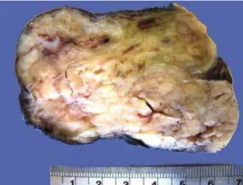
Histopathological indings included SFT with 13.8 x 8.5 cm, without necrotic foci and with 06/10 mitoses per high-power ields (HPF). Immunohistochemistry staining showed that tumor cells strongly expressed CD34 marker, with negative expression of desmin, S100 protein, cytokeratin 35BH11, beta-catenina, and calcitonin (Figures 2 and 3).
Figure 3. CD 34 immunostaining (immunoperoxidase reaction, 400X).
Figure 1. Tumor section surface in the thyroid gland. The tumor is irregular, with multinodular areas and white-tan color. The section surface is irm and curled.
Figure 2. Histological architecture with “patternless” growth pattern. Spindle cells are separated by thick hyalinized collagen with cracking artifact (arrowhead) and hemangiopericytoma-like vessels (arrow). Note the iniltrative tumor edge (hematoxilin and eosin, 200X). Inset: spindle cells with moderate atypia and mitotic igures (arrows) (hematoxilin and eosin, 1000X).
The patient was discharged from the hospital on the ifth postoperative day, and the tracheostomy tube was removed three weeks after the operation. She persisted with right vocal fold paralysis, with normal speech and respiration.
DISCUSSION
SFTs are rare ubiquitous neoplasms of mesenchymal origin. The etiology is unknown (10). It was described as a pleural-based lesion (1), but there were subsequent reports on other anatomic sites as well (3).
Cop
yright
© ABE&M t
odos os dir
eit
os r
eser
vados
.
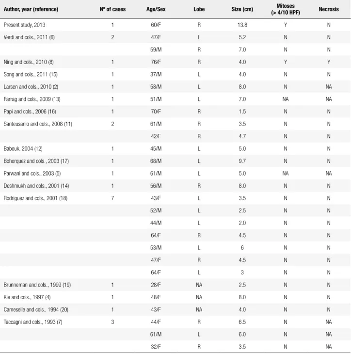
Table 1. Review of the literature of SFT of thyroid
Author, year (reference) Nº of cases Age/Sex Lobe Size (cm) Mitoses
(> 4/10 HPF) Necrosis
Present study, 2013 1 60/F R 13.8 Y N
Verdi and cols., 2011 (6) 2 47/F L 5.2 N N
59/M R 7.0 N N
Ning and cols., 2010 (8) 1 76/F R 4.0 Y Y
Song and cols., 2011 (15) 1 37/M L 4.0 N N
Larsen and cols., 2010 (2) 1 58/M L 8.0 N NA
Farrag and cols., 2009 (13) 1 51/M L 7.0 NA NA
Papi and cols., 2006 (16) 1 70/F R 1.5 N N
Santeusanio and cols., 2008 (11) 2 61/M R 3.5 N N
42/F R 4.7 N N
Babouk, 2004 (12) 1 45/M L 5.0 N N
Bohorquez and cols., 2003 (17) 1 68/M L 9.7 N N
Parwani and cols., 2003 (5) 1 61/M L 5.0 NA NA
Deshmukh and cols., 2001 (14) 1 56/M R 8.0 N N
Rodriguez and cols., 2001 (18) 7 43/F L 3.5 N N
52/M L 2.5 N N
44/M L 2.0 N N
64/F R 4.5 N N
53/M L 6 N N
47/F R 4.5 N N
64/F L 3 N N
Brunneman and cols., 1999 (19) 1 28/F NA 2.5 N N
Kie and cols., 1997 (4) 1 48/F NA 8.0 N N
Cameselle and cols., 1994 (20) 1 43/F NA 4.0 N N
Taccagni and cols., 1993 (7) 3 44/F R 6.5 N NA
61/M L 6.0 N NA
32/F R 3.5 N NA
HPF: high-power ields; SFT: solitary ibrous tumor; M: male; F: female; L: left; R: right; N: absence; Y: presence; NA: not available.
Less than 0.1% of all tumors appear on the head and neck area (2). The irst report of a SFT of the thyroid (SFT-T) was published in 1993 by Taccagni and cols.
(7). Since then, to our knowledge, only 26 cases were described in the English-language literature (6). A re-view of these articles is summarized in table 1.
According to cumulative data, the mean age for ap-pearance of SFT-T is 50 years (ranging from 28 to 68 years), with no difference in gender. The size of the
Cop
yright
© ABE&M t
odos os dir
eit
os r
eser
vados
.
Malignant solitary ibrous tumor of the thyroid
SFT-T can only be diagnosed after exclusion of oth-er thyroid tumors that exhibit spindle-cell morpholo-gy (12). Among the possible differential diagnosis are hemangiopericytomas, Riedel’s thyroiditis, medullary carcinomas, anaplastic carcinomas, sarcomas, leiomyo-mas, neuroibroleiomyo-mas, schwanoleiomyo-mas, and lymphomas (10).
Although FNAB is a standard procedure in the eva-luation of thyroid nodules, its eficacy in the diagnosis of SFT-T is limited (13). Only two reports showed a diagnosis of spindle-cell neoplasm using cytology com-bined with immunocytochemistry, but a deinitive con-irmation of SFT-T was not possible using this method alone (5,13). The FNAB of the present patient showed a benign lesion, most likely granulomatous thyroiditis. Immunocytochemistry was not performed in this case, since cytology demonstrated a benign lesion. Because of FNAB limitations, proper diagnosis can only be en-sured by combination of histopathological and immu-nohistochemistry evaluation.
Microscopically, SFT-T exhibits a unique morpholo-gy, often referred as a “patternless” pattern, with spindle cell proliferation, intermingled with hypercellular and hypocellular areas (14). At immunohistochemistry, the tumor cells revealed positivity for CD34, bcl-2, CD99, and vimentin, but not for epithelial markers (such as kera-tins, thyroglobulin), desmin, and S100 protein (10,11). Extra-pleural SFT-Ts are mostly benign neoplasms, and there are no distinct pathologic features to help differentiating benign from malignant lesions (3,15). Some characteristics suggesting a more aggressive be-havior were proposed in 1998 by Vallat-Decouvelaere and cols., and included (i) high cellularity, (ii) cytologi-cal atypia, (iii) higher frequency of mitoses (> 4/10 HPF), (iv) evidence of tumor necrosis or iniltrating margins (9). So far, there is only one report of malig-nant SFT-T, which already presented pulmonary me-tastasis when diagnosed (8). In the present case, even without distant metastasis, the tumor presented two of the above-cited criteria for malignancy (cytological atypia and > 4/10 mitoses per HPF). With these ind-ings, we believe that our case is the second report of malignant SFT-T.
The management of SFT-T is primarily surgical, which also provides a deinitive diagnosis (10,13). If there is no suspicion of malignancy, such as invasion of surrounding structures, a thyroid lobectomy may be suficient. In the present case, we did not ind any intra-operative sings of malignancy. Due to the preintra-operative right vocal fold paresis, combined with the
intraopera-tive inding of a normal left lobe, we chose to perform a right hemithyroidectomy. In such cases of preoperative vocal fold impairment, it is recommended that the sur-gical procedure starts by the exploration of the side of the neck that is ipsilateral to the paresis, as long as this does not affect the oncologic aspects of the operation (i.e., removal of the lobe containing the lesion). Even with an intact left RLN, the patient developed impor-tant dyspnea in the recovery room, and a tracheostomy was then performed. The patient received steroids in the postoperative period, and the tracheostomy tube was uneventfully withdrawn after 3 weeks.
TSH suppression therapy with hormones has no ra-tionale in the management of SFT-T, as the origin of tumor cells is stromal (8). The indication of chemo-therapy or external radiation chemo-therapy is controversial, even in malignant tumors (8,10).
Because of the rarity of these tumors, especially in cases of malignancy, not much is known about its be-havior or prognosis. Close follow-up after surgical re-section of lesions seems to be the best management.
In conclusion, this is the 27th reported case of
SFT-T in the English-language literature, being the second malignant tumor reported, and the larger lesion described so far.
Disclosure: no potential conlict of interest relevant to this article was reported.
REFERENCES
1. Klemperer P, Rabin CB. Primary neoplasm of the pleura: a report of ive cases. Arch Pathol. 1931;11:385-412.
2. Larsen SR, Godballe C, Krogdahl A. Solitary ibrous tumor arising in an intrathoracic goiter. Thyroid. 2010;20(4):435-7.
3. Chan JKC. Solitary ibrous tumor – everywhere, and a diagnosis in vogue. Histopathology. 1997;31:568-76.
4. Kie JH, Kim JY, Park YN, Lee MK, Yang WI, Park JS. Solitary i-brous tumor of the thyroid. Histopathology. 1997;30:365-8. 5. Parwani AV, Galindo R, Steinberg DM, Zeiger MA, Westra WH, Ali
SZ. Solitary ibrous tumor of the thyroid: cytopathologic indings and differential diagnosis. Diagn Cytopathol. 2003;28:213-6. 6. Verdi D, Pennelli G, Pelizzo MR, Toniato A. Solitary ibrous tumor
of the thyroid gland: a report of two cases with an analysis of their clinical and pathological features. Endocr Pathol. 2011;22:165-9. 7. Taccagni G, Sambade C, Nesland J, Terreni MR, Sobrinho-Simoes
M. Solitary ibrous tumour of the thyroid: clinicopathological, immunohistochemical and ultrastructural study of three cases. Virchows Arch A Pathol Anat Histopathol. 1993;422:491-7. 8. Ning S, Song X, Xiang L, Chen Y, Cheng Y, Chen H. Malignant
solitary ibrous tumor of the thyroid gland. Diagn Cytopathol. 2010;39:694-9.
Cop
yright
© ABE&M t
odos os dir
eit
os r
eser
vados
.
10. Papi G, Corrado S, Uberti ED, Roti E. Solitary ibrous tumor of the thyroid gland. Thyroid. 2007;17:119-26.
11. Santeusanio G, Schiaroli S, Ortenzi A, Mulè A, Perone G, Fadda G. Solitary ibrous tumor of thyroid: report of two cases with immunohistochemical features and literature review. Head Neck Pathol. 2008;2:231-5.
12. Babouk NL. Solitary ibrous tumor of the thyroid gland. Saudi Med J. 2004;25:805-7.
13. Farrag TY, Micchelli S, Tufano RP. Solitary ibrous tumor of the thyroid gland. Laryngoscope. 2009;119:2306-8.
14. Deshmukh NS, Mangham DC, Warield AT, Watkinson JC. Solitary ibrous tumor of the thyroid gland. J Laryngol Otol. 2001;115:940-2. 15. Song Z, Yu C, Song X, Wei L, Liu A. Primary solitary ibrous tumor of the thyroid – report of a case and a review of the literature. J Cancer. 2011;2:206-9.
16. Papi G, Corrado S, Ruggiero C, LiVolsi VA. Solitary ibrous tumor of the thyroid gland associated with papillary thyroid carcinoma. Thyroid. 2006;16:319-20.
17. Bohorquez CL, Gonzalez-Campora R, Loscertales MC, Escudero AG, Mezquita JC. Solitary ibrous tumor of the thyroid with cap-sular invasion. Pathol Res Pract. 2003;199:687-90.
18. Rodriguez I, Ayala E, Caballero C, De Miguel C, Matias-Guiu X, Cubilla AL, et al. Solitary ibrous tumor of the thyroid gland. Am J Surg Pathol. 2001;25:1424-8.
19. Brunnemann RB, Ro JY, Ordonez NG, Mooney J, El-Naggar AK, Ayala AG. Extrapleural solitary ibrous tumor: a clinicopathologic study of 24 cases. Mod Pathol. 1999;12:1034-42.