Differentiation of mouse embryonic stem cells and their hybrids during
embryoid body formation
Josane Mittmann
1, Irina Kerkis
2, Cinthia Kawashima
1, Marina Sukoyan
2, Enrico Santos
1and Alexandre Kerkis
21
Centro de Biociências e Biotecnologia, Universidade Estadual Norte Fluminense, Campos, RJ, Brazil.
2Departamento de Biologia, Instituto de Biociências, Universidade de São Paulo, São Paulo, SP, Brazil.
Abstract
We studied the karyotypes of three hybrid clones of mouse embryonic stem cells and murine splenocytes (two having near diploid and one having near tetraploid chromosome numbers) and the characteristics of their differentiation during the formation of embryoid bodies. The X chromosome originating from embryonic stem cells may be lost in hybrids with a near diploid chromosome number and reprogramming of the “somatic“ X may occur. The morphological data we obtained using light and electron microscopy revealed a correlation between the karyotype constitution of hybrid cells and their differentiation during the formation of embryoid bodies. At the beginning of development,the embryoid bodies derived from hybrid cells already showed an advanced degree of differentiation. The production of significant quantities of cartilage was typical for hybrid cells with near tetraploid chromosome numbers. The hybrid cells showed restricted pluripotent capacity and were already committed when they started to differentiate into embryoid bodies.
Key words:mouse ES-somatic cell hybrids, karyotype, pluriopotency, differentiation, embryoid bodies.
Received: May 2, 2000; accepted: July 3, 2000.
Introduction
Hybrids between pluripotent cells such as murine em-bryonic carcinoma cells (EC) and somatic cells (fibro-blasts, splenocytes, lymphocytes etc.) can be used to study the differentiation of hybrid cells. Chromosome sets in hy-brid cells can influence phenotype and differentiation ca-pacity, giving new characteristics to the cells (Kerkis and Zhdanova, 1992; Takagi, 1997). Hybrids of EC cells with differentiated somatic cells have the phenotype of EC pluri-potent cells and are able to differentiate into various tissues, derived from embryonic germ layers, and express some embryonic antigens (Miller and Ruddle, 1976; 1977; Litwack, and Croce, 1979; Andrews and Goodfellow, 1980; Roussetet al., 1983; Takagi et al., 1983; van der Kampet al., 1984; Subramanian, 1989).
The pluripotency of cells has been evaluated by sub-cutaneous injection into syngeneic mice, resulting in the formation of teratocarcinomas. During differentiation not all hybrid clones produce the same pattern of cellular diver-sity (Miller and Ruddle, 1976). The restriction of the plu-ripotency of some hybrid clones may depend on their
karyotype, but using an animal model it is difficult to deter-mine the influence of karyotype because the differentiation of cells occurs under the developmental control of the pa-rental organism.
Alternatively, the pluripotency of the cells can be ex-amined by their capacity to form embryoid bodies (Ebs), which have similar characteristics to the initial stages of embryo development. This model has been shown to be useful in studies of cell differentiation (for reviews see: Pedersen, 1994; Keller, 1995). Such cells permit the study of the differentiation of pluripotent cellsin vitrowithout the developmental influence of the animal environment. Under standardized culture conditions the observed differentia-tion can be attributed to the pluripotent cell genome.
Mouse embryonic stem cells (ES) are another type of pluripotent cell, which can be obtained from the inner cell mass of a normal embryo (Martin, 1981; Evans and Kaufman, 1981). Embryonic stem cells have been fused with differentiated mouse somatic cells (Matveevaet al., 1996) and human microcells (Tomizukaet al., 1997). The resulting hybrids had pluripotent characteristics in culture and could be used for the construction of transgenic (Tomizukaet al., 1997) and chimeric animals (Matveevaet al., 1998). Such hybrid cells can serve as a model for the www.sbg.org.br
study of the putative influence of the supplemental somatic chromosomes and other factors on differentiation.
In the study presented in this paper we used three mouse hybrid clones obtained by fusion of embryonic stem cells with splenocytes. We examined the karyotypes of these clones and investigated the influence of the karyo-types on the differentiation of these cells through the for-mation of embryonic bodies.
Materials and Methods
Embryonic stem cells and their hybrids
HM-1 cells, deficient in hypoxanthine phosphoribo-syl transferase (HPRT), were derived from HPRT-deficient strain 129 mice (Maginet al., 1992) and characterized as highly pluripotent (Magin et al., 1992; Selfridge et al. 1992). The HESS-1, HESS-2 and HESS-3 hybrid cells were isolated by the fusion of HM-1 cells with murine splenocytes of DD/c female, and characterized as pluripo-tents and HPRT positive by Matveevaet al. (1996, 1998).
We maintained the HM-1, HESS-1, HESS-2, HESS-3 cells in an undifferentiated state using a feeder layer of murine embryonic fibroblasts inactivated by mito-mycin C (10µg/mL for 3 h). For cell cultivation without a feeder layer, the medium described below was supple-mented with 103units/mL of murine leukemia inhibitory factor (mLIF, Sigma) and Petri dishes were previously treated with an aqueous solution of 0.1% gelatin.
The HM-1 and hybrid cells were cultivated in the al-pha-modification of Eagle’s medium (αMEM, Sigma) sup-plemented with 3.5g/L D(+)-glucose (Sigma), 10% (v/v) bovine fetal serum, 10-4 M β-mercaptoethanol, 50 U/mL penicillin and 50µg/mL streptomycin. For hybrid cell cul-tivationαMEM was supplemented with 100µM hypoxan-thine, 0.4µM aminopterin and 16 µM thymidine (HAT, Gibco), and the cells were passaged every 3 days onto a fresh feeder layer. Cultures were maintained in a humid at-mosphere containing 5% CO2at 37 °C.
For selection by 6-thioguanine, hybrid cells were plated in complete Eagles culture medium supplemented with 103units/mL mLIF and 30 µg/mL 6-thioguanine on gelatinized 60 mm dishes (3600-3800 cells/dish), the colo-nies being examined after 8-10 days of cultivation.
Embryoid body formation
Embryoid bodies were obtained by cultivation of ES cells and their hybrids in suspension according to the proto-col of Robertson (1987), using the same medium as that for undifferentiated cell culture, excluding mLIF andβ -mer-captoethanol. Embryonic stem and hybrid cells were trypsi-nized and the suspension left in a culture flask in the CO2
incubator for 15 min to eliminate fibroblasts, after which the suspension was transferred to another flask and incu-bated overnight. On the following day cell aggregates were separated by shaking, re-suspended in culture medium and placed into bacteriological dishes, the medium being changed every 2-3 days.
Alkaline phosphatase activity and cytogenetic analysis
The hybrid and HM-1 cells growing on microscope slides with feeder cells were washed in 0.1 M PBS, pH 7.4 and fixed with 4% paraformaldehyde in PBS for 15 min. Standard histochemical techniques were used for the detec-tion of alkaline phosphatase (AP) activity in the undifferen-tiated cells (Talbotet al., 1993).
For chromosome studies all cell lines were treated ac-cording to Hoganet al.,1994 and trypsin-Giemsa banding were obtained by the method of Seabright 1971. At least 250 different metaphases from each line were analyzed. Embryoid bodies were pre-treated with type III collagenase (Sigma).
Optical and electron microscopy studies of EBs
For optical microscopy embryoid bodies were fixed with 4% paraformaldehyde in 0.2 M PBS for one hour at room temperature and maintained at 4 °C overnight before being embedded in paraffin and cut into 3-5 µ sections which were then Gomori stained (Behmeret al., 1976).
For electron microscopy the embryoid bodies were fixed in a mixture of 2.5% gluteraldehyde, 4% paraformal-dehyde and 0.1 mM CaCl2in 0.1M cacodylate buffer (pH 7.4) for 1 h. Post-fixation was performed with 1% osmium tetroxide (OsO4) in the same buffer for 30 min, followed by embedding in Epon 812. After polymerization, sections were cut with a Reichert’s Ultracut ultramicrotome using a diamond knife. The sections were treated with an aqueous solution of uranyl acetate and lead citrate to enhance con-trast (Reynolds, 1963). The samples were analyzed using a Zeiss IN 900 electron microscope.
Results
Morphologic pattern of the hybrid cell cultures
Cytogenetic analysis
HM-1 cells were stable and had a normal diploid chromosome number of 2n = 40 (38A + XY), without any aneuploidy or structural alterations up to 30 passages in culture.
Analysis of HESS-1 cells after 25 passages showed that this hybrid was heterogeneous, with chromosome numbers from 2n = 40 upto 80, with a modal number of 71 chromosomes. Different patterns of sexual chromosomes were observed, the XXXY karyotype being frequently seen and the XXY karyotype rarely observed. The karyotypes with 41-43 chromosomes included trisomies for chromo-somes 1, 11, 14 and 16.
Karyotype analysis of HESS-2 cells after 38 passages showed a predominance of cells with 42 chromosomes. Al-most all metaphases had a pair of XY sex chromosomes. Among 47 metaphase spreads only seven (15%) had two X chromosomes, while 50% showed a chromosome frag-ment, probably derived from chromosome 5. Trisomy of chromosomes 1, 11, 12 or 16 was observed, Figure 1 show-ing the XY karyotype with trisomy 11 and the chromosome fragment.
The modal chromosome number of HESS-3 cells af-ter 39 passages was 41, including only one X chromosome. Cells with XXY or XX sex chromosomes were not ob-served, although trisomies for chromosomes 1, 11, 15 or 17 were detected (Figure 2).
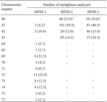
Cytogenetic analysis of embryoid bodies showed that the karyotypes of these bodies on the 7thday of their forma-tion had almost the same modal number as the hybrid clones in cell culture (Table 1).
Figure 1- GTG-banded karyotype of a HESS-2 cell with trisomy of chro-mosome 11 and a structural rearrangement (R) (Passage 38).
Figure 2- GTG-banded karyotype of a HESS-3 cell with trisomy of chro-mosome 1 and 17 (Passage 39).
Table I- Frequency of different chromosome numbers in 7-day embryoid bodies derived from HESS-1, HESS-2 and HESS-3 hybrid cells.
Chromosome number
Number of metaphases analyzed
HESS-1 HESS-2 HESS-3
40 - 46 (23.0)1 36 (18.0)1
41 2 (4.2)1 101 (50.5) 81 (40.5)
42 5 (10.4) 24 (12.0) 46 (23.0)
43 - 29 (14.5) 37 (18.5)
64 1 (2.1) -
-68 1 (2.1) -
-69 6 (12,5) -
-70 2 (4.2) -
-71 4 (8.3) -
-72 11 (22.9) -
-73 6 (12.5) -
-74 6 (12.5) -
-75 3 (6.2) -
-77 1 (2.1) -
-1
Origin of the X chromosome in the hybrid cell
Most HESS-2 and HESS-3 cells had only one X chro-mosome. To investigate the origin of this chromosome cells were treated with 6-thioguanine, a chemical which allows the selection of HPRT-deficient cells (Verma and Babu, 1995). HESS-3 cells were unable to grow in the presence of 6-thioguanine but were able to grow in the selective HAT medium (Table 2). This was a predictable result, since be-fore selection with 6-thioguanine the HESS-3 cells could not have lost the ‘somatic’ X chromosome due to the pre-cultivation in HAT medium. Therefore the frequency of cells with only one chromosome X of ‘embryonic origin’ in the HESS-3 hybrid cells was less than 2.6 x 10⁻⁴.
When the HESS-2 cells were repeatedly cultivated in the presence of HAT approximately 15% of the cells had two X chromosomes, as described above. It is conceivable that in the absence of selective pressure these cells lost one of their X chromosomes,i.e.the ‘somatic origin’ X chro-mosome could be preferentially lost, as the chrochro-mosome of the more differentiated partner of cell fusion (Ringertz and Savage, 1976). We cultivated HESS-2 cells for 27 days (11 passages) without HAT and then subjected them to selec-tion with 6-thioguanine (Table 2). None of the colonies of the hybrid cells grew in the culture medium with 6-thiogua-nine (a frequency below 2.7 x 10⁻⁴), whereas HAT-resistant colonies had a frequency of about 3 x 10⁻². These results show that in the absence of selective pressure (during about 50 cell divisions) the X chromosome of somatic origin (HPRT+) was not lost, instead the X chromosome originat-ing from the ES cells (HPRT+).
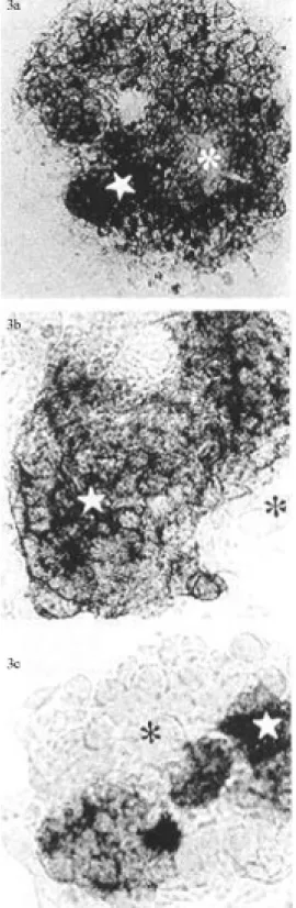
Alkaline phosphatase (AP) activity
Table 3 shows the percentage of AP positive cells in the HM-1 and hybrid cells. The largest numbers of AP posi-tive cells (about 94%, Figure 3a) were found in HM-1 cells but the HESS-3 line also had a large percentage (higher than 90%) of AP positive cells. HESS-2 had a smaller num-ber of AP positive cells (about 56%, Figure 3b) and the HESS-1 line the smallest number of AP positive cells (about 14%). In general, among the heterogeneous cells of the HESS-1 line only small cells were AP positive, whereas larger cells were weakly stained (Figure 3c).
Optical and electron microscopy of embryoid bodies
Comparative analysis of sections of EBs revealed a variety of differentiated cells in the hybrids and the HM-1 line. In the EBs originating from HM-1 cells, endodermal, cartilage-like and blood-like cells were marked positively at day 9 (Figure 4a). In EBs derived from HESS-2 and HESS-3 lines the formation of a large amount of cartilage matrix was observed in different parts of the EBs sections (Figure 4b). The inner part of the EBs showed a larger amount of cartilage matrix in sections obtained from HESS-1 cells (Figure 4c). On the periphery of the bodies a layer of the endodermal cells and islands of blood cells were also observed (Figures 4b and 4c).
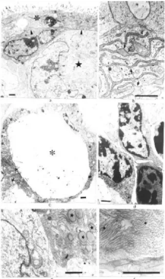
Electron microscopy of the EBs revealed distinctive differences in the EBs formed by hybrids and by HM-1 cells. At day 3, EBs derived from HM-1 had an outer layer of endodermal cells. These cells were at the beginning of their differentiation since they possessed few microvilli and few cytoplasmic organelles (Figure 5a). The central part of the EBs consisted of round undifferentiated cells with a large nuclei and cytoplasm with a small number of organelles (Figure 5b). On day 9, the outer layer of HM-1 derived EBs showed epithelium-like cells with numerous microvilli. These cells had junction structures of the des-mosome type and Reichert’s membrane was well devel-oped and the cystic cavity could be seen. In the inner part of the bodies muscular-like cells, adipose-like and haemato-poietic stem-like cells at various stages of differentiation (Figure 5c,g) were also found. On day 9, the HM-1 derived EBs showed the characteristic rhythmic contractions of heart muscle cells.
On day 3, EBs derived from hybrids presented an ad-vanced degree of differentiation. Embryoid bodies derived from HESS-2 and HESS-3 lines had an endodermal layer
Table II- Comparison of plating efficiency of the hybrid cells in the presence of 6-thioguanine and HAT (100µM hypoxanthine, 0.4µM aminopterin and 16µM thymidine).
Hybrid Passage number Number of cells 6-thioguanine resistant colonies HAT-resistant colonies
HESS-3 39 3800 0 ( 2.6 x 10⁻⁴)1 52 (1.4 x 10⁻²)1
HESS-2* 47 3600 0 ( 2.8 x 10⁻⁴) 115 (3.2 x 10⁻²)
1
Frequency.
*These cells were pre-cultivated in the absence of HAT for 27 days (11 passages).
Table III- Percentage of alkaline phosphatase positive cells in undiffer-entiated cultures of HM-1 cells and their hybrids.
Cell lines Total number of cells Number of AP- positive cells
HM-1 1106 950 (94.4)1
HESS-1 11484 1597 (13.9)
HESS-2 2320 1305 (56.3)
1
formed by cells already possessing multiple microvilli and an organelle-enriched cytoplasm. The start of Reichert’s membrane formation was initiate and cells resembling em-bryonic fibroblasts could be seen under this membrane (Figure 6a). The central part of individual EBs contained
morphologically undifferentiated cells. At a comparable stage of cultivation, EBs derived from HESS-1 hybrid cells were not very compact and the large spherical cells on their outer surface probably had near tetraploid chromosome Figure 4- Light microscopy of Gomori stained sections of nine-day old embryoid bodies derived from the hybrids and HM-1 cells: HM-1 (4a, 1500x). HESS-2 (4b) and HESS-1 (4c) both at 800x magnification. Carti-lage matrix (*), chondroblast-like cells (–), outer layer of endodermal cells (↑) and blood cell islands (è).
numbers. These cells had a cytoplasm rich in organelles, mainly granular endoplasmatic reticulum (Figure 6b).
From day 9 to 18, the outer layer of endodermal cells had a greater number of microvilli and their cytoplasms were filled with endoplasmatic reticulum. The cystic cavi-ties were well formed (Figure 6c) and areas resembling blood cell islands could be seen at the center of the EBs (Figure 6d). An important characteristic of differentiated hybrid cells was the production of a large number of poly-saccharide granules and a lot of extracellular collagen se-creted by chondroblasts or fibroblast-like cells (Figure 6e and 6f).
Embryoid bodies derived from hybrids lacked cells of the muscular type. To investigate whether these cells had
the capacity to differentiate into muscle cell, we allowed the EBs to developupto 30 days. Only a few HESS-2 de-rived EBs presented muscular contractions on the 10thto 15thday, the other hybrids not showing such contractions.
Discussion
We attempted to evaluate the influence of near dip-loid and near tetrapdip-loid karyotypes on thein vitro differen-tiation of hybrid cells obtained by fusion of somatic and Figure 5- Electron micrographs showing parts of HM -1 cell derived
embryoid bodies (EBs). A 3-day embryoid body (EB) showing the appear-ance of the outer layer of endodermal cells with a few microvilli (↑) and cytoplasm with only a few organelles (5a, 12000x). The same EB showing the central part formed by undifferentiated cells (★) (5b, 4300x). Figures 5c to 5g all show 9-day EBs: Reichert’s membrane (è) and the cystic cav-ity (*) (5c, 6300x); endodermal cells with a large number of microvilli (↑) and junction structures (5) (5d, 17600X); different types of cells can be seen in the inner part of EBs,e.g.adipose-like cells (A) (5e 12000x), mus-cle-like cells (M) and myofibers (↑) (5f, 1800x), haematopoietic stem-like cells (H) (5g, 3300x). Bar = 1µm.
embryonic genomes. If karyotype is influential, its effects would appear at the initial stages of embryo formation which is very sensitive and important for normal develop-ment. For this reason we chose Ebs as a model of early development, a choice which permitted us to study differ-entiation without any influence of the parental organism.
It is noteworthy that the intraspecific hybrids between the pluripotent and somatic cells usually have a near tetra-ploid chromosome number (Takagi, 1997). The hybrid cells used in our study were near diploid (HESS-2 and HESS-3) and near tetraploid (HESS-1) and our chromo-some analysis showed different trisomies, trisomies of chromosomes 1 and 11 being found in near diploid hybrids. These trisomies are probably typical of pluripotent cells, and have previously been described in the MESC embry-onic cell line (Crollaet al., 1990) and in embryonic carci-noma cells (McBurney and Rogers, 1982).
We found that the sex chromosome constitution in the HESS-2 line was predominantly XY, while in the HESS-3 line it was XO. This observation was unexpected since in the hybrid cells two X chromosomes should stay together because one X had the HPRT gene that was maintained by cultivation in HAT, while the other X belonged to the pluripotent embryonic cells. Indeed, it has been demon-strated by Ringertz and Savage (1976) that hybrids lose the chromosomes originating from differentiated, more slowly dividing, cells. Thus our results raised the question of the origin of the X chromosome in the hybrid cells, and to an-swer this question we used 6-thioguanine selection. In HESS-2 and HESS-3 lines, the segregated X chromosome was demonstrated to be of embryonic origin. Therefore, the segregation of the X chromosome of the more differenti-ated fusion partner during non-selective cultivation is not a rule in these hybrids.
During embryoid body formation the selection of cells with a diploid (2n = 40) karyotype is possible, but we did not detect selection for cells with a given chromosome number since the karyotypes of 7 day hybrid-derived em-bryoid bodies (EBs) maintained the same modal chromo-some number as the cells in monolayer culture.
In culture, HESS-1, HESS-2 and HESS-3 lines had, like other hybrids (q.v. review in Takagi, 1997), the pheno-type of pluripotent cells, the AP-reaction being considered as a marker of pluripotency (Resnicket al., 1992; Talbotet al., 1993). We found the highest number of AP-positive cells in the HESS-3 and HESS-2 cell lines, which most re-sembled the parental HM-1 line in morphological charac-teristics. The HESS-1 line was heterogeneous for the AP-reaction, HESS-1 AP-positive cells having near diploid chromosome numbers and AP-negative cells near tetra-ploid chromosome numbers.
In our experiments, hybrids showed the capacity to form EBsin vitro, even at late passages. The EBs formed by the hybrid cells could be considered as complex as those derived from the HM-1 line and the cystic-type EBs formed
by pluripotent cells (Martin and Evans, 1975; van der Kampet al., 1984; Doetschmanet al., 1985; Peaseet al., 1990).
The formation of blood cell islands, similar to the yolk sac, has been described in EBs derived from different ES cell lines (Doetschman et al., 1985; Hollands, 1988; Nicholset al., 1990; Chen, 1992; Bautchet al., 1996). We also observed haematopoietic-like cells in the EBs derived from hybrids and HM-1 cells. The formation of these cell types was related to the presence of visceral endoderm-like cells which we detected using electron microscopy. These cells possessed microvilli on their surface, cytoplasmatic vesicles and gapjunction-like structures. Bautch et al. (1996) observed the precursors of haematopoietic cells that were localized in the center of EBs, but they migrated to the periphery as soon as they became differentiated. Our elec-tron microscopy and histochemical analyses revealed cells with a high cytoplasm:nucleus ratio in the central area of EBs formed by the hybrids. These cells may be considered haematopoietic-like precursors.
Cells resembling skeletal and smooth muscle were seen during differentiation of the HM-1 line. The presence of the mesodermal cells showed that EBs obtained from this line follow the typical embryoid body pattern of devel-opment (Doetschman et al., 1985; Nichols et al., 1990; Bautchet al., 1996). The hybrid cells did not show this type of differentiation in EBs at the initial stages of develop-ment, suggesting restricted pluripotency.
Cells of ectodermal origin (e.g.nerve cells) were not identified in EBs derived from HM-1 cells and hybrids. The absence of nerve cells might be explained because cell dif-ferentiation took place in suspension, while nerve cells require a solid substrate and specific inductors for differen-tiation (Bainet al., 1995; Fraichardet al., 1995).
Studies of the influence of the karyotype on ES cell pluripotency have demonstrated that the normal karyotype may be a prerequisite for the efficient contribution of these cells to the germ line in transgenic and chimeric animals and for their ability to differentiatein vitrointo a wide spec-trum of cell types (Papaioannouet al., 1978; McBurney, Rogers, 1982; Peaseet al., 1990; Bronsonet al., 1995; Liu
et al., 1997; Suzukiet al., 1997). Our data shows that the ‘embryonic’ X chromosome may be lost in pluripotent hy-brids, but reprogramming of the ‘somatic’ X chromosome may still occur, thus allowing restricted pluripotency. Near diploid and near tetraploid hybrids did not differ as to their pluripotency, but appeared, however, to be restricted as compared to normal ES cells.
Acknowledgments
contribution to our laboratory. This work was supported by FENORTE and CAPES, Brazil.
References
Andrews PW, Goodfellow PN (1980) Antigen expression by so-matic cell hybrids of a murine embryonal carcinoma cell with thymocytes and L cell. Somat Cell Genet 6:271-284. Bain G, Kitchens D, Yao M, Huettner JE, Gottlieb DI (1995)
Em-bryonic stem cells express neuronal propertiesin vitro. Dev Biol 168 (2):342-357.
Bautch VL, Standford WL, Rapoport R, Russel S, Byrum RS, Futch TA (1996) Blood island formation in attached cultures of murine embryonic stem cells. Dev Dyn 205:1-12. Behmer OA, Tolosa EMC, Neto AGF (1976) Manual de técnicas
para histologia normal e patológica. EDART- São Paulo, Livraria Editora da Universidade de São Paulo, 25p. Bronson SK, Smithies O, Mascarello JT (1995) High incidence of
XXY and XYY males among the offspring of female chime-ras from embryonic stem cells. Proc Natl Acad Sci (USA), 92:3120-3123.
Chen U. (1992) Differentiation of mouse embryonic stem cells to lympho-hematopoietic lineages in vitro. Dev Immun 2:29-50.
Crolla JA, Brown D, Whittingham DG (1990) Spontaneous in-duction of an homologous Robertsonian translocation, Rb (11.11) in a murine embryonic stem cell line. Genet Res 55:107-110.
Doetschman T, Eistetter H, Katz M, Schmidt W, Kemler R (1985) Thein vitrodevelopment of blastocyst-derived embryonic stem cell lines: formation of visceral yolk sac, blood islands and myocardium. J Embryol Exptl Morph 87:27-45. Evans M, Kaufman M (1981) Establishment in culture of
pluripo-tential cells from mouse embryos. Nature 292:154-156. Fraichard A, Chassande O, Bilbaut G, Dehay C, Savatier P,
Samarut J (1995)In vitrodifferentiation of embryonic stem cells into glial cells and functional neurons. J Cell Sci 108:3181-3188.
Hogan B, Beddington R, Costantini F, Lacy E (1994) Manipu-lating the Mouse Embryo: A Laboratory Manual. Cold Spring Harbor Laboratory Press, New York, 332p. Hollands P (1988) Differentiation of embryonic haematopoietic
stem cells from mouse blastocysts grownin vitro. Develop-ment 102:135-141.
Keller GM (1995)In vitrodifferentiation of embryonic stem cells. Current Opinion in Cell Biology 7:862-869.
Kerkis AYu, Zhdanova NS (1992) Formation and ultrastructure of somatic cell hybrids. Electron Microscopy Rev 33:1-24. Litwack G, Croce CM (1979) Somatic cell hybrids between
toti-potent mouse teratocarcinoma and rat hepatoma cells. J Cell Physiol 101:1-8.
Liu X, Wu H, Loring J, Hormuzdi S, Disteche CM, Bornstein P, Jaenisch R (1997) Trisomy eight in ES cells is a common po-tential problem in gene targeting and interferes with germ line transmission. Dev Dyn 209:85-91.
Magin TM, McWhir J, Melton DW (1992) A new mouse embry-onic stem cell line with good germ line contribution and gene targeting frequency. Nucl Acid Res 20:3795-3796. Martin GR (1981) Isolation of a pluripotent cell line from early
mouse embryos cultured in medium conditioned by
terato-carcinoma stem cells. Proc Natl Acad Sci (USA) 78:7634-7638.
Martin GR, Evans MJ (1975) Differentiation of clonal lines of teratocarcinoma cells: formation of embryoid bodiesin vi-tro. Proc Natl Acad Sci (USA) 72:1441-1445
Matveeva NM, Shilov AG, Bayborodin SI, Filimonenko VV, Rolinskaya IV, Serov OL (1996) Hybrids between mouse embryonic and somatic cells retain pluripotency. Proc Acad Sci (Russia) 349:129-132.
Matveeva NM, Shilov AG, Kaftanovskaya NM, Maximovsky LP, Zhelezova AI, Golubitsa AN, Bayborodin SI, Fokina MM, Serov OL (1998)In vitroandin vivostudy of pluripotency in intraspecific hybrid cells obtained by fusion of murine em-bryonic stem cells with splenocytes. Mol Reprod Dev 50:128-138.
McBurney WM, Rogers BJ (1982) Isolation of male embryonal carcinoma cells and their chromosome replication patterns. Dev Biol 89:503-508.
Miller RA, Ruddle FH (1976) Pluripotent teratocarcinoma-thy-mus somatic cell hybrids. Cell 9:45-55.
Miller RA, Ruddle FH (1977) Teratocarcinoma x Friend erythro-leukemia cell hybrids resemble their pluripotent embryonal carcinoma parent. Dev Biol 56:157-173.
Nichols J, Evans EP, Smith AG (1990) Establishment of germ-li-ne-competent embryonic stem (ES) cells using differentia-tion inhibiting activity. Development 110:1341-1348. Papaioannou VE, Gardner RL, McBurney WM, Babinet C, Evans
MJ (1978) Participation of cultured teratocarcinoma cells in mouse embryogenesis. J Embryol ExpMorphol 44:93-104. Pease S, Williams RL (1990) Formation of germ-line chimeras from embryonic stem cells maintained with recombinant leukemia inhibitory factor. ExpCell Res 190:209-211. Pedersen RA. (1994) Studies ofin vitrodifferentiation with
em-bryonic stem cells. Reprod Fertil Dev 6:543-552.
Resnik JL, Bixler LS, Cheng L, Donovan PJ (1992) Long-term proliferation of mouse primordial germ cells in culture. Na-ture 359:550-551.
Reynolds ES (1963) The use of lead citrate of high pH for electron opaque stain in electron microscopy. J Cell Biol 17:208. Ringertz NR, Savage RE (1976) Cell hybrids. Acad. Press, New
York, 417 pp.
Robertson EJ 1987 Embryo-derived stem cell lines. In: Robertson EJ, editor. Teratocarcinomas and embryonic stem cells. A practical approach. IRL Press, Oxfordm pp. 71-112. Rousset J-P, Bucchi D, Jami J (1983) Hybrids between F9
nulli-potent teratocarcinoma and thymus cells produce multi-dif-ferentiated tumors in mice. Dev Biol 96:331-336.
Seabright M (1971) A rapid banding technique for human chro-mosome. Lancet 2:971-972.
Selfridge J, Pow AM, McWhir J, Magin TM, Melton DW (1992) Gene targeting using a mouse HPRT mini/HPRT-deficient embryonic stem cell system: inactivation of the mouse ERCC-1 gene. Somat Cell Mol Genet 18: 325-336. Subramanian V (1989) A malignant, stem cell-like somatic hybrid
between a mouse teratocarcinoma and rat ascitic hepatoma is differentiation competent. Cell Differ Dev 27:197-214. Suzuki H, Kamada N, Ueda O, Jishage K, Kurihara H, Terauchi
Talbot NC, Rexrod CE, Pursel V, Powell AM (1993) Alcaline phosphatase staining of pig and sheep epiblast cells in cul-ture. Mol Reprod Dev 36:139-147.
Takagi N, Yoshida MA, Sugawara O, Sasaki M (1983) Reversal of X-inactivation in female mouse somatic cells hybridized with murine teratocarcinoma stem cells in vitro. Cell 34:1053-1062.
Takagi N (1997) Mouse embryonal carcinoma cell - somatic hy-brids as experimental tools for the study of cell differentia-tion and X chromosome activity. Cancer Genet Cytogenet 93:48-55.
Tomizuka K, Yoshida H, Uejima H, Kudoh H, Sato K, Ohguma A, Hayasaka M, Honaoka K, Oshimura M, Ishida I (1997) Functional expression and germline transmission of a hu-mane chromosome fragment in chimaeric mice. Nature Ge-netics 16:133-143.
Van der KampAW, Roza-de Jongh EJ, Houwen RH, Magrane GG, van Dongen JM, Evans MJ (1984) Developmental characteristics of somatic cell hybrids between totipotent mouse teratocarcinoma and rat intestinal villus cells. Exp Cell Res 154 (1):53-64.