Serological and infection
statuses of dogs from a visceral
leishmaniasis-endemic area
Status
sorológico e de infecção
canina em área endêmica de
leishmaniose visceral
I Laboratório de Patologia de Moléstias Infecciosas/LIM-50. Departamento de Patologia. Faculdade de Medicina. Universidade de São Paulo. São Paulo, SP, Brasil
II Departamento de Clínica e Patologia. Faculdade de Medicina Veterinária e Zootecnia. Universidade Federal da Bahia. Salvador, BA, Brasil
III Departamento de Clínica, Cirurgia e Reprodução Animal. Faculdade de Medicina Veterinária. Universidade Estadual Paulista. Araçatuba, SP, Brasil
Correspondence: Márcia Dalastra Laurenti Departamento de Patologia Faculdade de Medicina – USP Av. Dr. Arnaldo, 455 1º andar sala 1209 Cerqueira César
01246-903 São Paulo, SP, Brasil E-mail: [email protected] Received: 11/5/2013 Approved: 4/21/2014
Article available from: www.scielo.br/rsp
ABSTRACT
OBJECTIVE:This study investigated the serological status of dogs living in a visceral leishmaniasis-endemic area and its correlation with the parasitological condition of the animals.
METHODS:Canine humoral responsewas evaluated using the sera of 134 dogs by enzyme-linked immunosorbent assay and immunohistochemistry to detect parasites in the skin, lymph node, and spleen of the animals. The
speciic antibodies investigated were IgG, IgG1, IgG2, and IgE.
RESULTS:According to the parasitological, laboratory, and clinical indings, the dogs were placed into one of four groups: asymptomatic with (AP+, n = 21) or without (AP-, n = 36) Leishmania tissue parasitism and symptomatic with
(SP+, n = 52) or without (SP-, n = 25)parasitism. Higher IgG and IgE levels
were positively correlated with the infection condition and parasite load, but not
with the clinical status. In all groups, total IgG was the predominant antibody, which occurred at the expense of IgG2 instead of IgG1. Most of the infected dogs tested positive for IgG (SP+, 98.1%; AP+, 95.2%), whereas this was not observed with IgE (SP+, 80.8%; AP+, 71.2%). The most relevant inding was
the high positivity of the uninfected dogs for Leishmania-speciic IgG (SP-,
60.0%; AP-, 44.4%), IgE (SP-, 44.0%; AP-, 27.8%), IgG1 (SP-, 28.0%; AP-, 22.2%), and IgG2 antibodies (SP-, 56.0%; AP-, 41.7%).
CONCLUSIONS:The serological status of dogs, as determined by any class or subclass of antibodies, did not accurately distinguish dogs infected with
L. (L.) infantum chagasi from uninfected animals. The inaccuracy of the serological result may impair not only the diagnosis, but also epidemiological investigations and strategies for visceral leishmaniasis control. This complex serological scenario occurring in a visceral leishmaniasis-endemic area highlights
the challenges associated with canine diagnosis and points out the dificulties
experienced by veterinary clinicians and coordinators of control programs.
DESCRIPTORS: Leishmaniasis, Visceral, diagnosis. Leishmaniasis, Visceral, epidemiology. Leishmania, immunology. Dogs.
Seroepidemiologic Studies. Endemic Diseases, veterinary.
Daniela Farias LaranjeiraI,II
Vânia Lúcia Ribeiro da MattaI
Thaíse Yumie TomokaneI
Mary MarcondesIII
Carlos Eduardo Pereira CorbetI
Visceral leishmaniasis (VL) is regarded worldwide as a
public health problem of increasing importance.15 The domestic dog is considered the main reservoir of the Leishmania pathogen, and from an epidemiological
point of view, canine visceral leishmaniasis (canVL)
is of considerable importance because canine disease precedes the occurrence of human cases, and a group of infected animals can serve as source of infection for the vector.5,7 In Brazil, the major prophylactic practi -ces for disease control include the systematic and early treatment of human cases, in addition to vector control and elimination of seropositive dogs.15
Serology is, by far, the most commonly used diagnos-tic tool in large surveys and in clinics. However, this method has been seriously criticized, as it frequently fails to recognize asymptomatic dogs,17 is not capable of differentiating infected animals from vaccinated ones,9
RESUMO
OBJETIVO: Foi investigado o status sorológico de cães, em área endêmica de leishmaniose visceral, e sua correlação com a infecção parasitológica dos animais.
MÉTODOS: A resposta humoral canina foi avaliada no soro de 134 cães pelo método
ELISA e pela imuno-histoquímica, para detectar parasitos na pele, linfonodo e baço desses animais. Os anticorpos especíicos investigados foram IgG, IgG1, IgG2 e IgE.
RESULTADOS: De acordo com os achados parasitológicos, laboratoriais e
clínicos, os cães foram alocados em um dos quatro grupos: assintomáticos com (AP+, n = 21) e sem (AP-, n = 36) parasitismo tecidual por Leishmania e
sintomáticos com (SP+, n = 52) ou sem (SP-, n = 25) parasitismo. Níveis mais elevados de IgG e IgE se correlacionaram positivamente com o status de infecção
e a carga parasitária, mas não com a condição clínica. Em todos os grupos, IgG total foi o anticorpo predominante, com maior concentração de IgG2 que IgG1. O anticorpo IgG foi positivo em proporção elevada nos animais infectados (SP+ 98,1%; AP+ 95,2%), mas não o IgE (SP+ 80,8%; AP+ 71,2%). O achado mais
relevante refere-se aos cães não infectados que apresentaram elevada positividade
para anticorpos IgG anti-Leishmania (SP- 60,0%; AP- 44,4%), IgE (SP- 44,0%;
AP- 27,8%), IgG1 (SP- 28,0%; AP- 22,2%) e IgG2 (SP- 56,0%; AP- 41,7%).
CONCLUSÕES: O status sorológico dos cães, determinado por qualquer classe ou subclasse de anticorpos, não distinguiu com acurácia cães infectados por
L. (L.) infantum chagasi daqueles não infectados. A imprecisão do resultado
sorológico pode prejudicar não só o diagnóstico, mas também as investigações
epidemiológicas e as estratégias para o controle da leishmaniose visceral.
Esse complexo cenário sorológico observado na área endêmica mostra quão desaiador é o diagnóstico canino, e aponta a diiculdade enfrentada pelos
médicos veterinários e coordenadores dos programas de controle.
DESCRITORES:Leishmaniose Visceral, diagnóstico. Leishmaniose Visceral, epidemiologia. Leishmania, imunologia. Cães. Estudos Soroepidemiológicos. Doenças Endêmicas, veterinária.
INTRODUCTION
and frequently shows cross-reactivity with sera from dogs infected with other pathogens.3 Thus, the resulting inaccuracies in diagnosing canVL has led to unneces-sary culling of dogs or even the maintenance of infec-ted dogs in areas of transmission, both of which
decre-ase the effectiveness of the Brazilian control program.1
Total IgG is the only anti-Leishmania antibody that is
routi-nely inspected. Other classes and IgG subclasses of antibo
-dies have been studied; however, these effects were geared
towards detecting markers related to clinical prognosis or vaccination status2,6,14 and evaluating the eficacy of treat -ments.10,24,25 Nevertheless, the practical use and the reliabi -lity of these markers for diagnostic purposes remain unclear.
METHODS
The investigation involved dogs from the municipality of Araçatuba, located in northwest Sao Paulo, Southeastern
Brazil, which is a region with high endemicity for canVL (12.0% - 42.0%) and reported its irst canine case in 1998.13
A total of 134 stray dogs were collected by the Centro de Controle de Zoonoses (Zoonosis Control Center) from
the streets of Araçatuba, SP, without previous serologi-cal inspection for leishmaniasis and ultimately destined to euthanasia for sanitary practices. These were male and female dogs between two and six years old and of various breeds and weights. The animals were
anestheti-zed with 25 mg/kg sodium thiopental (Cristália, Brazil),
and blood samples were drawn by cardiac puncture. The
sera were stored at -20°C. Necropsies were performed
following euthanasia using potassium chloride, and popli-teal lymph nodes and fragments of the spleen and skin
were collected and ixed in 10.0% buffered formalin.
This study was conducted in accordance with the ethical principles of animal experimentation adopted by the Colégio Brasileiro de Experimentação Animal and was approved
by the Ethics Committee of the Faculdade de Medicina of the Universidade de São Paulo, Brazil (number 706/04).
Immunohistochemistry was performed on parafin sections
of the popliteal lymph nodes, spleens, and skin from each dog.12 Briely, parafin-embedded sections were dewaxed and rehydrated. Antigen retrieval was conducted by
stea-ming the sections in a 10 mM citric acid solution (pH 6.0) for 30 min in a water bath at 95°C. Endogenous peroxi
-dase activity was quenched with 3.0% hydrogen peroxide, and unspeciic interactions were blocked with a solution of 60 g/L powdered skimmed milk diluted in distilled water. Immunolabeling was performed with a mouse anti- Leish-mania polyclonal antibody diluted 1/800 in 0.01 M phos
-phate-buffered saline (PBS) containing 1.0% bovine serum albumin at 4°C overnight. After washing, the sections were
incubated with a biotinylated secondary antibody and then
with a streptavidin-peroxidase complex from the LSAB kit (DakoCytomation, USA). Both incubations were performed at 37°C for 30 min. Color development was conducted for ive min at room temperature, using 3-3-diamenobenzidine (Sigma, USA) at 60 mg/100 mL 0.01 M PBS containing 1.0% hydrogen peroxide. The sections were counterstained
with hematoxylin, dehydrated, and mounted in resin. After a comparative analysis of all slides, tissue parasitism was
considered negative (-) when the sample did not contain any parasites in 20 ields, low (+) when a sample contai
-ned 1-10 amastigotes/ield, moderate (++) for 11-25 amas
-tigotes/ield, and high (+++) for more than 25 amastigotes/ ield, using the 40× objective.
To optimize the enzyme-linked immunosorbent assay
(ELISA), we tested different concentrations of crude and
soluble antigens ofL. (L.) infantum chagasi (MHOM/
BR/72/LD46), dilutions of control positive sera with low,
moderate, and high titers, protein A, alkaline phosphatase
conjugate, and levels of cut-offs. After checkerboard titra -tions, the best discriminative condition between positive
(n = 10) and negative controls (n = 30) for each isotype was deined, and we performed the ELISA as follows: a
suspension of stationary-phase promastigotes was
disrup-ted by freeze-thawing, sonicadisrup-ted once for 60 sec in ice bath, and then centrifuged at 10,000 g for 20 min. The superna
-tant was collected and microplates were coated with 10 µg/ mL of this soluble antigen in 0.1 M carbonate-bicarbonate buffer (pH 9.5) at 4°C overnight for IgG and with 20 µg/mL for IgG1, IgG2, and IgE. After blocking the wells with 10.0% powdered skim milk in 0.01 M PBS with 0.05% Tween (PBS-T), 100 µL of the diluted serum samples at ratios of 1:400 for IgG, 1:200 for IgG1 and IgG2, and 1:20 for IgE were added to each well and incubated at 37°C for 2 h for IgG detection or at 4°C overnight for IgG1, IgG2, and IgE. After washing with PBS-T, 100 µL of alkaline
phosphatase-labeled secondary antibodies at dilutions of
1:2,000 for IgG, 1:500 for IgG1 and IgG2, and 1:50 for IgE (Bethyl Laboratories, USA) were added to each well, and the plates were incubated at 37°C for 1h. After further washing, 100 µL/well of 1.0 mg/mL pNPP (Sigma, USA) in 0.1 M carbonate-bicarbonate buffer pH 9.5 was added, and the samples were incubated for 30 min at room tem
-perature. The reaction was stopped with 50 µL/well of 1 M NaOH, and absorbance was measured at a wavelength of 405 nm using an ELISA reader. The minimum level of detec
-tion (cut-off) was set at the mean optical density obtained
from the negative controls plus three standard deviations.
To measure the amount of cross-reactivity with L. (L.) infantum chagasi ELISA, sera of dogs with ehrlichio
-sis (n = 17), babesio-sis (n = 9), toxoplasmo-sis (n = 9), neosporosis (n = 6), Chagas disease (n = 6), toxocariasis (n = 9), and diroilariasis (n = 6) were tested.
The clinical signs assessed were as follows: lymphade-nopathy, splenomegaly, hepatomegaly, weight loss, skin
lesions (desquamation, alopecia, ulcers, and nodules), and
onychogryphosis. Serum biochemistry was conducted for all dogs. The animals were initially divided into two groups: symptomatic, including dogs displaying any clinical sign
compatible with canVL and/or biochemical abnormali -ties, and asymptomatic, composed with apparently
heal-thy animals with serum protein levels of < 8.5 mg/dL and
serum creatinine levels within normal limits, according
to the International Renal Interest Society (IRIS, 2006).a
Considering the parasitological, laboratory, and clinical
indings, the animals were inally classiied as follows: asymptomatic with (AP+) and without (AP-) detectable
parasitism by immunohistochemistry and symptomatic with
(SP+) or without (SP-) Leishmania visualization in tissues.
Statistical analysis was performed with SPSS for Windows
version 12.0. Kruskal-Wallis (Mann-Whitney Rank Sum test) was used to compare the anti-Leishmania antibody
levels (optical density values) and the parasite load between
groups. Spearman’s rank test (rs) was used to correlate
antibody levels with the degree of tissue parasitism and
clinical condition. The level of signiicance for all cases was set at p ≤ 0.05. Kappa test (κ) was used to quantify the agreement between IgG results and other isotypes.
RESULTS
Of the 134 dogs studied, 57 (42.5%) were asymptomatic and 77 (57.5%) were symptomatic (Table 1). The most
frequent clinical signs in the symptomatic population
included cutaneous lesions (84.4%), lymphadenomegaly (76.6%), splenomegaly (66.0%), weight loss (45.5%), onychogryphosis (28.6%), and hepatomegaly (27.3%). Tissue parasitism was detected in 21 (36.8%) of the asymptomatic cases (AP+ group) and in 52 (67.5%) of the symptomatic dogs (SP+ group). Leishmania was
not detected in 36 (63.2%) of the asymptomatic dogs (AP- group) and in 25 (32.5%) of those with clinical signs suggestive of canVL (SP- group) (Table 1). The
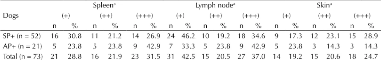
lymph nodes were the most common positive site in the
infected animals (n = 73; 100%), followed by the spleen, (n = 60; 82.2%), and skin (n = 47; 64.4%) (Table 2).
The degree of parasitism in all tissues did not differ
between AP+ and SP+ dogs (p > 0.05).
A high degree of variability in the levels of anti- Leish-mania antibody types and subtypes was observed
among dogs (Figure). Total IgG was the most abun -dant Leishmania-specific antibody (optical density
median = 0.61), followed by IgG2 (optical density median = 0.50), IgE (optical density median = 0.13),
and IgG1 (optical density median = 0.11) (Figure). In the infected groups, 20 (95.2%) AP+ and 51 (98.1%)
SP+ dogs tested positive for Leishmania-speciic IgG
antibodies (Table 3). In the uninfected dogs, positivity
for Leishmania-speciic IgG antibodies was observed in
both AP- (44.4%) and SP- (60.0%) animals (Table 3). The results examining the IgG2 showed high agree -ment (κ > 0.90), IgE a moderate to substantial agree -ment (κ < 0.79), and IgG1 a fair to moderate agreement (κ < 0.50) with those obtained for total IgG.
With respect to IgE antibodies, 71.4% (AP+) and 80.8% (SP+) of the infected dogs were positive. Uninfected dogs
also had Leishmania-speciic IgE in their sera, 27.8% in
the AP- group, and 44.0% in the SP- group (Table 3). The levels of IgG and IgE did not signiicantly differ
between the clinically symptomatic and asymptomatic groups (SP- versus AP-, and SP+ versus AP+; p > 0.05),
but the infection status showed higher levels of both
types of antibodies (p ≤ 0.05) (Figure). A correlation between total IgG and IgE levels (p ≤ 0.05) and the
amount of parasitism was observed in the lymph node (rs = 0.62 and rs = 0.55, respectively), spleen (rs = 0.56 and rs = 0.47, respectively), and skin (rs = 0.51 and rs = 0.52, respectively) and also IgG1, although in a weak fashion: lymph node (rs = 0.26), spleen (rs = 0.30), and skin (rs = 0.31). IgG2 showed no correlation.
Table 3. Percentage of positive results for each type of anti-Leishmania antibody detected in dogs from an endemic area,
according to their clinical and parasitological conditions.
Dogs Asymptomatic Symptomatic
IgG IgG1 IgG2 IgE IgG IgG1 IgG2 IgE
n % n % n % n % n % n % n % n %
Infected 20 95.2 13 61.9 20 95.2 15 71.4 51 98.1 37 71.2 50 96.2 42 80.8
Uninfected 16 44.4 8 22.2 15 41.7 10 27.8 15 60.0 7 28.0 14 56.0 11 44.0
Table 2. Frequencies of the parasite loads in the spleen, lymph node, and skin of symptomatic (SP+) and asymptomatic (AP+)
infected dogs in a visceral leishmaniasis-endemic area. Araçatuba, SP, Southeastern Brazil, 2006.
Dogs
Spleena Lymph nodea Skina
(+) (++) (+++) (+) (++) (+++) (+) (++) (+++)
n % n % n % n % n % n % n % n % n %
SP+ (n = 52) 16 30.8 11 21.2 14 26.9 24 46.2 10 19.2 18 34.6 9 17.3 12 23.1 15 28.9
AP+ (n = 21) 5 23.8 5 23.8 9 42.9 7 33.3 5 23.8 9 42.9 5 23.8 3 14.3 3 14.3
Total (n = 73) 21 28.8 16 21.9 23 31.5 31 42.5 15 20.5 27 37.0 14 19.2 15 20.6 18 24.7
SP+: symptomatic with parasitism dogs; AP+: asymptomatic with parasitism dogs
a Parasite load was graduated as: + low, ++ moderate and +++ high.
Table 1. Absolute number and percentage of infected and
uninfected dogs enrolled in the study, in relation to clinical status. Araçatuba, SP, Southeastern Brazil, 2006. (N = 134)
Clinical status Infected Uninfected Total
n % n % n %
Asymptomatic dogs 21 36.8 36 63.2 57 42.5
Symptomatic dogs 52 67.5 25 32.5 77 57.5
Cross-reactivity with the L. (L.) infantum chagasi ELISA
was detected with IgG antibodies present in the serum from two of nine animals diseased with babesiosis (22.2%), and with IgE from 1 out of 17 animals (5.9%) with ehr
-lichiosis, and 1 out of 9 (11.0%) with toxoplasmosis.
DISCUSSION
The non-speciic and varied clinical manifestations of
canVL, which can overlap with the symptoms of other canine infections, and the lack of external signs in the infected asymptomatic animals make the diagnosis
of leishmaniasis one of the most signiicant problems
concerning the disease, which in most cases may ren-der ineffective epidemiological surveillance and VL control measures.24
It is clear that veterinarians and practitioners of control pro -grams aim to diagnose a case of VL with certainty. Thus, we investigated the potential of employing serology for examining the levels and the positivity of anti-Leishmania
IgG (total and subclasses) and IgE antibodies in a canine
population living in a region with high canVL endemic-ity, taking into account their parasitological condition.
This study revealed four groups of dogs living in such region. There were asymptomatic animals infected with
L. (L.) infantum chagasi (AP+) or not (AP-), symptom
-atic dogs with conirmed tissue parasitism (SP+), and
dogs presenting signs related to canVL without detect-able Leishmania infection (SP-).
The sera of both asymptomatic and symptomatic dogs contained detectable levels of all antibodies inspected, even those without Leishmania tissue parasitism. IgG was
by far the most predominant Leishmania-speciic anti
-body and IgG2 was the major subclass present, a result
observed in previous reports,23,25 but not by Quinnell et al,18 who observed high levels of Leishmania-speciic
IgG1 in sick and asymptomatic parasite-positive dogs compared to the uninfected ones. In our study, all four groups were low responders for IgG1, including the SP+ dogs. Although IgG1 was higher in the SP+ dogs than in
the other groups, its level in asymptomatic infected
ani-mals (AP+) did not differ from those of the non-parasitized
dogs, excluding its possible use for diagnostic purposes.
Few studies have assessed IgE in canVL, but some have shown that high IgE production in symptomatic
dogs strongly correlates with active disease and high
OD
OD
AP-0.00 0.10 0.15 0.20 0.25 0.30 0.35
SP- AP+ SP+ Control
Cut-off: 0.11 a
IgE
AP-0.0 0.2 0.4 0.6 0.8 1.0
SP- AP+ SP+ Control
Cut-off: 0.26 IgG2
OD
AP-0.0 0.2 0.4 0.6 0.8 1.0 1.2
SP- AP+ SP+ Control
Cut-off: 0.17 a
IgG
OD
AP-0.1 0.2 0.3 0.6 0.7
SP- AP+ SP+ Control
Cut-off: 0.17 IgG1
a p ≤ 0.05
parasitism.6,19,20 In the present study, all groups pro -duced small amounts of Leishmania-speciic IgE anti
-bodies and were not signiicantly different from those of
symptomatic dogs and their asymptomatic counterparts (AP- versus SP-; AP+ versus SP+), which was similar
to the indings of Amorim et al.2 No correlation between
the levels of both IgG and IgE and the clinical status
was observed in the present study. However, correla-tion between these antibodies and infeccorrela-tion condicorrela-tion
was observed, speciically with the detection of amasti
-gotes in tissues (skin, lymph nodes, or spleen), and with
the levels of the parasite burden, which did not differ
between the asymptomatic (AP+) and sick dogs (SP+). In this investigation, 98.1% and 80.8% of the symp
-tomatic infected animals (SP+) tested positive for
Leishmania-speciic IgG and IgE antibodies, respec -tively, which demonstrate that serology adequately
relected the Leishmania infection status of these dogs,
especially with respect to IgG. Regarding the infected but asymptomatic dogs (AP+), 95.2% were positive
for Leishmania-speciic IgG, but IgE failed to detect
28.6% of these animals. Our major concern was that a
high percentage of the asymptomatic animals without
Leishmania tissue parasitism (AP-) also tested positive
for Leishmania-speciic IgG (44.4%), IgG subclasses
and IgE (27.8%). This inding may be attributable to
recent infection without the establishment of tissue parasitism. Another is that these dogs have suffered a transient infection, characterized by initial parasitolog-ical positivity that might have become and remained negative thereafter,16 and had already controlled the
Leishmania infection at the time we performed sera col-lection. Therefore, the Leishmania-speciic antibodies
detected in the AP- group could be the result of speciic
antibodies that were maintained in the bloodstream after recovery. However, cross-reactivity was not ruled out, which could also explain the anti-Leishmania antibod-ies found in the SP- group, with clinical signs related to canVL, although proven to be Leishmania-negative.
Leishmania antigens used in serological tests are recog-nized by antibodies present in the sera of dogs infected with a plethora of agents, including protozoans, bac-teria, fungi, intestinal and non-intestinal worms, and ectoparasites.3,8,11,17,21,22 Among the sera we tested,
cross-reactivity with IgG was observed in dogs with
babesiosis, and with IgE in those with ehrlichiosis and
toxoplasmosis. Thus, we may not exclude that SP- dogs were infected with other agents that can cause some clinical signs compatible with canVL, such as alope-cia or desquamation, sole signs presented in a portion of our symptomatic population, which highlights the importance of conducting a differential diagnosis, as evidenced by others.4,24
Among the four isotypes investigated, IgG would be the
only antibody that should be used for screening VL due
to its high positivity in infected dogs (AP+ and SP+),
but the overall results pointed that the use of serology as a sole diagnostic method is ineffective in determin-ing if dogs are actually infected with Leishmania, thus
requiring additional conirmatory tests.
With regard to the parasitological method used in this
study to deine infection status, immunohistochemistry
is a technique that could provide results that were com-parable to those obtained with conventional polymerase
chain reaction (PCR) and is sensitive enough to detect
low parasite load such as in samples from asymptomatic dogs.13 Moreover, to ensure precise immunohistochemi -cal parasitologi-cal diagnosis, the presence of parasites was examined in three different tissues, namely, skin, spleen, and lymph nodes, which are the most likely sites of high parasitism during a L. (L.) infantum cha-gasi infection.13,19 For those reasons, there is a strong probability that both groups in which no amastigotes were visualized in the tissues inspected, AP- and SP-, truly included only unparasitized animals.
In conclusion, we characterized the serological and
the infection statuses of dogs living in a VL-endemic region and demonstrated that serology, even using an
optimized technique with speciic L. (L.) infantum chagasi antigens, was not reliable method for dis-criminating between Leishmania-infected and unin-fected dogs through any of the four isotypes tested. This complex serological scenario that we observed
in the endemic area relects the dificulties experi -enced by veterinary clinicians and coordinators of control programs. Thus, to enhance the effectiveness of control measures, a re-evaluation of the canine serodiagnosis is necessary, especially in countries
1. Alves WA, Bevilacqua PD. Reflexões sobre a qualidade do diagnóstico da leishmaniose visceral canina em inquéritos epidemiológicos: o caso da epidemia de Belo Horizonte, Minas Gerais, Brasil,
1993-1997. Cad Saude Publica. 2004;20(1):259-65.
DOI:10.1590/S0102-311X2004000100043
2. Amorim IF, Freitas E, Alves CF, Tafuri WL, Melo MN, Michalick MS, et al. Humoral immunological profile and parasitological statuses of Leishmune vaccinated and visceral leishmaniasis infected dogs from an
endemic area. Vet Parasitol. 2010;173(1-2):55-63.
DOI:10.1016/j.vetpar.2010.06.021
3. Ferreira EC, Lana M, Carneiro M, Reis AB, Paes DV, Silva ES, et al. Comparison of serological assays for the diagnosis of canine visceral leishmaniasis in animals
presenting different clinical manifestations. Vet Parasitol.
2007;146(3-4):235-41. DOI:10.1016/j.vetpar.2007.02.015
4. Gomes YM, Paiva Cavalcanti M, Lira RA, Abath FG, Alves LC. Diagnosis of canine visceral leishmaniasis:
biotechnological advances. Vet J. 2008;175(1):45-52.
DOI:10.1016/j.tvjl.2006.10.019
5. Harhay MO, Olliaro PL, Costa DL, Costa CH. Urban parasitology: visceral leishmaniasis in
Brazil. Trends Parasitol. 2011;27(9):403-9.
DOI:10.1016/j.pt.2011.04.001
6. Iniesta L, Gállego M, Portús M. Immunoglobulins G and E responses in various stages
of canine leishmaniosis. Vet Immunol
Immunopathol. 2005;103(1-2):77-81. DOI:10.1016/j.vetimm.2004.08.011
7. Laurenti MD, Rossi CN, Matta VLR, Tomokane TY, Corbett CEP, Secundino NFC, et al. Asymptomatic
dogs are highly competent to transmit Leishmania
(Leishmania) infantum chagasi to the natural
vector. Vet Parasitol. 2013;196(3-4):296-300.
DOI:10.1016/j.vetpar.2013.03.017
8. Mancianti F, Pedonese F, Poli A. Evaluation of dot enzyme-linked immunosorbent assay (dot-ELISA) for the serodiagnosis of canine leishmaniosis as compared
with indirect immunofluorescence assay. Vet Parasitol.
1996;65(1-2):1-9. DOI:10.1016/0304-4017(96)00946-6
9. Marcondes M, Lima VM, Araújo MF, Hiramoto RM, Tolezano JE, Vieira RF, et al. Longitudinal analysis of serological tests officially adopted by the Brazilian Ministry of Health for the diagnosis of canine visceral leishmaniasis in dogs vaccinated with
Leishmune®. Vet Parasitol. 2013;197(3-4):649-52.
DOI:10.1016/j.vetpar.2013.07.013
10. Matta VL, Hoshino-Shimizu S, Dietze R, Corbett CEP. Detection of specific antibody isotypes and subtypes before and after treatment of American visceral
leishmaniasis. J Clin Lab Anal. 2000;14(1):5-12.
DOI:10.1002/(SICI)1098-2825(2000)14:1<5::AID-JCLA2>3.0.CO;2-F
11. Mettler M, Grimm F, Capelli G, Camp H, Deplazes P. Evaluation of enzyme-linked immunosorbent assays, an immunofluorescent-antibody test, and two rapid tests (immunochromatographic-dipstick and gel tests) for serological diagnosis of
symptomatic and asymptomatic Leishmania infections
REFERENCES
in dogs. J Clin Microbiol. 2005;43(11):5515-9.
DOI:10.1128/JCM.43.11.5515-5519.2005
12. Moreira MAB, Luvizotto MCR, Garcia JF, Corbett CEP, Laurenti MD. Comparison of parasitological, immunological and molecular methods for the diagnosis of leishmaniasis in dogs with different
clinical signs. Vet Parasitol. 2007;145(3-4):245-52.
DOI:10.1016/j.vetpar.2006.12.012
13. Nunes CM, Lima VM, Paula HB, Perri SH, Andrade AM, Dias FE, et al. Dog culling and replacement in an area endemic for visceral leishmaniasis
in Brazil. Vet Parasitol. 2008;153(1-2):19-23.
DOI:10.1016/j.vetpar.2008.01.005
14. Oliveira TMFS, Mineo TWP, Bason M, Day MJ, Machado RZ. IgG subclass profile of serum antibodies to Leishmania chagasi in naturally infected and
vaccinated dogs. Vet Parasitol. 2009;162(1-2):16-22.
DOI:10.1016/j.vetpar.2009.02.018
15. Palatnik-de-Sousa CB, Day MJ. One Health: the global
challenge of epidemic and endemic leishmaniasis. Parasit
Vectors. 2011;4:197. DOI:10.1186/1756-3305-4-197 16. Paradies P, Sasanelli M, Caprariis D, Testini G, Traversa
D, Lia RP, et al. Clinical and laboratory monitoring of
dogs naturally infected by Leishmania infantum. Vet J.
2010;186(3):370-3. DOI:10.1016/j.tvjl.2009.09.011
17. Porrozzi R, Santos da Costa MV, Teva A, Falqueto A, Ferreira AL, Santos CD, et al. Comparative evaluation of enzyme-linked immunosorbent assays based on crude and recombinant leishmanial antigens for serodiagnosis
of symptomatic and asymptomatic Leishmania infantum
visceral infections in dogs. Clin Vaccine Immunol.
2007;14(5):544-8. DOI:10.1128/CVI.00420-06
18. Quinnell RJ, Courtenay O, Garcez LM, Kaye PM, Shaw MA, Dye C, et al. IgG subclass responses in a longitudinal study of canine visceral leishmaniasis.
Vet Immunol Immunopathol. 2003;91(3-4):161-8. DOI:10.1016/S0165-2427(02)00311-2
19. Reis AB, Teixeira-Carvalho A, Vale AM, Marques MJ, Giunchetti RC, Mayrink W, et al. Isotype patterns of immunoglobulins: hallmarks for clinical status and tissue parasite density in Brazilian dogs naturally
infected by Leishmania (Leishmania) chagasi. Vet
Immunol Immunopathol. 2006;112(3-4):102-16. DOI:10.1016/j.vetimm.2006.02.001
20. Reis AB, Martins-Filho OA, Teixeira-Carvalho A, Giunchetti RC, Carneiro CM, Mayrink W, et al. Systemic and compartmentalized immune
response in canine visceral leishmaniasis. Vet
Immunol Immunopathol. 2009;128(1-3):87-95. DOI:10.1016/j.vetimm.2008.10.307
21. Rosário EY, Genaro O, França-Silva JC, Costa RT, Mayrink W, Reis AB, et al. Evaluation of enzyme-linked
immunosorbent assay using crude Leishmania and
recombinant antigens as a diagnostic marker for canine
visceral. Mem Inst Oswaldo Cruz. 2005;100(2):197-203.
DOI:10.1590/S0074-02762005000200015
leishmaniasis. J Clin Microbiol. 2010;48(5):1866-74. DOI:10.1128/JCM.02402-09
23. Solano-Gallego L, Riera C, Roura X, Iniesta L, Gallego M, Valladares JE, et al. Leishmania infantum-specific IgG, IgG1 and IgG2 antibody responses in healthy and ill dogs from endemic areas. Evolution in the course of infection and
after treatment. Vet Parasitol. 2001;96(4):265-76.
DOI:10.1016/S0304-4017(00)00446-5
24. Solano-Gallego L, Miró G, Koutinas A, Cardoso L, Pennisi MG, Ferrer L, et al. LeishVet guidelines for the
practical management of canine leishmaniosis. Parasit
Vectors. 2011;4:86. DOI:10.1186/1756-3305-4-86 25. Vercammen F, Fernandez-Perez FJ, Amo C,
Alunda JM. Follow-up of Leishmania infantum
naturally infected dogs treated with allopurinol: immunofluorescence antibody test, ELISA and
Western blot. Acta Trop. 2002;84(3):175-81.
DOI:10.1016/S0001-706X(02)00178-X
Article based on the doctoral thesis of Laranjeira DF, entitled: “Avaliação da imunidade humoral e celular em cães naturalmente
infectados com Leishmania (L.) chagasi e sua correlação com a transmissibilidade para o vetor”, presented to the Programa de
Pós-Graduação Patologia Experimental e Comparada of the Faculdade de Medicina Veterinária e Zootecnia of the Universidade de São Paulo, in 2008.
This study was supported by the Fundação de Amparo à Pesquisa do Estado de São Paulo (FAPESP – Process 04/07965-2).