Formation and Stabilization of Tubular Structures in
Multicomponent Membranes
Nataliya Bobrovska1, Wojciech Go´z´dz´1, Veronika Kralj-Iglicˇ2, Alesˇ Iglicˇ3*
1Institute of Physical Chemistry, Polish Academy of Sciences, Kasprzaka 44/52, 01–224 Warsaw, Poland,2Faculty of Health Studies, University of Ljubljana, Zdravstvena 5, SI-1000 Ljubljana, Slovenia,3Laboratory of Biophysics, Faculty of Electrical Engineering, University of Ljubljana, Trzˇasˇka 25, SI-1000 Ljubljana, Slovenia
Abstract
Influence of isotropic and anisotropic properties of membrane constituents (nanodomains) on formation of tubular membrane structures in two-component vesicle is numerically investigated by minimization of the free energy functional based on the deviatoric-elasticity model of the membrane. It is shown that the lateral redistribution and segregation of membrane components may induce substantial change in membrane curvature resulting in the growth of highly curved tubular structures.
Citation:Bobrovska N, Go´z´dz´ W, Kralj-Iglicˇ V, Iglicˇ A (2013) On the Role of Anisotropy of Membrane Components in Formation and Stabilization of Tubular Structures in Multicomponent Membranes. PLoS ONE 8(9): e73941. doi:10.1371/journal.pone.0073941
Editor:Yaakov Koby Levy, Weizmann Institute of Science, Israel
ReceivedApril 13, 2013;AcceptedJuly 25, 2013;PublishedSeptember 16, 2013
Copyright:ß2013 Bobrovska et al. This is an open-access article distributed under the terms of the Creative Commons Attribution License, which permits unrestricted use, distribution, and reproduction in any medium, provided the original author and source are credited.
Funding:The work of N.B. was realized within the International Ph.D. Projects Programme of the Foundation for Polish Science, co-financed from European Regional Development Fund within Innovative Economy Operational Programme ‘‘Grants for innovation’’. A.I. and V.K.I. were supported by Slovenian Research Agency grants J1-4109, J1-4136, J3-4108 and P2-0232. W.G. would like to acknowledge support by NCN grant No. 2012/05/B/ST3/03302. The funders had no role in study design, data collection and analysis, decision to publish, or preparation of the manuscript.
Competing Interests:The authors have declared that no competing interests exist.
* E-mail: [email protected]
Introduction
The shape of lipid bilayers, cellular or artificial, strongly depends on composition and lateral distribution of membrane components [1]. In cell membranes as well as in multicomponent artificial membranes the aggregation/segregation of membrane components may occur under different physiological or non-physiological conditions [2–5].
Except for the sake of simplicity there is no a priori reason to consider membrane constituents/nanodomains to be isotropic [6 7] instead of anisotropic, which actually represents a more general approach [2,5,8–12].
Not only proteins and/or protein-lipid complexes but also lipid molecules should be in general considered anisotropic [12–14]. Thermal rotational motion of lipids around their vertical axes may lead to wrong conclusion that the average (effective) intrinsic shape of lipid molecules is axisymmetric, i.e. isotropic. The membrane lipids have two tails and in general anisotropic headgroups. The rotational states in the curvature field of the membrane have different energy (except for the planar and spherical membranes). Averaging over rotational degrees of freedom gives effective anisotropic intrinsic shape of lipids [15]. When the membrane components are modeled as anisotropic, it is possible to explain formation of experimentally observed, transient, energetically stable, narrow necks (pores) connecting the fused vesicles to the target membrane. Such shapes may result from orientational ordering and lateral redistribution of membrane constituents/ nanodomains [16].
Coupling between the cell/liposome shape and non-homoge-neous lateral distribution [17] of membrane components may originate from the tendency of membrane components to find/
induce the optimal configuration (optimal membrane curvature) with respect to the intrinsic shape of membrane components. It was indicated in different theoretical and experimental studies [5,8–10,12,13,18–22]. that the generation and stability of the lipid bilayer tubes in the cellular and artificial multicomponent membrane systems in the absence ofelongated inner stiff supporters, e.g. microtubules [23–27] orexternal pulling forces, such as, optical tweezers [28,29] or motor proteins (kinesin, dynamin) [29,30], can be explained by the presence of membrane elements (nanodo-mains) and attached proteins with anisotropic properties. As for example, the membrane attached crescent shaped BAR domain proteins have clearly anisotropic shape and therefore their energy depend on their local orientation or statistically averaged local orientation, depending on the local curvature of the membrane [5,20,31]. The nanodomain can be a macromolecule which is partially or fully embedded into membrane bilayer (such as multi-anchor polymers [32]), membrane attached proteins plus inter-acting lipids, a small protein-lipid cluster or a small cluster of different kind of lipids etc. The membrane is then considered as the self-assembly of nanodomains. The area of a single nanodomain can be in general much larger than the area of a single lipid molecule. The intrinsic shape of a nanodomain and a single lipid can be modeled within the framework of the deviatoric elasticity model with the appropriate choice of two principal intrinsic curvaturesC1m andC2m [15,33].
structures with thin tubular protrusions having small spherical vesicles at their free tips (Fig. 1) induced by accumulation of anisotropic membrane components in tubular membrane regions.
Results and Discussion
The model vesicles are built up by two components (A and B) ant their shapes are obtained numerically by the direct minimi-zation of the free energy functional of the membrane under the constraints of constant vesicle surface areaSand volumeVand a constant number of A type constituents/nanodomains, i.e. at
constant total relative concentration of A component,
wtot~1=S Ð
wdS [24,34]. The dimensionless reduced volume is defined asv~V=Vs(the ratio of the volume of the vesicle to the
volume of a sphere Vs~4pR3s=3 with the same surface area)
where the radiusRsdefines the unit length. The calculations were
performed for vesicles with rotational symmetry, where the shape profile of the vesicle was described by the functionh(s) and the distribution of components on the vesicle surface by the function
w(s)(sis the arclength of the profile).h(s)andw(s)were calculated numerically by the minimization of the free energy functional Figure 1. (A) Scanning electron micrograph of membrane nanotubes of RT4 urothelial cancer cells. Some of the nanotubes have spherical vesicles at their free tips (indicated by the arrows).Bar = 10mm (adapted with permission from [49].) (B) Vesiculation in human red blood cells. Note the exovesicles located around the parent red blood cells. Tethers are not visible in the figure. Bar = 3mm (adapted with permission from [26]).
[7,24]. The minimization procedure and the detail description of
h(s)andw(s)is given in the Methods section.
In this work, we have investigated under what conditions the formation of thin tubular structures is favorable. The special examples of such systems observed in experiments (Fig. 1), i.e. the cells with thin tubular protrusions having small spherical vesicles at their free tips, are the main subject of this work, not thoroughly studied in our previous work [22].
The calculations were performed for different values of the bending rigidity for each component,kB
~30 kBT,kA~4kB. The
value of kB
is characteristic for lipid domains, the value of kA
characterizes the domains of lipid membranes with attached macromolecules, where the thickness of the membrane is relatively large [35–37]. In the model, the bending rigidity depends on the local concentration of macromolecules, k(w) [38]. The function describing the local bending rigidityk(w)is defined in the Methods section. It has been assumed that the surface area of the nanodomain, a0, was of order of 100 nm2. The chosen vesicle radius in the calculations was of the order of 250 nm.
When both components are isotropic, the vesicle is composed of small spherical beads connected by narrow passages, such as the first vesicle in Fig. 2. When one or two components are anisotropic we can obtain shapes in which thin tubular structures are formed. It is important to note that so far the formation of thin cylindrical protrusions which are attached to larger spherical vesicle has not been predicted in the models in which the anisotropy of the components is not taken into account [18,22].
In the membrane systems encountered in the nature a small number of membrane components (minority) is usually strongly anisotropic, while a much larger number of membrane compo-nents (majority) is considerably less anisotropic, or isotropic. The anisotropic BAR domain proteins attached to the bilayer membrane [31,39] are a typical example. For simplicity reasons we examine a single strongly anisotropic membrane component as a minority component, while the rest of the membrane is considered as composed of an isotropic component of a single type. We study the effect of concentration, reduced volume, and intrinsic mean curvature of an anisotropic membrane component on the formation of thin membrane tubular protrusions with a small vesicle at its tip. In the model, the local concentration of the components has an influence on the shape of the membrane, but also the curvature of the membrane determines the local distribution of the components [7,40]. Thus, the vesicle shape and the local concentration of the components is determined by these two effects. If the distribution of the components did not depend on the shape of the vesicles, the components would be uniformly mixed in order to maximize the entropy. It has to be noted that we do not consider phase separated mixtures [41]. The segregation of components on the vesicle surface is due to the
curvature gradients and the difference of the intrinsic curvatures of the constituents.
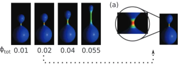
The cylindrical protrusions are formed when at least one component is anisotropic. Such behavior is demonstrated in Fig. 3, where the total concentration is varied from wtot~0:01 to wtot~0:055.
It is interesting to note that very small amount of the anisotropic component is enough to induce the formation of the cylindrical protrusion. Moreover, the length of the protrusion depends on the concentration. This is due to the separation of the components in the membrane, where the anisotropic components are located mainly in the tubular part which has a very small surface area compared to the rest of the vesicle. It is interesting to note, that total component segregation was observed forwtot~0:02. At this
concentration almost all anisotropic component was accumulated in the neck of the vesicle. The possibility of accumulation of the components in a small area may be important for some macromolecules, since they are biologically active only at sufficiently high concentration.
The calculations presented in Fig. 3 were performed for constant reduced volumev~0:8. For such a reduced volume we observe the shapes without up-down symmetry, but for smaller values of the reduced volume the shapes with up-down symmetry are stable, as presented in Fig. 4. Moreover, the smaller the volume the more mixed are the components, and the cylindrical protrusions are no longer stable.
The complete mixing was observed for small concentration of the anisotropic component,wtot~0:02, which for small reduced
Figure 2. The shapes of the vesicle composed of (iso+iso), (iso+aniso), and (aniso+aniso) components forv~0:8,wtot~0:02,
HA
m~8:0,H B m~4:2,k
A ~4kB
. doi:10.1371/journal.pone.0073941.g002
Figure 3. The vesicle shapes for different concentration,wtot, of anisotropic component, and forHB
m~4:2,v~0:8,H A
m~D
A m~8:0, kA
~4kB
. The inset (a) shows the configuration with almost total segregation of components forwtot~0:02. The anisotropic component is accumulated in the neck area.
doi:10.1371/journal.pone.0073941.g003
Figure 4. The vesicle shapes for different values of the reduced volume,v, and for (a)wtot~0:02,H
B
m~3:0(left),H B
m~4:2(right),
HA
m~D
A m~8:0,k
A ~4kB
and (b)wtot~0:055,HB
m~3:0(left),H B m~4:2 (right),HA
m~D
A m~8:0,k
A ~4kB
volume, vw0:65, results in pearl-like shapes with up-down symmetry. The increase of the concentration of anisotropic component stabilizes longer and wider tubular structures.
The anisotropy of one of the components is not however a sufficient condition for the formation of the tubular structures. We have also observed that cylindrical protrusions may be induced by changing the properties of the isotropic component. It is demonstrated in Fig. 5 that when the intrinsic mean curvature of the isotropic component is increased (for fixed reduced volume) the cylindrical protrusions are formed and their length increases with the increase of the intrinsic mean curvature of the anisoropic component.
In the systems in which the cylindrical tubules are created when the proteins (BAR domain proteins, epsin) are adsorbed at the membrane surface, the radius of the tubule is determined by the intrinsic curvature of the protein [42,43]. In Fig. 6 we show that there is a strict relation between the intrinsic curvature,DA
m, of the
anisotropic component and the radius of the tubule. For smaller values of the intrinsic curvature,DA
m, the cylindrical tubule is not
well developed yet. AtDA
m&9:45there is the transition to the well
developed cylindrical tubule. Apart from the values ofDA m in the
vicinity of the transition valueDA
m&9:45we can see that for the
well developed cylindrical tubes (for DA
mw10) the radius of the
tubular protrusion decreases linearly with increasing DA m. Thus,
the results of our theoretical calculations are in qualitative agreement with the experimental predictions showing that the membrane tubular protrusions induced by the membrane bound anisotropic molecules (such as highly anisotropic BAR domain-containing proteins [5,20,31]) with larger intrinsic curvature radius (corresponding to smaller DA
m in our notation) generally
have larger diameters than do those formed by the molecules characterized by smaller intrinsic curvature radius (i.e. largerDA
m)
[5,44].
Conclusions
We have shown that accumulation of anisotropic components may lead to the formation of thin tubular protrusions. The anisotropy of components is a necessary condition for creation of thestabletubular protrusions. When the components are isotropic such cylindrical structures may be created only when some external force is applied. For example when membrane is pushed by growing microtubules or pulled by molecular motors. The width of the tubes depends on the intrinsic curvatures of anisotropic components. When the membrane is composed of
isotropic components the stable protrusions which are created without any external force are built of a series of connected beads.
Methods: Theoretical model and parametrization
In the model the membrane is composed of two components A and B which can be either isotropic or anisotropic and are characterized by the intrinsic principal curvatures C1im, Ci2m (i~A,B). The free energy functional is composed of the (anisotropic) bending energy Fb and the free energy associated
with the entropy of mixingFmix:
F~FbzFmix, ð1Þ
where the membrane bending energy is given by [33,45,46]
Fb~ ð
A
k(w) (H{Hm(w))2z(D{Dm(w))2
dA, ð2Þ
whileFmixis [2,46]
Fmix~{
kBT
a0
ð
A
wlnwz(1{w) ln (1{w)
½ dA, ð3Þ
where kB is the Boltzmann constant, T is the absolute
temperature, a0 denotes the area of a single nanodomain, C1 and C2 are the membrane principal curvatures, k(w) is the bending rigidity, H~(C1zC2)=2 is the membrane mean curvature,D~DC1{C2D=2is the membrane curvature deviator, Dm~DC1m{C2mD=2 is the intrinsic nanodomain curvature
devi-ator andHm~(C1mzC2m)=2is the intrinsic nanodomain mean
curvature,wis the local relative concentration of the component A. The integral is taken over the whole surface of the vesicle membrane. The model parameters playing a crucial role in the vesicle shape transformations are: the total relative concentration (wtot) of the component A (the total relative concentration of the
component B is(1{wtot)), the bending rigidity of i-th component ki
, the nanodomain intrinsic mean curvature,
Hi m~(C
i 1mzC
i
2m)=2, and the nanodomain intrinsic curvature
deviator, Di m~DC
i 1m{C
i
2mD=2. The components can be either
anisotropic or isotropic. A component is considered as isotropic when its intrinsic deviatoric curvature is zero, Di
m~0. The
properties of anisotropic components are defined by setting the intrinsic devatoric and the mean curvature equalDi
m~H i m.
For simplicity we assume linear dependence of the bending rigidityk[47], the nanodomain intrinsic mean curvatureHmand
the deviator Dm on the local relative concentration of the
component A (w):
k(w)~(kA{kB)wzkB, ð4Þ
Hm(w)~(H A m{H
B m)wzH
B
m, ð5Þ
Dm(w)~(DAm{D B m)wzD
B
m: ð6Þ
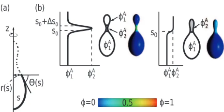
The contour of the vesicle is parametrized by the angle,h(s), which is the function of the arclength of the contour (s) and is defined by the tangent line to the vesicle profile and a horizontal line, which is perpendicular to the axis of rotation (see Fig. 7a) Figure 5. The vesicle shapes for different values of the mean
curvature of isotropic component, HB
m, and for v~0:8, wtot~0:055, HA
m~D
A m~8:0, D
B m~0, k
A ~4kB
: (a) HA
m~D
A ~6 and (b)HA
m~D
A m~8.
[24,34]. In this parametrization, the infinitesimal area element is given bydA~2pr(s), wherer(s)~Ð0scos (h(s0))ds0 is the distance from the rotation axis. The principal curvatures are given by C1~dh(s)=dsandC2~sin (h(s))=r(s)[34,48].
The ansatz for the local relative concentration of the component A has the form :
w~wð Þs~1 2(w
A 2{w
A
1) tanh½ ðjðs{s0ÞÞz
{tanhðjðs{s0{Ds0ÞÞzwA1,
ð7Þ
wheres0is the position of the boundary between the region rich in the component A and the region rich in the component B,jis the slope of the concentration profile ats0,Ds0is the distance between the inflection points of two hyperbolic tangents. The validity of Eq.(7) assumes the division of the vesicle surface into two regions, which are characterized by the minimal and maximal local concentrations of the component A,wA1 andwA2, respectively (see also Fig. 7 b)
In numerical calculation we have to find both the function
h(s) and the function w(s) for which the functional (1) is minimized. In the minimization procedure the function h(s) is expressed as a Fourier series. When the function h(s), in the form of the Fourier series is plugged into the equation (1), the functional minimization can be replaced by the minimization of the function of many variables. The functional (1) becomes the function of many variables which are the amplitudes in the Fourier series and the length of the shape profile. Since the functional is minimized with respect to the shape and the concentration profile, a few additional variables wA1, wA2, j, s0, and Ds0 which determine the concentration profile, are also used in the minimization [7,34,40].
Acknowledgments
The authors are grateful to dr. Sˇ. Perutkova for useful discussion.
Figure 6. The change of the radius of the tubular region of a vesicle as a function of the intrinsic curvature of the anisotropic component,DA
m, forv~0:8,wtot~0:04,H B m~4:2,D
B m~0,H
A
m~D
A m,k
A ~4kB.
The arrows in the lower panel indicate the position on the surface of the vesicle where the radius of the tubule was measured.
doi:10.1371/journal.pone.0073941.g006
Figure 7. (a) Schematic representation of the parametrization of the vesicle shape. s is the contour length, h(s) the angle between the tangent and the horizontal line, and r(s) the distance from the z-axis. (b) Color map shows the relative concentrations of anisotropic component, A, on the vesicle surface. Red color denotes high concentration of A component, blue color denotes high concentration of B component, green color denotes similar concentration of A and B component.wA
1andw
A
2 are the minimal
Author Contributions
Conceived and designed the experiments: AI VK-I. Performed the experiments: AI VK-I. Wrote the paper: NB AI WG VK-I. Theory: AI VK-I. Computer programs: NB WG.
References
1. Go´z´dz´ WT, Gompper G (2002) Phase behavior of two-component membranes. Colloids Surf A 208: 241–251.
2. Ha¨gerstrand H, Mrowczynska L, Salzer U, Prohaska R, Michelsenn AK, et al. (2006) Curvature dependent lateral distribution of raft markers in the human erythrocyte membrane. Mol Membr Biol 23: 277–288.
3. Risselada H, Marrink S, Mu¨ller M (2011) Curvature-dependent elastic properties of liquid-ordered domains result in inverted domain sorting on uniaxially compressed vesicles. Phys Rev Lett 106: 148102.
4. Sorre B, Callan-Jones A, Manneville J, Nassoy P, J-F J, et al. (2009) Curvature-driven lipid sorting needs proximity to a demixing point and is aided by proteins. PNAS 106: 5622–5626.
5. Baumgart T, Capraro BR, Zhu C, Das SL (2011) Thermodynamics and mechanics of membrane curvature generation and sensing by proteins and lipids. Annu Rev Phys Chem 62: 483–506.
6. Israelachvili J (1991) Intermolecular and Surface Forces. Academic Press, London.
7. Go´z´dz´ WT, Bobrovska N, Ciach A (2012) Separation of components in lipid membranes induced by shape transformation. J Chem Phys 137: 15101. 8. Helfrich W, Prost J (1988) Intrinsic bending force in anisotropic membranes
made of chiral molecules. Phys Rev A 38: 3065–3068.
9. Fournier J (1996) Nontopological saddle-splay and curvature instabilities from anisotropic membrane inclusions. Phys Rev Lett 76: 4436–4439.
10. Kralj-Iglicˇ V, Heinrich V, Svetina S, Zeks B (1999) Free energy of closed membrane with anisotropic inclusions. Eur Phys J B, 10: 5–8.
11. Kralj-Iglicˇ V, Iglicˇ A, Gomisˇcˇek G, Sevsˇek F, Arrigler V, et al. (2002) Microtubes and nanotubes of a phospholipid bilayer membrane. J Phys A: Math Gen 35: 1533–1549.
12. Perutkova S, Daniel M, Rappolt M, Pabst G, Dolinar G, et al. (2011) Elastic deformations in hexagonal phases studied by small-angle x-ray diffraction and simulations. Phys Chem Chem Phys 13: 3100–3107.
13. Rappolt M, Hodzic A, Sartori B, Ollivon M, Laggner P (2008) Conformational and hydrational properties during the Lb- to La- and La- to H||-phase transition in phosphatidylethanolamine. Chem Phys Lipids 154: 46–55.
14. Kulkarni C (2012) Lipid crystallization: from self-assembly to hierarchical and biological ordering. Nanoscale.
15. Kralj-Iglicˇ V, Babnik B, Gauger R, May S, Iglicˇ A (2006) Quadrupolar ordering of phospholipid molecules in narrow necks of phospholipid vesicles. J Stat Phys 125: 727–752.
16. Jorgacˇevski J, Fosˇnaricˇ M, Vardjan N, Stenovec M, Potokar M, et al. (2010) Fusion pore stability of peptidergic vesicles. Mol Membr Biol 27: 65–80. 17. Roux A, Cuvelier D, Nassoy P, Prost J, Bassereau P, et al. (2005) Role of
curvature and phase transition in lipid sorting and fission of membrane tubules. EMBO J 24: 1537–1545.
18. Kralj-Iglicˇ V, Iglicˇ A, Ha¨gerstrand H, Peterlin P (2000) Stable tubular microvesicles of the erythrocyte membrane induced by dimeric amphiphiles. Phys Rev E 61: 4230–4234.
19. Iglicˇ A, Ha¨gerstrand H, Veranicˇ P, Plemenitasˇ A, Kralj-Iglicˇ V (2006) Curvature induced accumulation of anisotropic membrane components and raft formation in cylindrical membrane protrusions. J Theor Biol 240: 368–373.
20. Sˇ Perutkova, Kralj-Iglicˇ V, Frank M, Iglicˇ A (2010) Mechanical stability of membrane nanotubular protrusions influenced by attachment of flexible rod-like proteins. J Biomech 43: 1612–1617.
21. Shlomovitz R, Gov NS, Roux A (2011) Membrane-mediated interactions and the dynamics ofdynamin oligomers on membrane tubes. New J Phys 13: 065008. 22. Kabaso D, Bobrovska N, Go´z´dz´ W, Gov N, Kralj-Iglicˇ V, et al. (2012) On the role of membrane anisotropy and BAR proteins in the stability of tubular membrane structures. J Biomech 45: 231–238.
23. D’Onofrio TG, Hatzor A, Counterman AE, Heetderks JJ, Sandel MJ, et al. (2003) Controlling and measuring the interdependence of local properties in biomembranes. Langmuir 19: 1618–1623.
24. Go´z´dz´ WT (2005) Influence of spontaneous curvature and microtubules on the conformations of lipid vesicles. J Phys Chem B 109: 21145–21149.
25. Davis D, Sowinski S (2008) Membrane nanotubes: dynamic long-distance connections between animal cells. Nat Rev Mol Cell Bio 9: 431–436.
26. Kralj-Iglicˇ V, Iglicˇ A, Bobrowska-Ha¨gerstrand M, Ha¨gerstrand H (2001) Tethers connecting daughter vesicles and parent red blood cell may be formed due to ordering of anisotropic membrane constituents. Colloids Surf A 179: 57– 64.
27. Li Y, Lipowsky R, Dimova R (2011) Membrane nanotubes induced by aqueous phase separation and stabilized by spontaneous curvature. PNAS 108: 4731– 4736.
28. Tian A, Baumgart T (2009) Sorting of lipids and proteins in membrane curvature gradients. Biophys J 96: 2676–2688.
29. Koster G, VanDuijn M, Hofs B, Dogterom M (2003) Membrane tube formation from giant vesicles by dyanmic association of motor proteins. PNAS 100: 15583– 15588.
30. Roux A, Cappello G, Cartaud J, Prost J, Goud B, et al. (2002) A minimal system allowing tubulation with molecular motors pulling on giant liposomes. PNAS 99: 5394–5399.
31. Kabaso D, Gongadze E, Elter P, van Rienen U, Gimsa J, et al. (2011) Attachment of rod-like (BAR) proteins and membrane shape. Mini Rev Med Chem 11: 272–282.
32. Tsafrir I, Caspi Y, Guedeau-Boudeville MA, Arzi T, Stavans J (2003) Budding and tubulation in highly oblate vesicles by anchored amphiphilic molecules. Phys Rev Lett 91: 138102.
33. Iglicˇ A, Babnik B, Gimsa U, Kralj-Iglicˇ V (2005) On the role of membrane anisotropy in the beading transition of undulated tubular membrane structures. J Phys A: Math Gen 38: 8527–8536.
34. Go´z´dz´ WT (2004) Spontaneous curvature induced shape transformation of tubular polymersomes. Langmuir 20: 7385–7391.
35. Christin Hiergeist, Reinhard Lipowsky (1996) Elastic properties of polymer-decorated membranes. J Phys II France 6: 1465–1481.
36. Nikolov V, Lipowsky R, Dimova R (2007) Behavior of giant vesicles with anchored DNA molecules. Biophysical Journal 92: 4356–4368.
37. Thakkar FM, Maiti PK, Kumaran V, Ayappa KG (2011) Verifying scalings for bending rigidity of bilayer membranes using mesoscale models. Soft Matter 7: 3963–3966.
38. In˜iguez Palomares R, Acun˜a Campa H, Maldonado A (2011) Effect of polymer on the elasticity of surfactant membranes: A light scattering study. Phys Rev E 84: 011604.
39. Zhao H, Pykalainen A, Lappalainen P (2010) I-bar domain proteins: linkinga actin and plasma membrane dynamics. Curr Op Cell Biol 23: 14–21. 40. Go´z´dz´ WT (2006) The interface width of separated two-component lipid
membranes. J Phys Chem B 110: 21981–21986.
41. Go´z´dz´ WT, Gompper G (1998) Composition-driven shape transformations of membranes of complex topology. Phys Rev Lett 80: 4213–4216.
42. Peter BJ, Kent HM, Mills IG, Vallis Y, Butler PJG, et al. (2004) Bar domains as sensors of membrane curvature: The amphiphysin bar structure. Science 303: 495–499.
43. Sorre B, Callan-Jones A, Manzi J, Goud B, Prost J, et al. (2012) Nature of curvature coupling of amphiphysin with membranes depends on its bound density. PNAS 109: 173–178.
44. Frost A, Perera R, Roux A, Spasov K, Destaing O, et al. (2008) Structural basis of membrane invagination by f-bar domains. Cell 132: 80717.
45. Iglicˇ A, Lokar M, Babnik B, Slivnik T, Veranicˇ P, et al. (2007) Possible role of flexible red blood cell membrane nanodomains in the growth and stability of membrane nanotubes. Blood Cells Mol Dis 39: 14–23.
46. Fosnaricˇ M, Iglicˇ A, Slivnik T, Kralj-Iglicˇ V (2008) Flexible membrane inclusions and membrane inclusions induced by rigid globular proteins. In: Advances in Planar Lipid Bilayers and Liposomes (A Leitmannova Liu, Ed) 7: 143–168.
47. Go´z´dz´ WT (2011) Transformation of lipid vesicles induced by diffusing macromolecules. J Chem Phys 134: 024110.
48. Helfrich W (1973) Elastic properties of lipid bilayers: Theory and possible experiments. Z Naturforsch 28c: 693–703.