Differential Requirement for
irf8
in
Formation of Embryonic and Adult
Macrophages in Zebrafish
Celia E. Shiau*¤, Zoe Kaufman, Ana M. Meireles, William S. Talbot*
Department of Developmental Biology, Stanford University School of Medicine, Stanford, California, United States of America
¤ Current address: Biosciences Division, Oak Ridge National Laboratory, Oak Ridge, Tennessee, United States of America
*wtalbot@stanford.edu(WST); shiauc@ornl.gov(CES)
Abstract
Interferon regulatory factor 8 (Irf8) is critical for mammalian macrophage development and innate immunity, but its role in teleost myelopoiesis remains incompletely understood. In particular, genetic tools to analyze the role of Irf8 in zebrafish macrophage development at larval and adult stages are lacking. We generatedirf8null mutants in zebrafish using TALEN-mediated targeting. Our analysis defines different requirements forirf8at different stages.irf8is required for formation of all macrophages during primitive and transient defini-tive hematopoiesis, but not during adult-phase definidefini-tive hematopoiesis starting at 5-6 days postfertilization. At early stages,irf8mutants have excess neutrophils and excess cell death inpu.1-expressing myeloid cells. Macrophage fates were recovered inirf8mutants after wildtypeirf8expression in neutrophil and macrophage lineages, suggesting thatirf8
regulates macrophage specification and survival. In juvenileirf8mutant fish, mature macro-phages are present, but at numbers significantly reduced compared to wildtype, indicating an ongoing requirement forirf8after embryogenesis. As development progresses, tissue macrophages become apparent in zebrafishirf8mutants, with the possible exception of microglia. Our study defines distinct requirement forirf8in myelopoiesis before and after transition to the adult hematopoietic system.
Introduction
Myeloid cells form the innate immune system that provides the immediate response to protect the host after infection and injury. Macrophages, monocytes, and granulocytes (including neu-trophils) are major myeloid cell types [1–4]. The proper formation of these myeloid cells dur-ing development and their continuous replenishment throughout life are essential to sustain the function of the immune system. As in mammals, hematopoiesis in zebrafish occurs in sev-eral waves [1,5,6]. This process begins with the primitive wave in the anterior lateral plate me-soderm of the zebrafish embryo, then transitions to the transient definitive wave in the
OPEN ACCESS
Citation:Shiau CE, Kaufman Z, Meireles AM, Talbot WS (2015) Differential Requirement forirf8in Formation of Embryonic and Adult Macrophages in Zebrafish. PLoS ONE 10(1): e0117513. doi:10.1371/ journal.pone.0117513
Academic Editor:Zilong Wen, Hong Kong University of Science and Technology, CHINA
Received:October 10, 2014
Accepted:December 28, 2014
Published:January 23, 2015
Copyright:This is an open access article, free of all copyright, and may be freely reproduced, distributed, transmitted, modified, built upon, or otherwise used by anyone for any lawful purpose. The work is made available under theCreative Commons CC0public domain dedication.
Data Availability Statement:All relevant data are within the paper and its Supporting Information files.
Funding:C.E.S. was supported by NIH NRSA fellowship 5F32NS067754 and ORNL Liane Russell Fellowship, Z.K. was supported by Stanford Undergraduate VPUE funds, and A.M.M. was supported by EMBO fellowship ALTF 1125–2011.
posterior blood island (PBI), which later becomes the caudal hematopoietic tissue (CHT). Sub-sequently, definitive blood development takes place in the CHT and the zebrafish aorta-gonad-mesonephros (AGM) analog. At later stages, definitive hematopoiesis moves to the pronephric kidney, which is the site of adult hematopoiesis [5,6]. More is known about the development of hematopoietic stem cells (HSCs) into different myeloid fates in the definitive wave than in the earlier waves of blood formation [2,3,6,7], although the mechanisms are not fully under-stood in either the early or late waves.
A major determinant of macrophage/monocyte fate during hematopoiesis is the transcrip-tion factor IRF8, a member of the interferon regulatory factor (IRF) family. IRF proteins con-tain a conserved N-terminal DNA binding domain that recognizes the interferon consensus sequence, and they regulate transcription of interferon genes during immune response [8]. In mammals, IRF8 is critical for myeloid development. IRF8 deficiency in human and mouse leads to a significant reduction of macrophage/monocyte development but an expansion of neutrophils and undifferentiated hematopoietic progenitor cells, reminiscent of myeloid leuke-mia [9–12]. In mouse, IRF8 is strongly expressed in mononuclear phagocytes (macrophages, monocytes, and dendritic cells)[3,13], and regulates differentiation of these phagocytes and granulocyte-macrophage progenitors [9,10,14]. In human, missense mutations disrupting transcriptional activity of IRF8 are linked with immunodeficiencies that severely reduce the numbers of dendritic cells and monocytes [11], indicating its role in human myeloid develop-ment. Furthermore, IRF8 is transcriptionally repressed in many acute and chronic myeloid leu-kemia (AML and CML, respectively) patients [15,16], suggesting a link between IRF8 activity and these diseases. In zebrafish, transient knockdown ofirf8by translation- and splice-blocking morpholinos eliminated the embryonic macrophage population and expanded the neutrophil population [17], but mutations in zebrafishirf8have not been available to enable long-term studies. Thus, IRF8 plays key roles in maintaining normal production of myeloid cell types, but the nature of its function in specification and maintenance of myeloid cells at dif-ferent times in development are not understood, and the extent of its functional conservation across species remains to be fully explored.
To investigate the function ofirf8at different stages of development to early adulthood in zebrafish, we createdirf8null mutations.irf8mutants are devoid of macrophages during em-bryogenesis, but have an increase of neutrophils and immature or apoptotic myeloid cells. As larval development progresses, some macrophages are present in the mutants, indicating that macrophages at different stages have distinct requirements for Irf8 function. The late-emerging macrophages inirf8mutants are likely derived from different progenitors from the embryonic macrophages (HSCs versus early myeloid), which coincides with the differential dependence onirf8. Our study provides new insights into the role ofirf8in myeloid cell fate regulation in zebrafish before and after transition to the adult hematopoietic system.
Results
irf8
null mutants lack microglia during development and are viable
Morpholino knockdown studies have indicated thatirf8plays a role in macrophage develop-ment in zebrafish [17], but mutations in the zebrafishirf8gene have not been available to allow analysis of a complete loss ofirf8function, or analysis at later stages. Furthermore, excess im-mature myeloid cells were observed in Irf8 mouse mutants but not reported in the zebrafish morpholino studies [17], so the degree of conservation of Irf8 function between zebrafish and mammals is not fully understood. To construct loss-of-function mutations inirf8, we used TALEN-mediated targeting to generate two new mutant alleles,irf8st95andirf8st96(Fig. 1A). Both alleles have frameshift mutations just 3’to the translational initiation codon, and both arepredicted null mutations (Fig. 1A). The zebrafish genome contains only one ortholog of mam-malian Irf8, and therefore these mutations are predicted to eliminate all functions of Irf8 in zebrafish.
To characterize the phenotypes ofirf8st95andirf8st96mutants, we examined microglia, the brain-resident macrophages previously shown to be disrupted by splice- and translation-blocking morpholinos againstirf8[17]. Homozygousirf8st95/st95andirf8st96/st96mutants, and transheterozygousirf8st95/st96mutants all lacked microglia at 3–6 dpf, as determined by neutral red staining [19] and expression ofapoe[20–22](Fig. 1A and B, and data not shown). We have focused our analysis onirf8st95, and theirf8-/-mutants pictured in the figures areirf8st95/st95 ho-mozygotes, unless noted otherwise.
Despite the loss of microglial cells during the embryonic and larval stages,irf8-/-mutants ap-peared morphologically normal as early larvae (Fig. 1C). Furthermore mostirf8-/-mutants were viable as adults at approximately 3 months postfertilization (Fig. 1C), although their sur-vival rate was reduced compared to heterozygous and wildtype siblings (S1 Fig.).
irf8
mutants have no macrophages but have excess neutrophils during
embryogenesis
To further characterize theirf8mutants, we examined the early development of myeloid cells at embryonic stages. In zebrafish, adaptive immunity begins after 3–4 weeks postfertilization [23], and macrophages and neutrophils are the main functional leukocytes in the embryo [18].
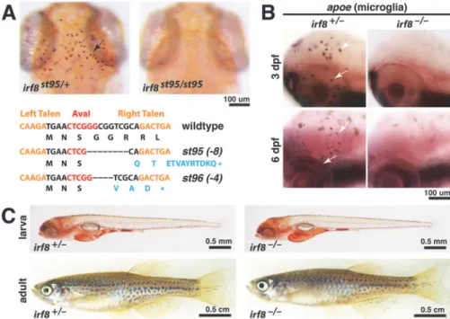
Fig 1. TALEN-inducedirf8mutationsst95andst96eliminate embryonic microglia but allow survival
to adulthood.(A) TALE nucleases target region nearirf8translational start site, creating frameshift
mutationsst95andst96, which introduced premature stop codons as shown (bottom). Top panels show representative neutral red staining for microglia inirf8st95/st95mutant that lacks all microglia, compared with a heterozygous sibling that has a wildtype microglial population (arrow). (B) Analysis ofapoeRNA expression by in situ hybridization shows presence of microglia inirf8sibling but no microglia inirf8mutants at 3 and 6 dpf. (C) Images of heterozygous and homozygous mutant larvae at 5 dpf, showing that the mutants have normal overall morphology. Images of the whole adult zebrafish were compiled from two tiled images of the same fish. At 3 months of age, theirf8mutant zebrafish grew to a similar size as its sibling. All images represent thest95allele. Scale bars are shown below each set of panels or for each individual panel.
We assessed the distribution of macrophages and neutrophils by examining the cell-type spe-cific markersmfap4andmpx, respectively. In comparison to wildtype and heterozygous sib-lings,irf8-/-mutants developed nomfap4-expressing macrophages (Fig. 2A), and had a large expansion ofmpx-expressing neutrophils (Fig. 2B and C). The increase in neutrophils was evi-dent throughout the embryo, and at stages as early as 1 dpf (Fig. 2B–D). Our genetic analysis validates the embryonic phenotypes observed previously inirf8morphants [17]. In addition, the finding thatirf8null mutants are viable as adults allows the analysis ofirf8function into adulthood.
Partial recovery of macrophages in
irf8
mutants coincides with
presumptive onset of hematopoiesis in the kidney
To address the possible roles ofirf8in macrophage development at later stages, we usedTg (mpeg1:EGFP)to examine GFP-labeled macrophages at stages up to 31 dpf inirf8-/-mutants and their siblings. These macrophages were examined in live fish up to 1 week old (7 dpf) and after fixation at later stages (Figs.3and4). We analyzed two regions where macrophages are
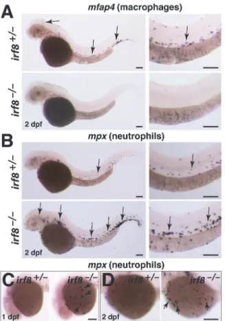
Fig 2.irf8mutant embryos have no macrophages but produce excessive neutrophils.(A)mfap4RNA
expression at 2 dpf shows a complete loss of macrophages inirf8mutant but abundant macrophages in heterozygous sibling (arrows). (B)mpxRNA expression at 2 dpf shows an overproduction of neutrophils in
irf8mutants (arrows, bottom panels) compared with the sibling (arrow, top panels). Right column in A and B shows a higher magnification image of the trunk region from the same embryos depicted in the left column. (C) Neutrophils can first be detected on the yolk sac by 1 dpf.irf8mutants have many yolk sac neutrophils (arrows) compared with sibling. (D) At 2 dpf,irf8mutants continue to have more neutrophils on the yolk sac (arrows). Overall,irf8mutants appear to have more neutrophils throughout the body. All scale bars are 100 um.
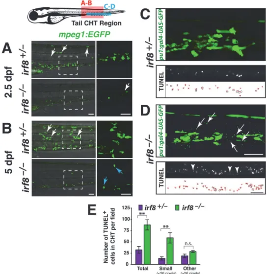
Fig 3.irf8mutants have immature myeloid cells and excess myeloid cell death during development in
the CHT.Diagram of a zebrafish larva showing the regions of analysis in the CHT: red box, region in A and B;
blue box, region in C and D; and dotted lines in blue box indicate area of TUNEL quantification in E. (A-B) Lateral view. Right panels, higher magnification of the dotted box shown on the left. Analysis at 2.5 dpf (A) and 5 dpf (B) shows that siblings have many macrophages strongly expressingmpeg1:EGFP; these cells have elaborate processes and complex morphologies, and some have migrated into other tissues (white arrows). By contrast,irf8mutants have cells weakly expressingmpeg1:EGFPthat appear immature and different from macrophages in siblings. A few strongly expressing cells are first detected in mutants at 5 dpf (B, blue arrows), indicating recovery of a few macrophages. (C-D) Early myeloid reporterpu.1:gal4-UAS-GFP
at 5 dpf shows abnormally small cellular specks restricted to the CHT in all mutants (D, arrows, n = 9/9 at 3 and 5 dpf) but not in the siblings (C, n = 5/5 at 3 and 5 dpf). Bottom, TUNEL labeling of apoptotic cells at 5 dpf. The smallpu.1reporter expressing cellular specks in the CHT are similar in size and appearance to small-sized dying cells labeled by TUNEL, which are shown in another 5 dpf stage-matchedirf8mutant larva (compare arrows in top panel with arrowheads in bottom panel from different larvae). TUNEL labeling (middle) and area traces of the TUNEL+ nuclei (bottom) are shown for each genotype. (E) Quantification of TUNEL assay as represented in C-D shows a significant increase in total dying cells in the CHT ofirf8
mutants (n = 3) compared with siblings (n = 6; p = 0.0039). This is largely accounted for by a significant increase in very small-sized TUNEL+ cells measuring less than 26 pixels in area (p = 0.0017). No significant difference was found in larger TUNEL+ cells (26 pixels in area, p = 0.16). Error bars represent S.E.M. Statistical significance was determined by two-tailed Student’s t-test.**, p<0.01;*, p<0.05; n.s., not significant; CHT, caudal hematopoietic tissue. All scale bars are 50 um and are the same for each set of panels.
normally widespread: first, the caudal hematopoietic tissue (CHT) in the ventral mesenchyme of the tail, which is the major location of new blood formation at larval stages; and second, the otic or ventral area of the head that is proximal to the pronephric kidney, the location of adult hematopoiesis (Figs.3and4).
Although there was a lack of maturemfap4-expressing macrophages at embryonic stages (Fig. 2), we observed small cells that weakly expressedmpeg1:EGFPat 2.5 dpf in the mutants. These cells were restricted to blood-forming regions in the tail—they did not disperse to other tissues, in contrast to wildtype macrophages (Fig. 3A). These cells may be immature myeloid cells, based on their location and weakmpeg1reporter expression, lack of detectablemfap4 ex-pression (Fig. 2), and the previous finding that Irf8 knockout mice have unchecked
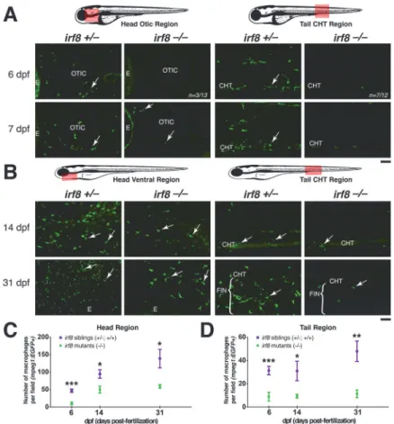
Fig 4. Partial recovery of macrophages inirf8mutants begins by ~7 dpf in the head region.Diagrams
of zebrafish showing region of quantification (pink box), which was taken using the same magnification and field size of view at all stages. The field of view in older stages covers a relatively smaller region of the head because the fish are larger (comparing B with A). Red dotted box shows the region of the fluorescent images. (A) Expression of macrophage reportermpeg1:EGFPat 6 and 7 dpf. Arrows show macrophages. At 6 dpf, mostirf8mutants have no macrophages in the CHT (n = 7/12), while most have a few macrophages in the head (n = 10/13); image shows the head region of a mutant at 6 dpf with no macrophages. Panels show the tail CHT region of theirf8mutants at 6 and 7 dpf with no macrophages; a few autofluorescent pigment cells are present after PTU treatment. (B) Distribution of macrophages expressingmpeg1:EGFPat 14 dpf and 31 dpf. Arrows point to macrophages. (C) Quantification of the number of macrophages per field in the head region over time (pink box in left diagram). (D) Quantification of the number of macrophages per field in the tail region over time (pink box in right diagram). At 6 dpf, n = 20 for siblings and n = 13 for mutants; at 14 dpf, n = 8 for siblings and n = 7 for mutants; at 31 dpf, n = 7 for siblings and n = 5 for mutants. Statistical significance was determined by two-tailed Student’s t-test. Error bars represent S.E.M.***, p<0.001;
**, p<0.01;*, p<0.05; CHT, caudal hematopoietic tissue; E, eye. All scale bars are 50 um.irf8-/-in this figure represents trans-heterozygousirf8st95/st96mutants.
proliferation of immature myeloid cells [9,12,24]. Alternatively, these small cells in the mu-tants may be dying myeloid cells or cell debris.
By 5 dpf, a few strongly labeledmpeg1-expressing macrophages were present in some ho-mozygousirf8mutants, but other mutants appeared to have no macrophages (Figs.3Band4). At this time, the mutants also showed significantly increased cell death in the CHT region (Fig. 3C and D), evidenced by an increase of very small TUNEL-positive cells (Fig. 3E). In addi-tion, we detected aberrant, small-sized or rounded cells and fragments expressing thepu.1 re-porter transgene in all mutants analyzed but not in the siblings (Fig. 3C–E), which is consistent with myeloid precursor cell death inirf8mutants. In summary,irf8mutants have abnormally small cells that weakly expressmpeg1:EGFP, and they have significantly increased cell death in thepu.1+ myeloid precursors.
By 7dpf,irf8mutants contained macrophages strongly expressingmpeg1:EGFPin the head and thymic region near the otic vesicle (Fig. 4A). These first-appearing macrophages in theirf8
mutants appear less mature than wildtype, being rounded with few or no processes (Fig. 4A). In the ventral head at 14 dpf and 31 dpf (Fig. 4B), the number of macrophages increased inirf8
mutants, but it remained less than half of that in wildtype and heterozygous siblings in the head and tail regions (Fig. 4C and D).
In our time course analysis ofmpeg1:EGFPexpression at different stages, it was clear that the number ofmpeg1:EGFPexpressing macrophages was more strongly reduced in the tail than in the head (Fig. 4C and D). The increased number of macrophages in the ventral head re-gion may reflect proximity to the pronephric kidney, which becomes the predominant site of blood formation starting at about the same time that the first macrophages became evident in
irf8mutants. The absence of macrophages inirf8mutants before 5–6 dpf indicates thatirf8is a key regulator of all macrophage populations in the embryo and early larva. The recovery that begins in mid-larval stages at ~7 dpf suggests that later precursor cells, such as the hematopoi-etic stem cells (HSCs) from the kidney marrow, may depend on a combination ofirf8and other factors. Despite this transition,irf8is essential to achieve normal macrophage numbers over the long-term, as the recovery of macrophages inirf8mutants is incomplete.
irf8
mutants recover macrophages but apparently lack microglia at
31 dpf
The presence of macrophages in the head ofirf8mutants at 14 dpf and later stages prompted us to determine whether microglia are also present at these stages. We examined microglia in sections of the midbrain at 31 dpf using antibodies against leukocyte marker L-plastin, which labels microglia in the uninjured adult zebrafish brain [25]. At 31 dpf, wildtype fish contained many cells expressing L-plastin with the elaborate morphologies characteristic of microglial cells (S2 Fig.). Inirf8mutants, we were unable to identify similar cells in the brain parenchyma, but instead observed only a few putative macrophages that were located in the ventricular zone, adjacent to vasculature or in the interstitial space between the brain and the head epithe-lium (S2 Fig.). These results suggest that microglia are absent inirf8mutants at stages up to 31 dpf, several weeks after other macrophages were evident inirf8mutants. Our analysis is consis-tent with previous studies showing that microglia originate from primitive macrophages at early stages but not from later-forming definitive macrophages or monocytes [26,27].
irf8
mutants have myeloid cells and neutrophils competent to become
macrophages after expression of wildtype
irf8
undifferentiated and dying myeloid cells (Fig. 3) in addition to increased neutrophils. To inves-tigate the possibilities thatirf8can regulate the specification and survival of macrophage pre-cursors and the fate decisions of neutrophil-macrophage prepre-cursors, we sought to restore macrophages inirf8mutants by expression of wildtypeirf8in the macrophage and neutrophil lineages, respectively. Regulatory sequences formpeg1,lyz,huc, andkrt4, as previously de-scribed [21], were used to expressirf8in macrophages, neutrophils, neurons, and skin cells, re-spectively (Fig. 5). These constructs also contain thecmlc2-GFPcassette to allow the analysis of animals with successful integration, as detected by strong GFP expression in the heart. We as-sessed microglial cells by neutral red staining at 4 dpf and the total macrophage population with the markermfap4at 2.5 dpf (Fig. 5). We analyzed the action of the transgenes in embryos
Fig 5. Specific expression ofirf8in macrophage or neutrophil lineage is sufficient to restore
macrophage fates inirf8mutants.(A) Neutral red staining for microglia at 4 dpf inirf8siblings and mutants
in control uninjected conditions or after tol2-mediated expression ofirf8driven by tissue specific regulatory sequences. Expression ofmpeg1:irf8orlyz:irf8was sufficient to restore some microglia inirf8mutants (arrows), but not expression in the neurons (huc) or skin (krt4). (B) Analysis of total macrophage population bymfap4RNA expression at 2.5 dpf after expression ofirf8in different tissues. Specific expression of
mpeg1:irf8orlyz:irf8was also sufficient to restore macrophages on yolk sac and embryo proper (arrows) in
irf8mutants. (C) Plot showing frequency of microglia rescue. (D) Plot showing frequency of macrophage recovery. Purple denotes>25 macrophages and green shows partial recovery of less than 25 but more than 1. Numbers below bar graphs representn, total number of embryos analyzed. sib,irf8+/+andirf8+/-; mut,
irf8-/-.
fromirf8heterozygous intercrosses in order to evaluate the effect ofirf8expression in mutant and wildtype siblings.
When wildtypeirf8expression was introduced transiently under the control of macro-phage-specific regulatory sequences formpeg1, we detected some recovery of macrophages by 2.5 dpf and microglia by 4 dpf inirf8mutants, which normally lack all macrophages and microglia at these stages (Fig. 5). The ability to rescue macrophages inirf8mutants using the
mpeg1:irf8transgene provides further evidence that the cells weakly expressing thempeg1 re-porter inirf8mutants are immature cells competent to become mature macrophages when wildtypeirf8is expressed.
To test the other possibility thatirf8can restore macrophage fate in the expanded neutro-phils inirf8mutants, we transiently expressed the wildtypeirf8coding sequence in neutrophils. Strikingly, ectopic expression ofirf8in neutrophils usinglyzregulatory sequences led to a par-tial recovery of microglia and macrophages in theirf8-/-mutants (Fig. 5). The rescue of macro-phages with thempeg1:irf8andlyz:irf8constructs appeared specific to the macrophage and neutrophil lineages, because the transgenes driving expression in skin and neurons did not res-cue macrophages or microglia inirf8mutants (Fig. 5). Interestingly, none of theirf8expression constructs appeared to affect the overall number and distribution of macrophages and micro-glia in wildtype andirf8heterozygotes (Fig. 5and data not shown). These experiments suggest that both immature myeloid and neutrophil populations inirf8mutants are capable of devel-oping into mature macrophages after expressing wildtypeirf8.
Discussion
Our study reveals that specification of all macrophages requiresirf8during a discrete early pe-riod of zebrafish hematopoiesis that includes the primitive and transient definitive waves in zebrafish (Fig. 6). The macrophage deficiency and overproduction of neutrophils and progeni-tor cells in zebrafishirf8mutants are similar to the phenotypes of humans and mice carrying loss-of-function mutations in Irf8 [10–12,30]. Also similar to Irf8 knockout mice and human patients homozygous for inactivating mutations of Irf8 [11,12], zebrafishirf8null mutants were viable. Zebrafishirf8mutants thereby provide a new model to understand the diverse roles ofirf8in development and innate immunity that will likely be relevant to mammalian Irf8.
Our analysis underscores different genetic controls of hematopoiesis at early and late stages. Despite the complete absence of macrophages inirf8mutants at early stages, some mature macrophages were present inirf8mutants by mid-larval stages. The recovery of macrophages inirf8mutants is more prominent in the head than in the tail through one month of age. This suggests that the late-emerging macrophages inirf8mutants may be derived from hematopoi-etic stem cells (HSCs) in the developing kidney, which is located near the ventral head. The dif-ferent origins of the macrophages at difdif-ferent stages (primitive myeloid cells early and HSCs later) may explain their differential dependence onirf8(Fig. 6). Kidney-derived HSCs may uti-lize a combination ofirf8and other factors to specify macrophages, whereas the early myeloid progenitors in the embryo all requireirf8to develop as macrophages, possibly reflecting a more homogenous population that requires the same genetic program for macrophage differ-entiation at early stages.
Our data in combination with the analysis ofirf8morphants [17] provide evidence thatirf8
granulocytes [13]. We show that driving expression ofirf8in the macrophage and neutrophil lineages inirf8mutants is sufficient to restore macrophage fate. Similarly, in mouse, induced Irf8 expression in Irf8-/-myeloid progenitorsin vitroallowed their differentiation into mature macrophages [10]. Althoughirf8expression in neutrophils does restore some macrophages in
irf8mutants, many excess neutrophils were not converted to macrophages. This likely reflects, in part, the mosaic expression ofirf8in these assays, but it is also possible that some of the ex-cess neutrophils arise as a secondary consequence of the loss of macrophages, increased mye-loid cell death, or both, and not from alterations of progenitor cell fates. Thus, some excess neutrophils may not be competent to convert to macrophages. In mammals, macrophage de-pletion can cause neutrophilia, possibly due to defective clearance of apoptotic cells after mac-rophage loss [31], and wounds depleted of macrophages are populated by large numbers of neutrophils [32].
The conservation of Irf8 in macrophage/monocyte specification seems clear, although the extent of the Irf8 requirement appears more far-reaching in zebrafish than in mammals. All macrophages and microglia are eliminated in theirf8mutant zebrafish embryo, whereasIrf8
deficient mice lose a subset of macrophages/monocytes and dendritic cells at embryonic and adult stages [14,33–36]. This includes a severe but incomplete reduction of the yolksac c-kit+ erthryomyeloid precursors that give rise to microglia [27]. Mouse Irf8 mutants also retain var-ied levels of other tissue macrophages including Langerhans cells [36]. At the time of macro-phage recovery, zebrafishirf8mutants do not appear to lack specific subpopulations of
Fig 6. Summary model for role ofirf8in macrophage ontogeny in zebrafish.irf8is required for development of all macrophages in the early
developmental period until 5–6 dpf (solid orange bar). Coinciding with the onset of the adult phase hematopoiesis in the kidney starting at ~7 dpf, hematopoiesis relies onirf8and other factors for macrophage specification and differentiation (dotted orange bar). RBI, rostral blood island; PBI, posterior blood island; CHT, caudal hematopoietic tissue; AGM, aorta-gonad-mesonephros analog; hpf, hours postfertilization; dpf, days postfertilization.
macrophages based on anatomical distribution, with the apparent exception of microglia. However, we cannot exclude the possibility that microglia arise at later stages, or that other subsets of macrophage/monocyte are lacking in zebrafishirf8mutants.
In summary, we show that Irf8 is essential for the development of all macrophage fates through the early waves of zebrafish hematopoiesis (primitive and transient definitive), but not adult-phase hematopoiesis (Fig. 6). Although the nature of its function changes over time, Irf8 is continuously required to allow normal production of macrophages in zebrafish. Besides its role in myeloid development, Irf8 has essential roles in host defense [37–39]. The generation of zebrafishirf8mutants thus provides a newin vivomodel to dissect the different functions of Irf8 in hematopoiesis and host defense.
Materials and Methods
Zebrafish lines and embryos
Embryos from wildtype (TL, AB/TU, and WIK), transgenic (Tg(lyz:EGFP)[28],Tg(mpeg1:
EGFP)[29], andTg(pu.1:Gal4-UAS-EGFP)[22]), andirf8st95andirf8st96heterozygous intercross backgrounds were raised at 28.5°C, and staged by established standards [40]. Embryos were treated with 0.003% 1-phenyl-2-thiourea (PTU) in methylene blue embryo water to inhibit pig-mentation. For in situ hybridization and TUNEL staining, zebrafish embryos and larvae were fixed immediately at the indicated time points of analysis using 4% paraformaldehyde/PBS for overnight fixation at 4°C. For fluorescent imaging, zebrafish embryos and larvae up to 7 dpf were imaged in the living animals at the time of analysis, and older larvae up to 31 dpf were im-aged after fixation; they were briefly anesthetized using 0.02% MS-222 (tricaine) prior to over-night fixation in 4% paraformaldehyde/PBS. All euthanasia and procedures followed the protocols approved by the Stanford Institutional Animal Care and Use Committee.
TALEN-targeting to create
irf8
mutations
A pair of transcription activator-like effector nucleases (TALENs) was designed to target a 50
region ofirf8near the translational start site that encompasses a unique AvaI restriction site (seeFig. 1A). The web tool TAL Effector-Nucleotide Targeter 2.0 (TALE-NT 2.0; https://tale-nt.cac.cornell.edu) was used to design the TALENs. Recognition sequences for the left and
right TALENs are, respectively: 50-TGAAGTAAAGGTCTACAAGA-30and 50
-TATAAGC-CACTGTTTCAGTC-30
. The PCR/Golden Gate cloning protocol for creating the TALENs was used [41]. 400–800 pg of TALE nuclease mRNA (transcribed by Sp6 mMessage mMachine Kit, Ambion) was injected into 1–2 cell stage embryos and raised at 28.5°C. To identify founders, injected fish were crossed and a subset of the F1 progeny was assayed for the presence of a mu-tation in theirf8locus based on disruption of the AvaI site; when a mutation was identified the remaining progeny were raised to adulthood. Individual F1 adults carrying anirf8mutation disrupting the AvaI site were further analyzed by sequencing, which enabled identification of two specific alleles,st95andst96, that contain frameshift mutations. To genotypest95andst96, a PCR fragment ofirf8exon1 was amplified using the following primers: 50
-ACATAAGGCG-TAGAGATTGGACG-30and 50-GAAACATAGTGCGGTCCTCATCC-30, followed by a digest
with AvaI on the PCR product.
Neutral red assay
followed by 1–2 water changes, and then analyzed 0.5–24 hours later using a dissecting microscope.
Whole mount RNA in situ hybridization, TUNEL assay and
immunostaining
In situ hybridization on whole zebrafish embryos and larvae from 20 hpf to 3 dpf was per-formed using standard methods. Antisense riboprobes used were:lyz,mfap4, andapoeb(or
apoe), as described [21]. TUNEL assay (In situ Cell Death Detection Kit, TMR Red, Roche) was performed as previously described [21]. Subsequent immunostaining was performed using the anti-L-plastin (LCP) antibody [42] at 1:250–500 dilution, followed by DAPI staining. For the cell death analysis, 5 dpf larvae fromirf8heterozygous intercrosses were processed for TUNEL and L-plastin immunostaining, and genotyped. Images of the sibling and mutant larvae were taken from the same anatomical region caudal to the end of yolk extension on an upright Zeiss Axio Imager.M2 microscope using the 20x (NA 0.8) objective. Most TUNEL signals were con-centrated in the CHT of the larval tail; the images were cropped to analyze TUNEL signals only in the CHT using ImageJ. The anatomical region is illustrated inFig. 3diagram. Similar thresh-old levels were set for all images and adjusted to accurately reflect the TUNEL signals to mea-sure area of the nuclei labeled by TUNEL (Fig. 3C,D). Representative tracing of the TUNEL signals to determine area of each labeled nucleus is shown inFig. 3C,Dbelow the actual fluo-rescent image of the TUNEL staining. Total number and number of small and other sized TUNEL-positive cells were quantified.
Time-lapse and fluorescent imaging
For live imaging, embryos were embedded in 1.5% low melting point agarose on glass slides. Fluorescent images were taken on a spinning-disk confocal microscope (Perkin Elmer Ultra-VIEW VoX imaging system using the Zeiss Axio Observer Z.1 microscope base with the Yoko-gawa CSU-X1 scanner unit and Hamamatsu EM-CCD C9100–13 camera), unless noted otherwise. The Volocity Acquisition suite was used for 3-dimensional multichannel recording. The following objectives were used: EC Plan-NeoFluar 10x/0.3 Ph1 DIC, Plan-Apochromat 25x/0.8W, and C-Aprochromat 63x/1.2W Korr. Images were analyzed using the Volocity soft-ware and ImageJ, and processed using Adobe Photoshop CS5. Static images of live and fixed larvae for analysis of macrophage numbers inFig. 4were taken on a Zeiss LSM 5 Pascal confo-cal microscope using the 20x (NA 0.75) objective. ImageJ Cell Counter tool was used to count the number of cells.
Cryosectioning for brain microglia analysis
Larvae were fixed overnight in 4% paraformaldehyde/PBS and washed three times in PBS with 0.1% tween. Post-fixed larvae were equilibrated to 30% sucrose, embedded in O.C.T. medium, and snap frozen in dry ice-ethanol bath. Samples were sectioned at 12–14μm in the head re-gion, and subsequently imaged on an upright Zeiss Axio Imager.M2 microscope using the 10x/ 0.45 and 40x/0.95 objectives.
Expression constructs and embryo injection
for selection. Each plasmid expressing a transgene was co-injected at 12–25 pg along with 50–100 pg of Tol2 transposase mRNA (transcribed by Sp6 mMessage mMachine Kit, Ambion) at 1–4 cell stage. Injected embryos were selected for further analysis based on strong expression of GFP in the heart by the cardiac reportercmlc2:EGFP.
Supporting Information
S1 Fig. Kaplan-Meier plot showing survival of homozygousirf8mutant zebrafish into adulthood past 3 months of age.A survival study of the progeny from heterozygousirf8 inter-cross showed survival of all wildtypeirf8+/+progenies (n = 10/10), nearly all heterozygous
irf8+/-fish (n = 16/17), and some homozygousirf8-/-mutants (n = 3/5) at 100 dpf. The geno-types of the fish were determined by fin clip assay at 30 dpf, and survival was monitored there-after, until 100 dpf. Between these stages, 60% of the homozygous mutants survived in this analysis.
(TIFF)
S2 Fig. Thin brain sections reveal many microglial cells in wildtype but not inirf8mutants, which appear to have only macrophages at 31 dpf.(A) The midbrain optic tectum, where many microglia normally reside, was the region of analysis. (B) Higher magnification of the brain region indicated in dotted box in A. 12–14 um cryosections were taken from wildtype
irf8+/+and heterozygousirf8st95/+fish (n = 4) to compare withirf8st95/st9mutants (n = 3) at 31 dpf. Immunostaining for L-plasin (with DAPI as a counterstain) was used to identify macro-phages in relation to all cell bodies in the sections. Microglia were identified by their L-plastin expression, elaborate morphology with fine processes, and location in the parenchyma (white arrows). Cells in or adjacent to the interstitial space, vasculature, and ventricular zone were likely macrophages. Autofluorescence in the green channel helped to define the vasculature and distinguish actual L-plastin signal from vasculature autofluorescence in the red channel.
irf8mutants had a few L-plastin positive cells in the interstitial region and near the ventricular zone that lack fine processes (blue arrows), suggesting that these cells were not microglia. OT, midbrain optic tectum; DC, diencephalon. All scale bars are 50 um.
(TIFF)
Acknowledgments
We are grateful to M. Barna and S. Kim for sharing microscopes. We thank D. Lyons and H. Sidik for critical reading of the manuscript, members of our laboratory for insightful discus-sions, and T. Reyes and C. Hill for fish care.
Author Contributions
Conceived and designed the experiments: CES WST. Performed the experiments: CES ZK AM. Analyzed the data: CES AM WST. Wrote the paper: CES AM WST.
References
1. Bennett CM, Kanki JP, Rhodes J, Liu TX, Paw BH, et al. (2001) Myelopoiesis in the zebrafish, Danio rerio. Blood 98: 643–651. PMID:11468162
2. Richards DM, Hettinger J, Feuerer M (2013) Monocytes and macrophages in cancer: development and functions. Cancer Microenviron 6: 179–191. doi:10.1007/s12307-012-0123-xPMID:23179263
4. Rosenbauer F, Tenen DG (2007) Transcription factors in myeloid development: balancing differentia-tion with transformadifferentia-tion. Nat Rev Immunol 7: 105–117. PMID:17259967
5. Stachura DL, Traver D (2011) Cellular dissection of zebrafish hematopoiesis. Methods Cell Biol 101: 75–110. doi:10.1016/B978-0-12-387036-0.00004-9PMID:21550440
6. Chen AT, Zon LI (2009) Zebrafish blood stem cells. J Cell Biochem 108: 35–42. doi:10.1002/jcb.22251 PMID:19565566
7. Terry RL, Miller SD (2014) Molecular control of monocyte development. Cell Immunol. doi:10.1016/j. cellimm.2014.12.004PMID:25594139
8. Taniguchi T, Ogasawara K, Takaoka A, Tanaka N (2001) IRF family of transcription factors as regula-tors of host defense. Annu Rev Immunol 19: 623–655. PMID:11244049
9. Scheller M, Foerster J, Heyworth CM, Waring JF, Lohler J, et al. (1999) Altered development and cyto-kine responses of myeloid progenitors in the absence of transcription factor, interferon consensus se-quence binding protein. Blood 94: 3764–3771. PMID:10572090
10. Tamura T, Ozato K (2002) ICSBP/IRF-8: its regulatory roles in the development of myeloid cells. J Inter-feron Cytokine Res 22: 145–152. PMID:11846985
11. Hambleton S, Salem S, Bustamante J, Bigley V, Boisson-Dupuis S, et al. (2011) IRF8 mutations and human dendritic-cell immunodeficiency. N Engl J Med 365: 127–138. doi:10.1056/NEJMoa1100066 PMID:21524210
12. Holtschke T, Lohler J, Kanno Y, Fehr T, Giese N, et al. (1996) Immunodeficiency and chronic myeloge-nous leukemia-like syndrome in mice with a targeted mutation of the ICSBP gene. Cell 87: 307–317. PMID:8861914
13. Wang H, Yan M, Sun J, Jain S, Yoshimi R, et al. (2014) A Reporter Mouse Reveals Lineage-Specific and Heterogeneous Expression of IRF8 during Lymphoid and Myeloid Cell Differentiation. J Immunol. PMID:25552543
14. Aliberti J, Schulz O, Pennington DJ, Tsujimura H, Reis e Sousa C, et al. (2003) Essential role for ICSBP in the in vivo development of murine CD8alpha + dendritic cells. Blood 101: 305–310. PMID: 12393690
15. Schmidt M, Nagel S, Proba J, Thiede C, Ritter M, et al. (1998) Lack of interferon consensus sequence binding protein (ICSBP) transcripts in human myeloid leukemias. Blood 91: 22–29. PMID:9414265
16. Pogosova-Agadjanyan EL, Kopecky KJ, Ostronoff F, Appelbaum FR, Godwin J, et al. (2013) The prog-nostic significance of IRF8 transcripts in adult patients with acute myeloid leukemia. PLoS One 8: e70812. doi:10.1371/journal.pone.0070812PMID:23967110
17. Li L, Jin H, Xu J, Shi Y, Wen Z (2011) Irf8 regulates macrophage versus neutrophil fate during zebrafish primitive myelopoiesis. Blood 117: 1359–1369. doi:10.1182/blood-2010-06-290700PMID:21079149
18. Rhodes J, Hagen A, Hsu K, Deng M, Liu TX, et al. (2005) Interplay of pu.1 and gata1 determines myelo-erythroid progenitor cell fate in zebrafish. Dev Cell 8: 97–108. PMID:15621533
19. Herbomel P, Thisse B, Thisse C (2001) Zebrafish early macrophages colonize cephalic mesenchyme and developing brain, retina, and epidermis through a M-CSF receptor-dependent invasive process. Dev Biol 238: 274–288. PMID:11784010
20. Svahn AJ, Graeber MB, Ellett F, Lieschke GJ, Rinkwitz S, et al. (2013) Development of ramified micro-glia from early macrophages in the zebrafish optic tectum. Dev Neurobiol 73: 60–71. doi:10.1002/ dneu.22039PMID:22648905
21. Shiau CE, Monk KR, Joo W, Talbot WS (2013) An Anti-inflammatory NOD-like Receptor Is Required for Microglia Development. Cell Rep 5: 1342–1352. doi:10.1016/j.celrep.2013.11.004PMID:24316075
22. Peri F, Nusslein-Volhard C (2008) Live imaging of neuronal degradation by microglia reveals a role for v0-ATPase a1 in phagosomal fusion in vivo. Cell 133: 916–927. doi:10.1016/j.cell.2008.04.037PMID: 18510934
23. Novoa B, Figueras A (2012) Zebrafish: model for the study of inflammation and the innate immune re-sponse to infectious diseases. Adv Exp Med Biol 946: 253–275. doi:10.1007/978-1-4614-0106-3_15 PMID:21948373
24. Tsujimura H, Nagamura-Inoue T, Tamura T, Ozato K (2002) IFN consensus sequence binding protein/ IFN regulatory factor-8 guides bone marrow progenitor cells toward the macrophage lineage. J Immu-nol 169: 1261–1269. PMID:12133947
26. Ginhoux F, Greter M, Leboeuf M, Nandi S, See P, et al. (2010) Fate mapping analysis reveals that adult microglia derive from primitive macrophages. Science 330: 841–845. doi:10.1126/science.1194637 PMID:20966214
27. Kierdorf K, Erny D, Goldmann T, Sander V, Schulz C, et al. (2013) Microglia emerge from erythromye-loid precursors via Pu.1- and Irf8-dependent pathways. Nat Neurosci 16: 273–280. doi:10.1038/nn. 3318PMID:23334579
28. Hall C, Flores MV, Storm T, Crosier K, Crosier P (2007) The zebrafish lysozyme C promoter drives my-eloid-specific expression in transgenic fish. BMC Dev Biol 7: 42. PMID:17477879
29. Ellett F, Pase L, Hayman JW, Andrianopoulos A, Lieschke GJ (2011) mpeg1 promoter transgenes di-rect macrophage-lineage expression in zebrafish. Blood 117: e49–56. doi: 10.1182/blood-2010-10-314120PMID:21084707
30. Wang H, Lee CH, Qi C, Tailor P, Feng J, et al. (2008) IRF8 regulates B-cell lineage specification, com-mitment, and differentiation. Blood 112: 4028–4038. doi:10.1182/blood-2008-01-129049PMID: 18799728
31. Gordy C, Pua H, Sempowski GD, He YW (2011) Regulation of steady-state neutrophil homeostasis by macrophages. Blood 117: 618–629. doi:10.1182/blood-2010-01-265959PMID:20980680
32. Goren I, Allmann N, Yogev N, Schurmann C, Linke A, et al. (2009) A transgenic mouse model of induc-ible macrophage depletion: effects of diphtheria toxin-driven lysozyme M-specific cell lineage ablation on wound inflammatory, angiogenic, and contractive processes. Am J Pathol 175: 132–147. doi:10. 2353/ajpath.2009.081002PMID:19528348
33. Schiavoni G, Mattei F, Sestili P, Borghi P, Venditti M, et al. (2002) ICSBP is essential for the develop-ment of mouse type I interferon-producing cells and for the generation and activation of CD8alpha(+) dendritic cells. J Exp Med 196: 1415–1425. PMID:12461077
34. Tailor P, Tamura T, Morse HC, Ozato K (2008) The BXH2 mutation in IRF8 differentially impairs dendrit-ic cell subset development in the mouse. Blood 111: 1942–1945. PMID:18055870
35. Qi CF, Li Z, Raffeld M, Wang H, Kovalchuk AL, et al. (2009) Differential expression of IRF8 in subsets of macrophages and dendritic cells and effects of IRF8 deficiency on splenic B cell and macrophage compartments. Immunol Res 45: 62–74. doi:10.1007/s12026-008-8032-2PMID:18663414
36. Schiavoni G, Mattei F, Borghi P, Sestili P, Venditti M, et al. (2004) ICSBP is critically involved in the nor-mal development and trafficking of Langerhans cells and dernor-mal dendritic cells. Blood 103: 2221–2228. PMID:14615368
37. Turcotte K, Gauthier S, Malo D, Tam M, Stevenson MM, et al. (2007) Icsbp1/IRF-8 is required for innate and adaptive immune responses against intracellular pathogens. J Immunol 179: 2467–2476. PMID: 17675508
38. Marquis JF, LaCourse R, Ryan L, North RJ, Gros P (2009) Disseminated and rapidly fatal tuberculosis in mice bearing a defective allele at IFN regulatory factor 8. J Immunol 182: 3008–3015. doi:10.4049/ jimmunol.0800680PMID:19234196
39. Yoshida Y, Yoshimi R, Yoshii H, Kim D, Dey A, et al. (2014) The transcription factor IRF8 activates integrin-mediated TGF-beta signaling and promotes neuroinflammation. Immunity 40: 187–198. doi: 10.1016/j.immuni.2013.11.022PMID:24485804
40. Kimmel CB, Ballard WW, Kimmel SR, Ullmann B, Schilling TF (1995) Stages of embryonic develop-ment of the zebrafish. Dev Dyn 203: 253–310. PMID:8589427
41. Sanjana NE, Cong L, Zhou Y, Cunniff MM, Feng G, et al. (2012) A transcription activator-like effector toolbox for genome engineering. Nat Protoc 7: 171–192. doi:10.1038/nprot.2011.431PMID:22222791
42. Redd MJ, Kelly G, Dunn G, Way M, Martin P (2006) Imaging macrophage chemotaxis in vivo: studies of microtubule function in zebrafish wound inflammation. Cell Motil Cytoskeleton 63: 415–422. PMID: 16671106