http://dx.doi.org/10.1590/S1984-46702015000100004
Since thoracic limbs are not only used in locomotion, but also in prey capture and grooming and mating behavior, their morphology can be a good predictor of numerous eco-logical variables, such as the size and kind of prey, the variety of movements, the role in supporting body mass (ANDERSSON
2004a, FABRE et al. 2013a) and the habitat (DAVIS 1964, EWER et al. 1973, TAYLOR 1989, POLLY 2007, MEACHEN-SAMUELS & VAN
-VALKENBURGH 2009, FABRE et al. 2013a, et al. 2013b, MELORO et al.
2013, MARTÍN-SERRA et al. 2014). Together with cranio-dental
data, data on thoracic limbs are also used to extrapolate the predatory behavior of extinct species (IWANIUK et al. 1999, ANDERSSON & WERDELIN 2003). Most ecomorphology studies have prioritized the osteological characteristics of the humerus, whereas the shape and other features of the radio-ulnar joint remain largely unstudied (FABRE et al. 2013a, b, 2014).
More-over, the muscular arrangement is rarely taken into account. Knowledge about muscular disposition associated with some biomechanical findings can better elucidate the function of some important bone structures that would otherwise be ne-glected (JULIK et al. 2012). Several studies in this field have
bi-ases because of misunderstanding of the muscular topography. Thus, the addition of quantitative and qualitative data on
muscular anatomy would contribute to improve the capacity to characterize forelimb morphology in the context of loco-motion, grasping ability and dexterity of the species (IWANIUK
et al. 2001, FABRE et al. 2013b). By homology, soft tissue
infor-mation from extant species can help making well-founded or even speculative inferences about extinct species (WITMER 1995). The musculoskeletal system forms an arrangement based on levers in which the joints act as fulcra (HERMANSON 2013).
The mechanical benefits of its configuration depend on the positions of the muscle attachments (relative to the fulcrum) and the usage of the load. A muscle attached close to a ful-crum is less powerful than a comparable muscle inserted at a greater distance, although the former produces its effects faster. This reflects a conflict between the requirements of speed and power (DYCE et al. 2010).
The brachioradialis muscle (formerly called the supina-tor longus) usually consists of a narrow muscular band situ-ated at the flexor angle of the humerus-radius-ulna joint (Fig. 1) (BUDRAS et al. 2012, HERMANSON 2013). It is positioned between
the superficial and deep layers of antebrachial fascia and ad-heres to the surface of the deep fascia’s leaflet (MILLS 2003, DYCE
et al. 2010, HERMANSON 2013), together with the cephalic vein
Occurrence and morphometrics of the brachioradialis muscle in wild
carnivorans (Carnivora: Caniformia, Feliformia)
Paulo de Souza Junior
1,4, Lucas M.R.P. dos Santos
1,
Daniele M.P. Nogueira
1,
Marcelo Abidu-Figueiredo
2& André L.Q. Santos
31Laboratório de Anatomia Animal, Universidade Federal do Pampa. Rodovia BR-472, km 585, Caixa postal 118, 97500-970 Uruguaiana, RS, Brazil.
2Departamento de Biologia Animal, Universidade Federal Rural do Rio de Janeiro. Rodovia BR-465, km 07, 23890-000 Seropédica, RJ, Brazil.
3Laboratório de Ensino e Pesquisa em Animais Silvestres, Universidade Federal de Uberlândia. Avenida Amazonas 2245, 38405-302 Uberlândia, MG, Brazil.
4Corresponding author. E-mail: paulosouza@unipampa.edu.br
and the superficial branch of the radial nerve (SAINT CLAIR 1986,
SEBASTIANI & FISHBECK 2005), therefore being the most cranial
and superficial muscle of the craniolateral group of the fore-arm (BOHENSKY 2002, SEBASTIANI & FISHBECK 2005).
In domestic carnivorans, this muscle has its origin at the proximal extremity of the humeral lateral supracondylar crest, immediately proximal and superficial to the extensor carpi radialis muscle (SCHWARZE 1984, MILLS 2003, LIEBICH et al. 2011,
HERMANSON 2013). The muscle extends cranially over the proxi-mal part of the extensor carpi radialis muscle, crosses the fore-arm medially, and extends distally in the groove between the extensor carpi radialis muscle and the radius (SCHWARZE 1984,
LIEBICH et al. 2011, HERMANSON 2013). It ends in the periosteum of the radius at the level of the third or fourth distal parts, by a thin aponeurosis (BOHENSKY 2002, MILLS 2003, BUDRAS et al.
2012, HERMANSON 2013). Some authors describe its insertion into
the styloid process of the radius (LEACH 1976, SEBASTIAN & FISHBECK
2005, LIEBICH et al. 2011, MOORE et al. 2013, ERCOLI et al. 2014). The function of the brachioradialis muscle is to perform the craniolateral rotation of the radius (supination) (BOHENSKY
2002, SEBASTIANI & FISHBECK 2005, HERMANSON 2013).
Supination is a movement of flipping the distal radius over the distal ulna, rotating the radius craniolaterally around its long axis (ANDERSSON 2004b). The movement starts with the
contraction of forearm muscles (supinator and brachioradialis), transmitting external rotation also to the manus. Cursorial mammals often have restricted pronation-supination, whereas scansorial mammals can usually completely supinate the manus (POLLY 2007). Thus, the brachioradialis functionally belongs to
the group of muscles that act on the radio-ulnar joint, and is expected to be well developed only in carnivorans. In domes-tic ungulates, this muscle is vestigial or absent due to the re-duced or lost capacity of movement between these two bones (POLLY 2007, LIEBICH et al. 2011).
In domestic dogs, the occurrence and antimeric distri-bution of the brachioradialis muscle has been well documented by WAKURI & KANO (1966), SANTOS JUNIOR et al. (2002), and PESTANA
et al. (2009). There are no reports in the literature, as far as we know, about the occurrence of the brachioradialis muscle in wild carnivorans, although there are studies with variable con-texts containing references to the presence of the muscle in Carnivora (DAVIS 1964, ARLAMOWSKA-PALIDER 1970, JULIK et al. 2012, SÁNCHEZ et al. 2013, ERCOLI et al. 2014).
Carnivorans form a successful and functionally diverse clade, with close to 300 living species (EWER 1973, WILSON &
MITTERMEIER 2009, HUNTER 2011). Despite this diversity, the ac-cumulated knowledge of myological variation within the or-der is still incomplete (MACALISTER 1873a, MACKINTOSH 1875,
WINDLE & PARSONS 1897, HALL 1926, 1927, HOWARD 1973, LEACH
1976, FISCHER et al. 2009, MOORE et al. 2013, ERCOLI et al. 2014).
Hence, these animals provide a good model for this study, as they represent one of the most successful cases of repeated and independent evolution of similar morphologies in a great range of ecologies (ANDERSSON 2004a, b, 2005, NOWAK 2005, SATO et al.
2009, 2012, SLATER et al. 2012, FABRE et al. 2013a, 2014, SAMUELS
et al. 2013, MARTÍN-SERRA et al. 2014). The locomotor range of
movements of carnivorans includes, to varying extents, climb-ing, diggclimb-ing, running and swimming (ANDERSSON & WERDELIN
2003). Furthermore, carnivorans species show different degrees of supination and some species cannot even use the forelimbs for grappling with or handling prey (EWER 1973, ANDERSSON & WERDELIN 2003). We hypothesized that the life style of carnivorans, including cursoriality and food procurement strat-egies, should be reflected in changes in brachioradialis muscle arrangement. This muscle is expected to be frequently found and to be relatively larger in species that need to rotate the forelimbs. Thereby, the aim of this study is to verify the occur-rence, anatomo-functional arrangement and sexual dimor-phism of the brachioradialis muscle in wild carnivoran species, thus contributing to studies in ecomorphology.
MATERIAL AND METHODS
This study was carried out with 29 carnivorans cadavers of Canidae: Cerdocyon thous (Linnaeus, 1766) (three males and six females) and Lycalopex gymnocercus (G. Fischer, 1814) (four males and one female); Mustelidae: Galictis cuja (Molina, 1782)
1 2
(three males and two females) and Lontra longicaudis (Olfers, 1818) (one female); Procyonidae: Procyon cancrivorus (G.[Baron] Cuvier, 1798) (one male and two females) and Nasua nasua
(Linnaeus, 1766) (one female); and Felidae: Leopardus geoffroyi
(d’Orbigny & Gervais, 1844) (four females) and Leopardus colocolo (Molina, 1782) (one female). These specimens were collected dead from highways in the southwest region of the state of Rio Grande do Sul (Pampa biome) between July 2012 and November 2013 (IBAMA/SISBIO authorization number 33667-1). Only adult individuals were included, based on in-spection of permanent dentition.
After collection, the specimens were fixed in a formalde-hyde solution (50%) and conserved in opaque polyethylene tanks with the same solution at 10% for at least 14 days, until they were dissected. The skin and fascia of the thoracic limbs were carefully removed and the superficial intrinsic muscles identi-fied. The brachioradialis muscle, whenever present, was dissected until its origin and insertion were exposed. Then measurements were taken (Fig. 2) of the length of the brachioradialis muscle from its origin until its insertion (ML) and the length of the forearm from the olecranon tuberosity until the radiocarpian joint (FL). Also, lateral-medial width of the brachioradialis muscle (MW) and the lateral-medial diameter of the forearm (FD) were obtained at the level of their middle thirds. The measurements were performed by a single examiner using a digital pachymeter (resolution 0.01mm, accuracy ±0.02 mm, ZAAS Precision Amatools®). Thereafter, two ratios were calculated: MW/FD and ML/FL. The MW/FD ratio was calculated to reflect the relative proportion of the functional participation of the brachioradialis muscle in the forearm region of the specimens. The ML/FL ratio represents the proportion of muscle length in relation to the forearm length and can be associated with added speed during contraction. The 29 specimens are deposited in the Laboratory of Animal Anatomy of the Universidade Federal do Pampa, Uruguaiana, RS, Brazil. The deposit numbers of the specimens analyzed are available in the appendix.
Descriptive statistical data (mean, standard deviation, vari-ance and coefficient of variation) were calculated. The ratios were compared among species and families using analysis of variance (one-way ANOVA) and significant differences between the means were determined by using the Tukey test at 99% probability. In species with enough samples for comparison of the ratios bet-ween genders (C. thous, G. cuja and P. cancrivorus), the t-test was performed at 99% probability. These tests were executed by the BioEstat 5.3® program. Photomacrographs were taken with a Sony Cybershot DSC-TF1® camera with 16.1 MP and the images were treated with the Photoscape® v.3.5 software.
RESULTS
Except for two specimens of L. gymnocercus, in which one male only had the muscle on the right antimere and a female only on the left, along with a female of C. thous that did not
have the muscle in any antimere, all the other specimens (26) had the brachioradialis muscle in both antimeres. Therefore, among all 58 thoracic limbs analyzed, 54 presented the muscle (Table I).
The results of the MW/FD and ML/FL ratios grouped by family (Canidae, Mustelidae, Procyonidae and Felidae) are pre-sented in Table II and in Figs. 3 and 4.
In canids, felids and procyonids, the brachioradialis muscle originated on the lateral supracondylar crest of the humerus and its insertion was on the medial surface of the radial distal extremity (medial styloid process) in every specimen analyzed (Figs. 5-12), except one male specimen of L. gymnocercus, in which Figures 3-4. Box-plots showing the mean ± SD of the MW/FD ratio (3) and ML/FL ratio (4) grouped by families of carnivorans analyzed.
3
the right brachioradialis muscle was shortened and joined to the middle third of the extensor carpi radialis muscle. In mustelids, the muscle originated from the caudal surface of the humeral neck and caudomedially to the brachial muscle point of origin, and also was inserted in the styloid process of the radius, being very fleshy in its course. In procyonids, especially in P. cancrivorus, the muscular part was restricted until the middle third of the forearm, where it narrowed into a thin insertion tendon (Fig. 7). In every specimen it was the most superficial muscle in the forearm, transiting near the cephalic vein, and was innervated by branches of the radial nerve.
The t-test (p < 0.01) for comparison of means of the MW/ FD ratios revealed similarity between genders in C. thous (p = 0.2219), G. cuja (p = 0.7273) and P. cancrivorus (p = 0.0986). The same test for comparison of means of the ML/FL ratios revealed similarity between genders in C. thous (p = 0.4026) and G. cuja
(p = 0.1743) and difference in P. cancrivorus (p = 0.0024).
DISCUSSION
The presence in nearly all the specimens assessed in this study reflects the functional relevance of the brachioradialis muscle to the order Carnivora. Its occurrence is expected in species that require significant mobility in the radius-ulnar joint, especially in external rotation (supination) of the hand
(paw) and forearm, which does not happen, for instance, in ungulates (NICKEL et al. 1986, LIEBICH et al. 2011). According to
SALADIN (2010), the brachioradialis muscle also acts as a
syner-gist in the flexion of the humerus-radio-ulnar joint, but by itself it is not able to generate enough strength because its in-sertion is far from the fulcrum.
Only among canids were individuals identified that did not present the muscle unilaterally or bilaterally (one C. thous
and two L. gymnocercus). Besides this, in one male specimen of L. gymnocercus the brachioradialis muscle was not long enough to be inserted in the radius, joining the extensor carpi radialis muscle in a clearly accessory position. The absence or presence, unilaterally or bilaterally, of the brachioradialis muscle has also been reported in C. familiaris by WAKURI & KANO (1966), SANTOS JUNIOR et al. (2002) and PESTANA et al. (2009).
Considered together, these three studies reveal that the brachioradialis muscle is absent in 46 to 62% of dogs, appear-ing unilaterally in 15 to 20% of cases. In runnappear-ing dogs of the greyhound breed, the muscle was not found in ten individu-als dissected by WILLIAMS et al. (2008). These findings corrobo-rate the well-recognized observation that canids have lost some of the ability to supinate their manus (EWER 1973, ANDERSSON
& WERDELIN 2003). Therefore, in a superficial analysis
consid-ering the canids, the brachioradialis muscle seems to be more frequent in wild ones than in domestic ones, perhaps because
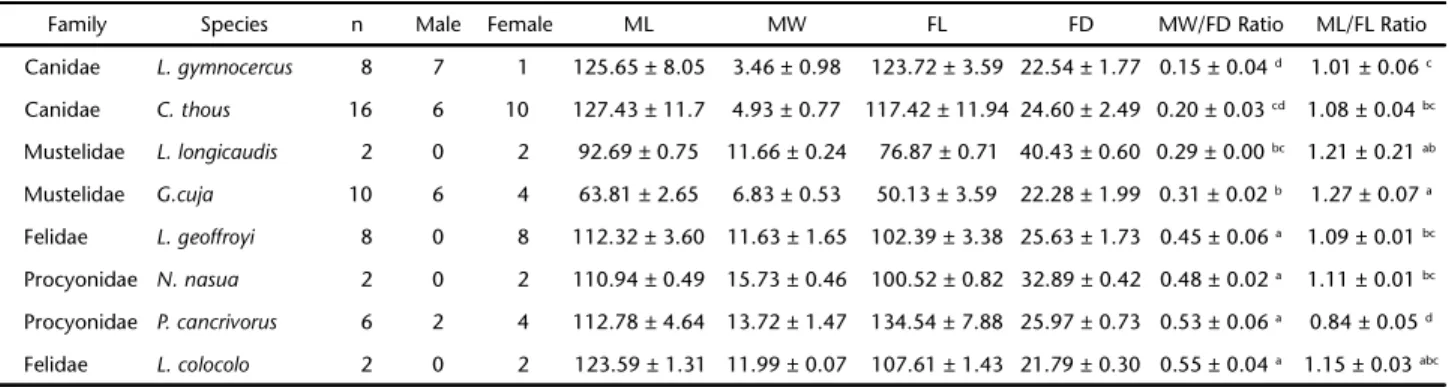
Table I. Means and standard deviations (mm) of the measurements and MW/FD and ML/FL ratios obtained from the forelimbs (n = 54) of carnivorans specimens that presented the brachioradialis muscle.
Family Species n Male Female ML MW FL FD MW/FD Ratio ML/FL Ratio
Canidae L. gymnocercus 8 7 1 125.65 ± 8.05 3.46 ± 0.98 123.72 ± 3.59 22.54 ± 1.77 0.15 ± 0.04 d 1.01 ± 0.06 c
Canidae C. thous 16 6 10 127.43 ± 11.7 4.93 ± 0.77 117.42 ± 11.94 24.60 ± 2.49 0.20 ± 0.03 cd 1.08 ± 0.04 bc
Mustelidae L. longicaudis 2 0 2 92.69 ± 0.75 11.66 ± 0.24 76.87 ± 0.71 40.43 ± 0.60 0.29 ± 0.00 bc 1.21 ± 0.21 ab
Mustelidae G.cuja 10 6 4 63.81 ± 2.65 6.83 ± 0.53 50.13 ± 3.59 22.28 ± 1.99 0.31 ± 0.02 b 1.27 ± 0.07 a
Felidae L. geoffroyi 8 0 8 112.32 ± 3.60 11.63 ± 1.65 102.39 ± 3.38 25.63 ± 1.73 0.45 ± 0.06 a 1.09 ± 0.01 bc
Procyonidae N. nasua 2 0 2 110.94 ± 0.49 15.73 ± 0.46 100.52 ± 0.82 32.89 ± 0.42 0.48 ± 0.02 a 1.11 ± 0.01 bc
Procyonidae P. cancrivorus 6 2 4 112.78 ± 4.64 13.72 ± 1.47 134.54 ± 7.88 25.97 ± 0.73 0.53 ± 0.06 a 0.84 ± 0.05 d
Felidae L. colocolo 2 0 2 123.59 ± 1.31 11.99 ± 0.07 107.61 ± 1.43 21.79 ± 0.30 0.55 ± 0.04 a 1.15 ± 0.03 abc
Values followed by different letters in the same column show statistically significant differences according to the Tukey test (p < 0.01). Brachioradialis muscle length (ML); brachioradialis muscle width (MW); length of the forearm (FL); diameter of the forearm (FD).
Table II. Descriptive statistics regarding the MW/FD and ML/FL ratios obtained for the thoracic limbs (n = 54) of carnivorans specimens grouped by families (SD) Standard deviation, (CV) Coefficient of variation.
Family MW/FD ratio
ML/FL ratio
n Mean Variance SD CV (%) Minimum Maximum n Mean Variance SD CV (%) Minimum Maximum
Procyonidae 8 0.51a 0.0036 0.06 11.62 0.45 0.62 12 1.27 a 0.0050 0.07 5.62 1.19 1.44
Felidae 10 0.47a 0.0048 0.07 14.58 0.38 0.58 10 1.11 b 0.0008 0.02 2.48 1.08 1.17
Mustelidae 12 0.30b 0.0005 0.02 7.44 0.28 0.35 24 1.06 b 0.0031 0.05 5.28 0.88 1.16
Canidae 24 0.18c 0.0020 0.45 24.22 0.11 0.28 8 0.91 c 0.0171 0.13 14.38 0.79 1.11
the former animals retain a greater need to perform supina-tion movements to capture prey. Nevertheless, the unilateral and even bilateral absence of this muscle in some individuals suggests that it may no longer perform a useful function and instead is a rudimentary muscle that is on its way out. The apparent absence of this muscle in greyhound dogs could re-flect the increased intensity of artificial selection to eliminate a useless muscle, since it can interfere with a desirable run-ning ability in wild canids.
After examining some specimens, W.J. Gonyea (unpubl. data) noted that the brachioradialis muscle was present in ar-boreal and fossorial taxa and absent in cursorial ones. In sup-port of this hypothesis, the muscle was not identified in the cursorial canids Canis latrans (Say, 1823) (n = 1), Urocyon cinereoargenteus (Schreber, 1775) (n = 4) and Vulpes vulpes
(Linnaeus, 1758) (n = 5) (S.A. Feeney unpubl. data). The ap-pearance of the brachioradialis muscle in almost all cursorial canids C. thous (n = 8/9) and L. gymnocercus (n = 5/5) analyzed Figures 5-12. Photomacrographs showing the comparative anatomic arrangement of the brachioradialis muscle (white arrow) in the forearm region of: (5) Cerdocyon thous, right forearm; (6) Lycalopex gymnocercus, left forearm; (7) Procyon cancrivorus, right forearm; (8)
Nasua nasua, right of forearm; (9) Leopardus geoffroyi, left forearm; (10) Leopardus colocolo, right forearm; (11) Lontra longicaudis, left forearm; (12) Galictis cuja, left forearm. Scale bars: 20 mm.
5 6
9
7 8
in this study contrasts with previous observation (W.J. Gonyea, unpubl. data). Until there are more investigations, we can pro-pose that the presence of this muscle in canids may be a charac-teristic shared by Neotropical species with close phylogenetic relationship rather than just a morphofunctional issue. Sup-porting this hypothesis, the presence of the brachioradialis muscle was also mentioned by VAZ et al. (2011) in an adult female Atelocynus microtis (Sclater, 1883) and in an adult male
C. thous. Indeed, C. thous, L. gymnocercus and A. microtis in-habit the Neotropics and have the same number of chromo-somes (74) (PESSUTTI et al. 2001), unlike C. latrans, U.
cinereoargenteus and V. vulpes (S.A. Feeney, unpubl. data). Despite disagreement over the cursorial canids, observa-tions of the presence of the muscle in arboreal and fossorial carnivorans are compatible with the findings of this study. In fact, the mustelids, procyonids and felids analyzed here have scansorial, arboreal and fossorial habits (REIS et al. 2010, HUNTER
2011). Mustelids and procyonids in particular can display great ability to perform different kinds of movements such as grasp-ing, swimming and food manipulation (MCCLEARN 1992, IWANIUK
et al. 1999, FABRE et al. 2013b), and their lack of cursorial
adap-tation means that none of them have lost the ability to supi-nate their paws, in contrast to other carnivorans such as canids and some hyaenids (IWANIUK et al. 1999, POLLY 2007, FABRE et al.
2014). Although with different purposes and small samples, some other studies have mentioned the occurrence of this muscle in species of these families, for instance in the mustelids
Aonyx cinerea (Illiger, 1815) (MACALISTER 1873b), Lutra lutra
(Linnaeus, 1758) (WINDLE & PARSONS 1897), Enhydra lutris
(Linnaeus, 1758) (HOWARD 1973), Martes pennanti (Erxleben,
1777) (n = 1) (S.A. Feeney, unpubl. data) and G. cuja (n = 3) (ERCOLI et al. 2014); in the procyonid Procyon lotor (Linnaeus,
1758) (n = 2) (S.A. FEENEY, unpubl. data); and in the felids Puma concolor (Linnaeus, 1771) (n = 2) (CONCHA et al. 2004), Acinonyx
jubatus (Schreber, 1775) (n = 8) (HUDSON et al. 2011), Leopardus
pardalis (Linnaeus, 1758) (n = 1) (JULIK et al. 2012) and Panthera
onca (Linnaeus, 1758) (n = 2) (SÁNCHEZ et al. 2013).
The superficial location in the forearm, the proximity of the cephalic vein and innervation by branches of the radial nerve were common to all specimens analyzed, resembling the description of domestic carnivores (SEBASTIANI & FISHBECK 2005,
BUDRAS et al. 2012).
The MW/FD ratio was calculated to reflect the relative proportion of its participation in the forearm region of the specimens. This way, when comparing species and/or families, those with the highest ratios should be the ones in which the brachioradialis muscle has greater relative contribution through the group of antebrachium muscles. Although calculation of physiological cross-section area (PCSA) has been used to esti-mate the maximum isometric force of muscles (WILLIAMS et al.
2008), in this study we considered this determination to be less important since it generates an absolute value which is highly influenced by the body size of the individuals. For
ex-ample, the brachioradialis of a crab-eating fox (C. thous) would exhibit both higher absolute PCSA and maximum isometric force values than the muscle in a lesser grison (G. cuja). Howe-ver, the muscle is proportionally weaker in the former. Since the brachioradialis has parallel fibers, determination of the ratio between cross section widths (MW) to the forearm diameter would give a more proportional estimate of functional rele-vance, at least in its topographic region (forearm). In fact, the results of MW/FD ratio reflected observations from the usage of the forelimbs in each species.
The significantly lower MW/FD ratio in canids compared to the other three families used in this study can be explained by the fact that canids are essentially terrestrial and have de-veloped a highly specialized lifestyle among carnivorans. In more cursorial taxa, the functional adaptations prioritize, among other aspects, the movements of the limbs in the sagit-tal plane, disfavoring supination or pronation (EWER 1973). In
contrast to felids, canids have a limited ability to subdue and grapple with other animals by using their forelimbs. Instead they engage in sustained pursuit predation, an activity whose success depends on the number of animals participating in the hunt (ANDERSSON 2005). In addition, canids rarely climb or manipulate prey to a higher extent. These habits are corre-lated to a less functional, or even absent, brachioradialis muscle. In the mustelids G. cuja and L. longicaudis the MW/FD ratio, significantly higher only than in the canids, compara-tively reflects greater recruitment of the muscle to help in spe-cific swimming movements and fossorial habits. At times, they also use their thoracic limbs to drag prey out of the water (REIS
et al. 2010).
The felids and procyonids of this study showed the high-est MW/FD ratios, with no differences among them. The reper-toire of manual movements of these two families is more complex because, besides being fast runners, they are also able to swim, climb trees and remain balanced at tall heights (REIS
et al. 2010, HUNTER 2011). Undeniably, scansorial habits and
grasping requires accurate three-dimensional movements, which demand morphofunctional adaptations and higher re-cruitment and precision in muscular contraction (EWER 1973,
FABRE et al. 2013b). Procyonids even use their hands to
pre-cisely bring food into their mouths, requiring a greater capac-ity for supination (PARANAÍBA et al. 2012). Species showing well-developed grasping ability potentially have a wide range of pronation-supination movements, which can confer greater mobility to the forearm and the hand (FABRE et al. 2013b). This
was the case of P. cancrivorus, which exhibited a high MW/FD ratio. In felids, essentially carnivores, rotation movements are even more necessary to capture, overwhelm and manipulate prey extensively (HUDSON et al. 2011).
rota-tion of the distal porrota-tion of the limb. The brachioradialis muscle presents a correlation not only with the forelimb usage in carnivorans. ANDERSSON (2004b) stated that manipulation and locomotion are conflicting functions, since elbow-joint mor-phology supports a division between grapplers (i.e., ambushers) and nongrapplers (i.e., pursuers). Joints of the former are rela-tively wide, while in the latter they are relarela-tively narrow and box-like with pronounced stabilizing features. Concerning fore-paw dexterity, IWANIUK et al. (2001) considered that manus and
carpal shape and myology may play a more critical role than manus proportions. According to them, behavioral observa-tions also suggest that manus proporobserva-tions correlated more closely with locomotion than non-locomotory forepaw usage. The ML/FL ratio represents the proportion of muscle length in relation to the forearm length. This ratio was signifi-cantly higher in mustelids. Longer muscles, especially with par-allel fibers (which is the case of the brachioradialis), have more sarcomeres in series, which means added speed during contrac-tion (KARDONG 2011). Therefore, in the mustelids one can
as-sume that the brachioradialis muscle has a higher shortening speed than in the other families, an aspect that may be func-tionally important during swimming. This relatively longer length is a reflection of a more proximal level of origin in the humerus than in the other families, in other words, on the cau-dal surface of the humeral neck instead of the lateral supracondy-lar crest. Strengthening this hypothesis, in procyonids the ratio was significantly lower, because these animals have the slowest movements among the analyzed families. This shorter length was determined by the fact that its muscular part extends only until the forearm’s middle third. This trait was also verified in two specimens of P. lotor (S.A. Feeney, unpubl. data).
From a mechanical point of view, it can be supposed that the arrangement of the mustelid brachioradialis muscle raises the power leverage, creating a low power ratio, which increases the strength and becomes important in fossorial habits. In fe-lids, in contrast, the origin at a more distal level makes the power leverage lower, creating a higher power ratio, which increases speed, an important aspect for chasing prey. This confirms the findings of HUDSON et al. (2011) for A. jubatus, that the internal architecture with long fibers of the brachioradialis muscle is an adaptation that allows the muscle to contract at high speeds and extensively rotate the radio-ulnar joint.
In the mustelids of the subfamily Lutrinae A. cinerea, L. lutra and E. lutris (MACALISTER 1873b, WINDLE & PARSONS 1897,
HOWARD 1973), the brachioradialis muscle has its origin
proxi-mal to the humeral diaphysis, which also happened with L. longicaudis, a member of the same subfamily analyzed in this study. The origin at a proximal level could be a synapomorphy of Lutrinae. In the Lontra canadensis (Schreber, 1777), a repre-sentative species of the most basal lineage of otters, FISHER (1942)
described the brachioradialis muscle as originating from the proximal region of the humerus and running to the lateral
su-pracondylar crest. From an evolutionary perspective, this con-dition may reflect an intermediate position between the mustelids of the ferret type (Mustelinae) and lineages like otters (Lutrinae). Based on Bayesian inference methods, KOEPFLI et al.
(2008) stated these two subfamilies diverged in the late Miocene (10 MYA) during the first burst of diversification among Mustelidae.
Among the mustelids of Mustelinae, in the five speci-mens of ferrets (G. cuja) dissected in this study, the muscle extended from the proximal part of the humerus to the lateral supracondylar ridge. However, in six ferrets of the same spe-cies dissected by ERCOLI et al. (2014), the muscle had proximal origin in one-half (similar to the findings of this study) and in the other half the origin was restricted to the supracondylar crest. In four individuals of the species M. pennanti, the origin of the muscle occurred just proximally to the lateral humeral supracondylar ridge (S.A. Feeney, unpubl. data).
In fact, a great number of morphologic characteristics shared between the subfamilies Lutrinae and Mustelinae can be understood as favorable for both aquatic habits and loco-motion in tunnels (ERCOLI et al. 2014). This peculiar
arrange-ment, elongated and wide, of the brachioradialis muscle in this family reinforces this observation. Furthermore, a more proxi-mal origin means the levers are more equilibrated. Thus it can be assumed that the muscle also acts as an important flexor of the humerus radio-ulnar joint.
SCHWARZE (1984) and DYCE et al. (2010) reported that the muscle is small and almost never identified in dogs, especially in small ones. In L. gymnocercus, a canid slightly smaller than
C. thous, the averages of the MW/FD and ML/FL ratios were also lower (significantly at 95% probability). However, the body size should not be a condition to predict the occurrence or functional relevance of the brachioradialis muscle in carnivorans, since small specimens such as G. cuja and felines exhibited a bilaterally well developed muscle.
In species with enough samples for comparison of the ratios between genders (C. thous, G. cuja and P. cancrivorus), only ML/FL ratio was significantly lower (p = 0.024) in female (0.81) than male (0.92) P. cancrivorus. Though it would be de-sirable to study a larger number of specimens, this may reflect the need for more developed skills for pray chasing in males.
ACKNOWLEGMENTS
We acknowledge grants received from the Scholarships for Academic Development Program (PBDA) of Universidade Federal do Pampa and from the Young Talents for Science Pro-gram awarded to the second and third authors, respectively.
LITERATURE CITED
ANDERSSON K (2004a) Predicting carnivoran body mass from a weight-bearing joint. Journal of Zoology 262(2): 161-172. doi:10.1017/S0952836903004564
ANDERSSON K (2004b) Elbow-joint morphology as a guide to
forearm function and foraging behaviour in mammalian carnivores. Zoological Journal of the Linnean Society 142: 91-104. doi: 10.1111/j.1096-3642.2004.00129.x
ANDERSSON K (2005) Were there pack-hunting canids in the
Tertiary, and how can we know? Paleobiology 31(1): 56-72. doi:10.1666/0094-8373(2005)031<0056:WTPCIT>2.0.CO;2 ANDERSSON K, WERDELIN L (2003) The evolution of cursorial
carnivores in the Tertiary: implications of elbow joint morphology. Proceeding of the Biological Society 270
(Suppl.): S163-S165. doi:10.1098/rsbl.2003.0070
ARLAMOWSKA-PALIDER A (1970) Morphological Studies on the Main
Branches of the Radial Nerve in Mammals. Acta Theriologica 15(2): 185-197.
BOHENSKY F (2002) Photo manual and dissection Guide of the Cat. New York, Square One Publishers, 167p.
BUDRAS KD, MCCARTHY PH, FRICKE W, RICHTER R, HOROWITZ A, BERG R
(2012) Anatomia do Cão, Texto e Atlas. Barueri, Manole, 219p. CONCHA I, ADARO L, BORRONI C, ALTAMIRANO C (2004) Consideraciones
anatómicas sobre la musculatura intrínseca del miembro torácico del puma (Puma concolor). International Journal of Morphology 22(2): 121-125. doi: 10.4067/S0717-95022004000200004 DAVIS DD (1964) The giant panda: a morphological study of
evolutionary mechanisms. Fieldiana: Zoology Memors 3: 1-339.
DYCE KM, SACK WO, WENSING CJG (2010) Tratado de Anatomia Veterinária. São Paulo, Elsevier, 856p.
ERCOLI MD, ÁLVAREZ A, STEFANINI MI, BUSKER F, MORALES MM (2014) Muscular Anatomy of the Forelimbs of the Lesser Grison (Galictis cuja), and a Functional and Phylogenetic Overview of Mustelidae and Other Caniformia. Journal of Mammalian Evolution 22(1): 57-91. doi: 10.1007/s10914-014-9257-6 EWER RF (1973) The Carnivores. New York, Cornell University
Press, 494p.
FABRE AC, CORNETTE R, PEIGNÉ S, GOSWAMI A (2013a) Influence of
body mass on the shape of forelimb in musteloid carnivorans.
Biological Journal of the Linnean Society 110: 91-103. doi: 10.1111/bij.12103
FABRE AC, CORNETTE R, SLATER G, ARGOT C, PEIGNÉ S, GOSWAMI A, POUYDEBAT E (2013b) Getting a grip on the evolution of
grasping in musteloid carnivorans: a three-dimensional
analysis of forelimb shape. Journal of Evolutionary Biology 26: 1521-1535. doi: 10.1111/jeb.12161
FABRE AC, GOSWAMI A, PEIGNÉ S, CORNETTE R (2014) Morphological integration in the forelimb of musteloid carnivorans.
Journal of Anatomy 225: 19-30. doi: 10.1111/joa.12194 FISHER EM (1942) The Osteology and Myology of the California
River Otter. Stanford, Stanford University Press, 66p. FISHER RE, ADRIAN B, BARTON M, HOLMGREN J, TANG SY (2009) The
phylogeny of the red panda (Ailurus fulgens): evidence from the forelimb. Journal of Anatomy 215 (6): 611-635. doi:10.1111/j.1469-7580.2009.01156.x
HALL ER (1926). The muscular anatomy of three mustelid mammals, Mephitis, Spilogale, and Martes. University of California Publications in Zoology 30(2): 7-39.
HALL ER (1927) The muscular anatomy of the American badger
(Taxidea taxus). University of California Publications in Zoology 30(8): 205-219.
HERMANSON JW (2013) The Muscular System, p. 185-280. In: EVANS
HE, DELAHUNTA A (Eds) Miller’s Anatomy of the Dog.
Missouri, Elsevier, 872p.
HOWARD LD (1973) Muscular anatomy of the fore-limb of the
sea otter (Enhydra lutris). Proceedings of the California Academia of Science 39(4): 411-500.
HUDSON PE, CORR SA, PAYNE-DAVIS RC, CLANCY SN, LANE E, WILSON
AM (2011) Functional anatomy of the cheetah (Acinonyx jubatus) forelimb. Journal of Anatomy 218(4): 375-385. doi: 10.1111/j.1469-7580.2011.01344.x
HUNTER L (2011) Carnivores of the world. Princeton, Princeton University Press, 240p.
IWANIUK AN, PELLIS SM, WHISHAW IQ (1999) The relationship
between forelimb morphology and behaviour in North American carnivores (Carnivora). Canadian Journal of Zoology 77(7): 1064-1074. doi: 10.1139/z99-082
IWANIUK AN, PELLIS SM, WHISHAW IQ (2001) Are long digits
correlated with high forepaw dexterity? A comparative test in terrestrial carnivores (Carnivora). Canadian Journal of Zoology 79(5): 900-906. doi: 10.1139/z01-058
JULIK E, ZACK S, ADRIAN B, MAREDIA S, PARSA A, POOLE M, STARBUCK A, FISHER RE (2012) Functional Anatomy of the Forelimb Muscles
of the Ocelot (Leopardus pardalis). Journal of Mammalian Evolution 19(4): 277-304. doi:10.1007/s10914-012-9191-4 KARDONG KV (2011) Vertebrados: anatomia comparada,
fun-ção e evolufun-ção. São Paulo, Roca, 913p.
KOEPFLI KP, DEERE KA, SLATER GJ, BEGG C, BEGG K, GRASSMAN L,
LUCHERINI M, VERON G, WAYNE RK (2008) Multigene phylogeny
of the Mustelidae: resolving relationships, tempo and biogeographic history of a mammalian adaptive radiation.
BMC Biology6(10): 122. doi: 10.1186/1741-7007-6-10 LEACH D (1976) The forelimb musculature of marten (Martes
ame-ricana Turton) and fisher (Martes pennanti Erxleben). Canadian Journal of Zoology 55(1): 31-41. doi:10.1139/z77-00 LIEBICH HG, MAIERL J, KÖNIG HE (2011) Membros Torácicos ou
LIEBICH HG (Eds) Anatomia dos Animais Domésticos: Tex-to e Atlas Colorido. Porto Alegre, Artmed, 787p.
MACALISTER A (1873a) The muscular anatomy of the civet and tayra. Proceedings of the Royal Irish Academy Academy Series 2: 506-513.
MACALISTER A (1873b) On the anatomy of Aonyx. Proceedings of the Royal Irish Academy Series 2: 539-547.
MACKINTOSH BA (1875) Notes on the myology of the coati mondth (Nasua narica and N. fusca) and common marten (Martes foina). Proceedings of the Royal lrish Academy Series 2: 48-55.
MARTÍN-SERRA A, FIGUEIRIDO B, PALMQVIST P (2014) A Three-Dimen-sional Analysis of Morphological Evolution and Locomotor Performance of the Carnivoran Forelimb. PLoS ONE 9: e85574. doi:10.1371/journal.pone.0085574
MCCLEARN D (1992) Locomotion, Posture, and feeding behavior
of kinkajous, coatis, and raccoons. Journal of Mammalogy 73 (2): 245-261.
MEACHEN-SAMUELS JA, VAN-VALKENBURGH B (2009) Forelimb
indicators of prey-size preference in the Felidae. Journal of Morphology 270 (6): 729-744. doi: 10.1002/jmor.10712 MELORO C, ELTON C, LOUYS J, BISHOP LC, DITCHFIELD P (2013) Cats
in the forest: predicting habitat adaptations from humerus morphometry in extant and fossil Felidae (Carnivora).
Paleobiology 39(3): 323-244. doi:10.1666/12001
MILLS P (2003) Comparative Animal Anatomy. Queensland,
Gatton Desktop Publishing, 316p.
MOORE AL, BUDNY JE, RUSSEL AP, BUTCHER MT (2013) Architectural specialization of the intrinsic thoracic limb musculature of the American badger (Taxidea taxus). Journal of Morphology 274(1): 35-48. doi: 10.1002/jmor.20074
NICKEL R, SCHUMMER A, SEIFERLE E, FREWEIN J, WILKENS H, WILLE K
(1986) The Locomotor System of Domestic Mammals.
Berlin, Verlag Paul Parey, 515p.
NO WA K RM (2005) Walker’s Carnivores of the World.
Baltimore, The Johns Hopkins University Press, 328p. PARANAIBA JF, HELRIGLE C, ARAÚJO EG, PEREIRA KF (2012) Aspectos
morfológicos da mão e pé de Procyon cancrivorus. Natureza On line 10 (4): 165-169. Available online at: http:// www.naturezaonline.com.br/natureza/conteudo/pdf/ 03_ParanaibaJFetal_165_169.pdf. [Accessed: 31 July 2014] PESSUTTI C, SANTIAGO MEB, OLIVEIRA LTF (2001) Order Carnivora,
Family Canidae (Dogs, foxes and maned wolves), p. 279-290. In: FOWLER ME, CUBAS ZS (Eds) Biology, Medicine and Surgery of South American Wild Animals. Ames, Iowa State University Press, 550p.
PESTANA FM, SILVA BX, CHAGAS MA, BABINSKI MA, ABIDU-FIGUEIREDO
M (2009) Distribuição antimérica do músculo braquiorradial em cães sem raça definida. Revista de Ciências da Vida 29(1): 55-59.
POLLY PD (2007) Limbs in mammalian evolution, p. 245-268. In:
Hall BK (Ed.) Fins into Limbs: Evolution, Development, and Transformation. Chicago, University of Chicago Press, 344p.
REIS NR, PERACCHI AL, FREGONEZI MN, ROSSANEIS BK (2010) Mamí-feros do Brasil: guia de identificação. Rio de Janeiro, Technical Books, 560p.
SAINT CLAIR LE (1986) Músculos do carnívoro, p. 1416-1444. In:
GETTY R (Ed.) Anatomia dos Animais Domésticos. Rio de
Janeiro, Guanabara Koogan, 2048p.
SALADIN KS (2010) Anatomy and Physiology: The Unity of Form and Function. New York, McGraw Hill, 1248p. SAMUELS JX, MEACHEN JA, SAKAI SA (2013) Postcranial morphology
and the locomotor habits of living and extinct carnivorans.
Journal of Morphology 274(2): 121-146. doi:10.1002/ jmor.20077
SÁNCHEZ HL, SILVA LB, RAFASQUINO ME, MATEO AG, ZUCCOLILLI
GO, PORTIANSKY EL, ALONSO CR (2013) Anatomical study of
the forearm and hand nerves of the domestic cat (Felis catus), puma (Puma concolor) and jaguar (Panthera onca). Anato-mia Histologia Embryologia 42 (2): 99-104. doi: 10.1111/ j.1439-0264.2012.01170.x
SANTOS JUNIOR I, RODRIGUES CA, CAMPOS A, SANTOS D (2002)
Pre-sença do músculo braquiorradial em cães. Bioscience Journal 18(1): 79-83.
SATO JJ, WOLSAN M, MINAMI S, HOSODA T, SINAGA MH, HIYAMA K, YAMAGUCHI Y, SUZUKI H (2009) Deciphering and dating the red panda’s ancestry and early adaptive radiation of Musteloidea. Molecular Phylogenetics and Evolution 53(3): 907-922. doi:10.1016/j.ympev.2009.08.019
SATO JJ, WOLSAN M, PREVOSTI FJF, D’ELÍA G, BEGG C, BEGG K, HOSODA T, CAMPBELL KL, SUZUKI H (2012) Evolutionary and biogeographic history of Weasellike carnivorans (Musteloidea). Molecular Phylogenetics and Evolution 63(3): 745-757. doi:10.1016/ j.ympev.2012.02.025
SCHWARZE E (1984) Compendio de Anatomia Veterinaria.
Zaragoza, Editora Acribia, 318p.
SLATER GJ, HARMON LJ, ALFARO ME (2012) Integrating fossils with
molecular phylogenies improves inference of trait evolution.
Evolution 66(12): 3931-3944. doi:10.1111/j.1558-5646.2012.01723.x
SEBASTIANI AM, FISHBECK DW (2005) Mammalian anatomy: the cat. Colorado, Morton Publishing Company, 184p. TAYLOR ME (1989) Locomotor adaptations by carnivores, p.
382-409. In: GITTLEMAN JL (Ed.) Carnivore Behavior, Ecology, and Evolution. Cornell, Comstosck Publiching Associates, 620p.
VAZ MGR, LIMA AR, SOUZA ACB, PEREIRA LC, BRANCO E (2011)
Es-tudo morfológico dos músculos do antebraço de cachorro-do-mato-de-orelhas-curtas (Atelocynus microtis) e cachorro-do-mato (Cerdocyon thous). Biotemas 24(4): 121-127. doi:10.5007/2175-7925.2011v24n4p121
WAKURI H, KANO Y (1966) Anatomical studies on the brachioradial
muscle in dogs. Acta Anatomica Nipponica 41: 222-231. WILLIAMS SB, WILSON AM, RHODES L, ANDREWS J, PAYNE RC (2008)
greyhound (Canis familiaris). Journal of Anatomy 213(4): 361-372. doi: 10.1111/j.1469-7580.2008.00962.x
WILSON DE, MITTERMEIER RA (2009) Handbook of the Mammals of the World. Barcelona, Lynx Edicions, vol. 1, 727p. WINDLE BCA, PARSONS FG (1897) On the myology of the terrestrial
Carnivora. Part I: muscles of the head, neck, and fore-limb.
Submitted: 11 August 2014
Received in revised form: 25 December 2014 Accepted: 5 February 2015
Editorial responsibility: Carolina Arruda Freire
Appendix. The specimens analyzed are deposited in the Laboratory of Animal Anatomy of Universidade Federal do Pampa, Uruguaiana, RS, Brazil, with the following numbers:
Cerdocyon thous: 5139, 5144, 5267, 5273, 5275, 5717, 8503, 8504, 8505
Lycalopexgymnocercus: 5134, 5261, 5269, 5274, 5603
Procyon cancrivorus: 5136, 5268, 8517
Galictis cuja: 5142, 5146, 5599, 5714, 5720
Leopardus geoffroyi: 5138, 5145, 5147, 5150
Nasua nasua: 8527
Lontra longicaudis: 8507
Leopardus colocolo: 5137
Proceedings of Zoological Society of London 65: 370-409. WITMER LM (1995) The extant phylogenetic bracket and the