www.ramb.org.br
Revista da
ASSOCIAÇÃO MÉDICA BRASILEIRA
Original article
Congenital pancreas malformations: a clinical case report
☆
Ana Bento*, Hamilton Baptista, Fernando Oliveira
Universidade de Coimbra, Coimbra, Portugal
A R T I C L E I N F O
Article history:
Received 01 November 2011 Accepted 02 August 2012
Keywords:
Congenital pancreas malformations Pancreatitis
Dorsal agenesis of the pancreas Pancreas divisum
Annular pancreas
☆Study conducted at Universidade de Coimbra, Coimbra, Portugal
*Corresponding author at: Hospitais da Universidade Coimbra, Cirurgia B, Praceta Mota Pinto, 3000-075, Coimbra, Portugal E-mail address: [email protected] (A.Bento)
0104-4230/$ – see front matter © 2013 Elsevier Editora Ltda. All rights reserved. A B S T R A C T
Objective: This study aimed to review the congenital malformation known as agenesis of the dorsal pancreas (ADP) and other pancreatic birth defects, based on a rare and exemplary clinical case of pancreatic malformations. The intent was to review the latest information published in the national and international literature on pancreatic birth defects, and to investigate the diversity of clinical presentations of ADP and other congenital pancreas abnormalities. The purpose was to identify which situations have therapeutic indication, the most appropriate time to institute treatment, and the currently available medical or surgical treatment of pancreatic congenital malformations.
Results: ADP is a very rare malformation that occurs during organogenesis. In the last decades, a large volume of embryological and genetic information has been obtained, helping to understand the causes of pancreatic malformations, which must be studied and understood as a whole.
Conclusion: Pancreatic malformations are infrequently studied causes of acute and chronic pancreatitis in adults. The possibility of pancreatic malformations should always be considered in patients with acute or chronic pancreatitis with no evident cause.
© 2013 Elsevier Editora Ltda. All rights reserved.
Palavras-chave:
Malformações congênitas pancreáticas
Pancreatite
Agenesia dorsal do pâncreas Pâncreas divisum
Pâncreas anular
Malformações congênitas do pâncreas: um caso clínico
R E S U M O
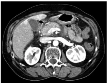
Fig. 1 – Computed tomography image of a patient with pancreatic dorsal agenesis and acute pancreatitis. Absence of pancreas body and tail.
Introduction
The interest regarding the research and study of congenital malformations of the pancreas is not merely academic, nor does it concern simple peculiarities of anatomical development; rather, it is intimately linked with clinical situations, where rapid identification is necessary to achieve a timely diagnosis and to implement an effective therapy. The concern related to this theme comes from a peculiar clinical case, described in this article, which required a review of the entire subject.
The patient presented acute pancreatitis, caused by a rare congenital malformation of the pancreas, called agenesis of the dorsal pancreas (ADP).
Knowledge regarding all pancreatic malformations and their clinical presentations is essential for differential diagnosis and treatment. Pancreatic birth defects occur during embryogenesis.1-5
ADP, also known as congenital short pancreas, is very rare; approximately 23 cases of complete ADP have been reported in the international literature between 1913 and 2007,6 and 54
cases were reported in the last 100 years.7,8 This is the first case
described in Portugal. The absence of the pancreas tail and body, as well as the absence of the dorsal pancreatic duct is synonymous with ADP. This malformation was first described in 1911 during an autopsy and was associated with diabetes, approximately ten years before the discovery of insulin.7
Pancreas divisum (PD) is the most common of all pancreatic congenital malformations. By definition, it presents as a pancreas with two ducts and independent drainage orifices. Nevertheless, in the current medical language, the designation of PD comprises a set of anatomical variants with dissimilar clinical presentations, prognoses, and treatments.
In a clinical case study, the authors describe a 59-year-old female patient, who was attended to at the emergency department complaining of continuous epigastric pain, with dorsal irradiation that had lasted for 48 hours. There was no nausea or vomiting, and no other complaints. She had no history of disease, did not consume alcohol or drugs, and the family history was unremarkable. At the physical examination, she presented a painful abdomen on palpation of the epigastric region, no muscular defense and no palpable masses.
Laboratory assessment at admission presented: hemoglobin 12.1 g/dL (reference values from 11.7 to 16 g/dL), leukocytes 7.2 x 10³ μL (5x10³ - 13x103 uL), glucose 90 mg/dL (90-109
mg/dL), LDH 278 U/L (< 240 U/L), AST 20 U/L (< 31 U/L), and
amylase 2076 U/L (22-80 U/L). She underwent an abdominal ultrasound, which showed simple hepatic cysts without any other alterations. The pancreas could not be assessed due to gas interposition.
She was admitted and medical treatment was initiated. After 48 hours, she presented pain improvement without fluid retention, and laboratory assessment after 48 hours showed hemoglobin of 10.9 g/dL, calcium of 8.6 mg/dL (reference value: 8.8-10.6 mg/dL), albumin of 3.5 g/dL (3.5-5.2 g/dL), and blood urea nitrogen of 9 mg/dL (7.94-20.9 mg/dL). Computed tomography (CT) showed absence of tail and body of the pancreas, and an enlarged and swollen head of the pancreas with a computed tomography severity index (CTSI) of 2. ADP was demonstrated by the CT images (Fig. 1). The patient had complete resolution of the pancreatitis, with good clinical evolution and response to medical treatment.
Resultados: A ADP é uma malformação muito rara que surge durante a organogênese. Nas últimas décadas, foi produzido um volume importante de informação genética e embriológica que ajuda a compreender as causas das malformações pancreáticas. As malformações pancreáticas têm de ser estudadas e compreendidas no seu conjunto.
Conclusão: A malformação pancreática é uma causa de pancreatite aguda e crônica no adulto, pouco estudada. A possibilidade da existência de malformações pancreáticas deve estar sempre presente em doentes com pancreatite aguda ou crônica sem causa evidente.
© 2013 Elsevier Editora Ltda. Todos os direitos reservados.
The patient was asymptomatic when she was discharged, after seven days of hospitalization; a CT scan performed four weeks after hospital discharge showed a slightly enlarged pancreatic head, with no edema, effusion, or pseudocysts, and absence of pancreatic body and tail. After one year, the patient remains asymptomatic and has no diabetes.
Pancreas embryology
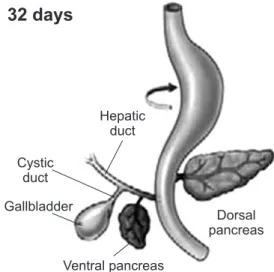
Understanding the embryological origin of the pancreas is crucial for an assessment of the anatomic variants of the pancreatic ducts. The pancreatic excretory ducts mark the division between dorsal and ventral pancreas, two areas of a single organ, divided from their origin. The pancreas emerges between the sixth and eighth week of embryonic life. It originates from the endoderm, as do other gastrointestinal organs. It develops from two evaginations, dorsal and ventral, of the primitive duodenum. These two evaginations subsequently merge. The fusion is more or less complete, and the pancreas forms a single organ. The ventral evagination of the primitive duodenum arises from a liver diverticulum, having a direct connection with the common biliary duct. The duct that originates from the ventral evagination is known as the duct of Wirsung. The primitive duodenum, during its early development, rotates to the right, and the ventral evagination follows this rotation movement, passing through the posterior duodenum and merging with the dorsal evagination (Fig. 2). This allows the merging of the ventral duct (duct of Wirsung) with the dorsal duct (duct of Santorini) at the head of the pancreas. The ventral evagination originates the lower portion of the pancreas head and the processus uncinatus, while the dorsal evagination originates the body and tail of the pancreas.
The existing literature emphasizes the importance of the Hedgehog (Hh) gene in the embryological development and
tissue differentiation of the pancreas. Disorders in Hh gene activity can result in congenital pancreas anomalies and pancreatic diseases in adults, particularly diabetes mellitus, chronic pancreatitis, and pancreatic carcinoma.9-15
It appears that certain levels of Hh activity are necessary for the formation of the pancreas, and that gene activity deregulation results in moderately severe alterations in organ morphogenesis and function.11 Reduced activity of the Hh gene
is associated with severe birth defects of the pancreas and intestine in rats and in humans.11
The evaginations of the primitive duodenum merge and grow to form a complex network of ducts that constitute the organ in its most primitive form. The adult pancreas is a heterogeneous organ consisting of two cell types, the exocrine acinar cell compartment and the endocrine compartment, which consists of cells located in the islets of Langerhans.11
The islets of Langerhans, which produce key hormones for the regulation of glucose levels in the blood, consist of four different cell types: insulin-producing β cells; glucagon-producing α cells; somatostatin-producing δ cells; and pancreatic polypeptide-producing P cells. However, cell distribution is not uniform and is closely related to the organ’s dorsal and ventral origin. The body and tail have a greater density of α and β cells, whereas there is a prevalence of P cells in the head.
Pancreas anatomy
The pancreatic duct, as well as the anatomical description of the organ and its connection with digestion, was first described by a German anatomist, Johann Wirsung (1642). Later, Giovani Santorini (1681-1737) described in detail the pancreatic excretory duct system.3 The pancreas is an organ with a
retroperitoneal location, placed in an oblique position between the duodenal arc and the spleen.16 The pancreatic ducts are
divided into main (Wirsung) and accessory (Santorini). The duct of Wirsung runs through the body and tail of the pancreas in a medial location. The duct of Santorini drains directly into the duodenum through an accessory papilla in 60% of the population. In up to 30% of cases, the duct of Santorini ends as a blind accessory duct, with no exit to the duodenum, or does not exist.
Pancreas congenital malformations
The complex process of pancreas morphogenesis, during which two evaginations of the primitive duodenum give origin to the organ, explains the existence of several variants of pancreatic congenital malformations, such as ADP; annular pancreas; pancreas divisum; pancreatic duct malformations, of which the common bile duct syndrome is the most common; ectopic pancreas; and congenital pancreatic cyst.
Agenesis of the dorsal pancreas (ADP)
Also known as congenital short pancreas, it was first described in 1911.7 It is a rare congenital malformation, in which there is
32 days
Hepatic duct
Cystic duct
Gallbladder
Dorsal pancreas
Ventral pancreas
Fig. 2 – The primitive duodenum, during its early
only one pancreatic duct system, with no duct of Santorini or body and tail of the pancreas. Partial agenesis of the pancreas or pancreatic hypoplasia occurs in approximately one to 2/10,000 patients, and is often associated with other congenital malformations.17 In ADP, there is a regression in the dorsal
evagination during embryogenesis. According to the degree of immaturity of the dorsal pancreas, there can be total agenesis, hypogenesis of body and tail, or hypogenesis only of the tail of the pancreas.9
Fifty-four cases of complete and incomplete ADP have been described over the last 100 years. The publication of reports on some families with ADP indicates that there may be autosomal dominant transmission of the malformation,13,18
and an association with the HNF1β gene has been described.17
Annular pancreas
It is a rare condition in which the second part of the duodenum is surrounded by a ring of pancreatic tissue. This malformation is diagnosed in the first weeks of life, and few cases have been reported in adults.
Pancreas divisum (PD)
It is the absence of fusion or incomplete fusion of the ventral and dorsal pancreas, mainly of the drainage ducts (Wirsung’ and Santorini). PD is the most common congenital malformation of the pancreas. By definition, it is a pancreas with two separate ducts and independent drainage orifices.
Common bile duct syndrome
It is a congenital malformation of the pancreatic and biliary ducts, whose clinical relevance results from the anomalous pancreaticobiliary junction, also called common duct syndrome.
Ectopic pancreas
It is defined as pancreatic tissue in an aberrant location, with no connection to the main organ.
Congenital pancreatic cyst
It is a rare disease, particularly in adults. The cysts are located in the body and tail of the pancreas and have no communication with the pancreatic duct.13
Clinical manifestations
The incidence of pancreatitis associated with pancreatic abnormalities is approximately 0.1%.19 Patients with ADP
are mostly asymptomatic, and the remaining present with abdominal pain and pancreatitis, and 52% have diabetes mellitus.6,9,18,19 Approximately 5% experience pancreatitis
symptoms at least once; some patients report steatorrhea and signs of pancreatic exocrine failure. The association with diabetes mellitus is frequent because the islet cells of
the pancreas are located in the organ’s body and tail. The onset of pancreatitis in these patients is due to morphological alterations in the pancreatic ducts. It may present alone or associated with other malformations.7,13 It is necessary to
differentiate agenesis of the dorsal pancreas from the pseudo-agenesis of the pancreas; the latter results from the atrophy of the pancreas body and tail by self-digestion secondary to chronic pancreatitis.17,20 It is also crucial to differentiate the
diagnosis ADP from carcinoma of the pancreas head, which obstructs the pancreatic ducts and provides a false clinical picture of ADP.17,18,20
In this context it is necessary to obtain a careful medical history, to assess serum amylase levels, and to perform imaging studies by computed axial tomography and magnetic resonance pancreatogram.6,21
Complementary diagnostic examinations
The diagnosis of a pancreatic malformation and identification of its type is achieved with imaging studies, since the analytical study of patients can only provide indirect pancreatopathy information. A CT is useful in the differential diagnosis of pancreatic malformations, but this test has limitations for the detailed study of the pancreatic ducts.6 Endoscopic retrograde
cholangiopancreatography (ERCP) allows for the performance of a pancreatography, which discloses details of pancreatic duct morphology and of some pancreatic malformations. However, it is an invasive examination that uses contrast, requires exposure to radiation, and is sometimes associated with complications.6
MRCP is a noninvasive test that allows the study of pancreatic duct morphology; its diagnostic accuracy can be improved with three-dimensional reconstructions or dynamically with secretin injection.1,19,22-26 ERCP and MRCP show agreement in
the diagnosis of congenital pancreaticobiliary malformations in approximately 70% of cases.5,7 The use of S-MRCP (with
secretin injection) allows for a greater diagnostic accuracy, has no associated complications, and avoids the risks of ERCP.
Treatment
T r e a t m e n t i s r e s e r v e d f o r s y m p t o m a t i c p a t i e n t s . Recommendation of a low-fat diet and diabetes control is the first line of treatment. The use of analgesics for pain control is recommended, as well as the administration of pancreatic enzymes, which reduce pancreatic secretion and promote pain improvement.5,26 Patients with ADP associated with
pancreatitis and pancreatic duct alterations may benefit from sphincterotomy.9
Surgical therapy has relatively better results and lower incidence of restenosis. In spite of the surgical trauma, complication rates in surgical sphincteroplasty of the minor papilla vary from 4.2% to 10%, lower than that of the endoscopic papillotomy, which is approximately 14.6%.5 Surgical treatment
aims to treat recurrent acute pancreatitis, treat pancreatic chronic pain, and prevent pancreatitis complications.
Conclusion
Pancreatic malformation is a seldom-studied cause of acute and chronic pancreatitis in adults. The incidence of pancreatitis associated with pancreatic malformations is about 0.1%.17 ADP is a very rare malformation. The case presented
here is unusual and illustrates the importance of considering this type of abnormality as the etiology of acute pancreatitis.
Conflict of interest
All authors declare to have no conflict of interest.
R E F E R E N C E S
1. Varshney S, Johnson CD. Pancreas divisum. Int J Pancreatol. 1999;25:135-41.
2. Cameron JL. Current surgical therapy. 9th ed. Philadelphia:
Elsevier; 2011. p. 481-5.
3. Gomes MLT. Pâncreas um órgão único com duas partes [Thesis]. Porto: Universidade do Porto; 1992.
4. Brunicardi FC, Hunter J, Dunn D, Pollock RE, Andersen D. Schwartz’s principles of surgery. New York: McGrawHill; 2004. p. 1221-5.
5. Fischer JE. Mastery of surgery. 5th ed. Philadelphia: Lippincott
Williams & Wilkins; 2006. p. 1224-32.
6. Vural M, Pasaoglu L, Hatipoglu HG. Agenesis of the dorsal pancreas. World J Gastroenterol. 2008;14:2915-6.
7. Schnedl WJ, Piswanger C, Soelkner C, Wallner SJ, Krause R, Lipp RW. Agenesis of the dorsal pancreas and associated diseases. Dig Dis Sci. 2009;54:481-7.
8. Joo YE, Kang HC. Agenesia of the dorsal pancreas. A case report and review of literature. Korean J Intern Med. 2006;21:236-9.
9. Juan D, Xu GQ, Xu P, Jin EY, Liu Q, Li YM. Congenital short pancreas. Chinese Med J (Engl). 2007;120:259-62.
10. Haumaitre C, Barbacci E, Jenny M, Ott MO, Gradwohl G, Cereghini S. Lack of TCF2/vHNF1 in mice leads to pancreas agenesis. Proc Natl Acad Sci. 2005;102:1490-5.
11. Lau J, Kawahira H, Hebrok M. Hedgehog signaling in pancreas development and disease. Cell Mol Life Sci. 2006;63:642-52. 12. Burdick JS, Horvath E. Management of pancreas divisum.
Curr Treat Option Gastroenterol. 2006;9:391-6.
13. Cano DA, Hebrok M. Pancreatic development and disease. Gastroenterology. 2007;132:745-62.
14. Valerie M. Schwitzgebel VM. Programming of the pancreas. Mol Cell Endocrinol. 2001;185:99-108.
15. Onge ST, Wehr P, Gruss P. Pancreas development and diabetes. Curr Opin Genet Dev. 1999;9:295-300. 16. Skandalakis JE, Skandalakis PJ. Surgical anatomy and
technique. New York: Springer; 2009. p. 381-91. 17. Rastogi R, Kumar R, Bhargava S, Rastogi V. Isolated
pancreatic hypoplasia: a rare but significant radiological finding. Saudi J Gastroenterol. 2009;15:289-90.
18. Wolfgang J, Schnedl WJ, Reisinger EC, Scheiber P, Pieber TR, Lipp RW, et al. Complete and partial agenesis of the dorsal pancreas within one family. Gatrointest Endosc. 1995;42:485-7. 19. Delhaye M, Matos C, Devière J. Acute relapsing pancreatitis.
Congenital variants: diagnosis, treatment; outcome. JOP J Pancreas. 2001;2:373-81.
20. Radi JM, Gaubert R, Cristol-Gaubert R, Baecker V, Travo P, Prudhomme M. A 3D reconstruction of pancreas development in the human embryos during embryonic period (Carnegie stages 15-23). Surg Radiol Anat. 2010;32:11-5. 21. Gold RP. Agenesis and pseudo-agenesis of the dorsal
pancreas. Abdom Imaging. 1993;18:141-4.
22. Kamisawa K, Tu Y, Tsuruta K, Okamoto A, Kamata N. MRCP of congenital pancreaticobiliary malformation. Abdom Imaging. 2007;32:129-33.
23. Nicaise N, Pellet O, Metens T, Devière J, Braudé P, Struyven J, et al. Magnetic resonance cholangiopancreatography: interest of IV secretin administration in the evaluation of pancreatic ducts. Eur Radiol. 1998;8:16-22.
24. Delhaye M, Matos C, Arvanitakis M, Devière J. Pancreatic ductal system obstruction and acute recurrent pancreatitis. World J Gastroenterol. 2008;14:1027-33.
25. Uygur-Bayramiçil O, Dabak R, Kiliçoglu D, Dolapçioglu C, Oztas D. Dorsal pancreatic agenesis. J Pancreas. 2007;8:450-2. 26. Karcaaltincaba M. CT differentiation of distal pancreas fat