O
RIGINALA
RTICLE ©Revista Brasileira de FisioterapiaEffect of cathodal high-voltage electrical
stimulation on pain in women with TMD
Efeito da estimulação elétrica de alta voltagem catódica sobre a dor em mulheres
com DTM
Natalia C. M. C. Gomes1, Kelly C. S. Berni-Schwarzenbeck2, Amanda C. Packer1, Delaine Rodrigues-Bigaton1,2
Abstract
Background: Pain is the main symptom of patients with temporomandibular disorder (TMD). Objective: To evaluate the effect of cathodal high-voltage electrical stimulation (HVES) on pain intensity in women with TMD. Methods: Twenty women with TMD (24.25±8.90 years old) participated in the study. They were divided into experimental group (EG, n=10), which received 10 applications of HVES, and placebo group (PG, n=10), which received sham treatment with disconnected HVES equipment. For the sample selection, we used the Research Diagnostic Criteria for Temporomandibular Disorder (RDC/TMD). Pain level was evaluated using a visual analog scale (VAS) applied prior to and after the tenth application of HVES. Data were analyzed using the Wilcoxon signed-rank test and the Mann-Whitney test. Results: Ten applications of HVES reduced pain intensity in the EG (p=0.01). In the PG, there was no significant difference
(p=0.20). After the application of HVES, no difference was found (p=0.65) between the groups. Conclusion: The cathodal HVES was effective in reducing pain in women with TMD. Trial Registration RBR-4bk94x.
Keywords: physical therapy; clinical trial; temporomandibular joint disorders; electrical stimulation.
Resumo
Contextualização: A dor é o principal sintoma dos pacientes com disfunção temporomandibular (DTM). Objetivo: Avaliar o efeito da estimulação elétrica de alta voltagem catódica (EEAV) sobre a intensidade da dor em mulheres com DTM. Métodos: Participaram do estudo 20 mulheres (24,25±8,90 anos) com DTM, divididas em grupo experimental (GE n=10), no qual as mulheres receberam dez aplicações de EEAV, e grupo placebo (GP n=10), no qual foi aplicada a EEAV, porém com o aparelho desligado. Para seleção da amostra, utilizou-se o critério de diagnóstico em pesquisa para DTM (RDC/TMD) e, para avaliação da dor, utilizou-se a Escala Visual Analógica (EVA) aplicada antes do início do tratamento (pré-tratamento) e após a décima aplicação da EEAV (pós-tratamento). Os
dados foram analisados pelos testes Wilcoxon das ordens assinaladas e Mann-Whitney. Resultados: As dez aplicações de EEAV promoveram redução da intensidade da dor no GE (p=0,01); no GP, não se observou diferença significativa (p=0,20). Comparando-se
os grupos após a aplicação da EEAV, não se notou diferença (p=0,65). Conclusão: A EEAV catódica é efetiva para redução da dor em mulheres com DTM. Registro de Ensaio Clinico RBR-4bk94x.
Palavras-chave: fisioterapia; ensaio clínico; transtornos da articulação temporomandibular; estimulação elétrica.
Received: 10/01/2010 – Revised: 04/05/2011 – Accepted: 08/25/2011
1 School of Health Sciences, Universidade Metodista de Piracicaba (UNIMEP), Piracicaba, SP, Brazil
2 Department of Morphology, School of Dentistry of Piracicaba, Universidade Estadual de Campinas (UNICAMP), Piracicaba, SP, Brazil
Correspondence to: Delaine Rodrigues Bigaton, Rua Dom João Bosco, 139, Apto 11, Vila Rezende, CEP 13405-137, Piracicaba, SP, Brazil, e-mail: [email protected]
Introduction
Temporomandibular disorder (TMD) is characterized by functional and pathological changes affecting the tem-poromandibular joint (TMJ), masticatory muscles, and other structures of the stomatognathic system1. It presents as joint sounds, muscle and/or joint pain, headaches, diffi-culty chewing, limited and/or abnormal jaw movement2. An epidemiological study carried out by Gonçalves et al.3 found that the most prevalent symptoms of TMD in the Brazilian urban population are joint sounds followed by joint and muscle pain, with higher frequency in women.
In contrast, Cooper and Kleinberg4 reported that pain is one of the main complaints of patients with TMD. The au-thors conducted a retrospective analysis of data from 4528 patients with TMD assessed by a single evaluator (dentist) over 25 years. Of the 4528 patients, 96.1% reported pain, and of that total, 79.3% had headaches, 75% had temporo-mandibular dysfunction or discomfort, and 82.4% had ear dysfunction or discomfort.
he musculoskeletal conditions of the mandibular and cervical regions found in TMD are the major causes of non-dental pain in the orofacial region5. Such conditions have a major impact on the quality of life of individuals afected by TMD6. It is known that, in TMD, the high intensity of pain is associated with decreased blood low to muscles, however treatments that improve blood low are efective in relieving muscle pain7,8.
Among the therapeutic procedures used by physical therapists to treat TMD are acupuncture9, jaw exercises10, massage11, manual therapy12, laser13, and transcutaneous electrical nerve stimulation (TENS)14,15. Another resource used in physical therapy is high-voltage electrical stimu-lation (HVES). With this current, it is possible to perform numerous treatments because it has a single-phase wave, thus it can be effective in controlling and absorbing acute edemas, accelerating dermal and subdermal tissue repair, and controlling pain16.
For the administration of HVES, both positive (cathodal) and negative (anodic) polarity can be used. Anodic HVES promotes protein denaturation, reduction in the mast cells of wounds, and stimulation of new capillary growth. In con-trast, the application of cathodal HVES stimulates tissue granulation, reduces edema, promotes the proliferation of fibroblasts, and increases blood flow16,17. Although the phys-iological effect of each of the poles is well-established, the clinical effects of the polarity in humans have not been well defined. Nevertheless, among the effects of HVES, the most important are pain relief and increased blood flow, which can be obtained with both poles18.
Most HVES experiments performed in humans so far have focused on the circulatory and regenerative efects; however, analgesia can also be obtained with the use of this current, as reported by the studies of Rodrigues-Bigaton et al.15, Almeida19, and Schwarzenbeck20. hese authors evaluated the efect of ten applications of anodic HVES on pain15,19,20, on the electromyographic (EMG) signal of the masticatory muscles, and on the clinical characteristics of TMD19,20.
Almeida19 and Schwarzenbeck20 observed that anodic HVES improved the classification and severity of TMD, changing the clinical characteristics of the disease. Re-garding the EMG signal, Almeida19 noted improvement in muscle activity evaluated at rest and in isometric condi-tions. In contrast, Schwarzenbeck20 noted improvement in the muscle activation pattern assessed by means of isotonic activity. According to these authors, the clinical benefits ob-tained with ten applications of anodic HVES are due to the current’s circulatory and analgesic actions.
Although the aforementioned studies used HVES to treat pain in individuals with TMD, the efect of HVES on the pla-cebo group has not been described in the literature, therefore the presence of this group must be taken into consideration, particularly for HVES application due to the lack of studies on the clinical applicability of this resource for analgesia. In addi-tion, it is important to emphasize that, in the current literature, only HVES applications with positive polarity were found, thus it is crucial to study the efect of negative polarity on pain, given that analgesia can be obtained with both polarities18.
Another factor that justifies the importance of this study is that, as shown in other studies15,19,20, HVES has beneficial effects on TMD treatment and, if its effectiveness is actually proven, this therapeutic resource can be incorporated into the clinical practice of physical therapists who treat TMD. Although HVES is more cost-effective and yields good re-sults with shorter treatment periods than galvanic current21 and TENS15, its use is restricted in Brazil, and one of the rea-sons for this is the lack of publications on its application21.
Considering that HVES is indicated for analgesia, we hy-pothesized that cathodal HVES contributes directly to pain reduction in women with TMD. Therefore, the goal of the present study was to evaluate the effect of cathodal HVES on pain in women with TMD.
Methods
Study design
he study design is a randomized, double-blind clini-cal trial, approved by the Research Ethics Committee of
Universidade Metodista de Piracicaba (UNIMEP), Piracicaba, SP, Brazil, under protocol number 21/08. All participants signed the informed consent form. he participants and eval-uators were blinded, therefore the participants did not know which group they belonged to, and the evaluators did not know whether the participant belonged to the experimental group (EG) or to the placebo group (PG).
Sample
Patients were recruited from the waiting list of the UNIMEP Physical Therapy Clinic and from the university community.
Sample loss
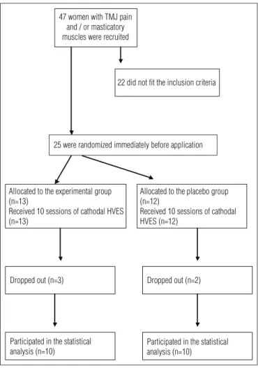
For the sample selection, we used the Research Diagnos-tic Criteria for Temporomandibular Disorders (RDC/TMD). Forty-seven women with pain in TMJ and/or mastica-tory muscles were selected. Of these, 22 were excluded because they did not meet the inclusion criteria for the present study.
he sample size (n) was determined by means of sample calculation based on standard deviation values obtained by a Visual Analog Scale (VAS), which provided a measure of pain intensity in centimeters. he sample calculation was performed using GraphPad StatMate, version 1.01i, a power of 80%, and alpha=0.05. he sample size was calculated to be 18 participants, divided between the EG and the PG.
We selected 25 women with a diagnosis of TMD, con-firmed by RDC/TMD, axis I. They were randomly divided into two groups: EG (n=13), aged between 17 and 32 years (22.50±7.07 years), in which the participants received ten applications of cathodal HVES; and PG (n=2), aged between 17 and 44 years (26±10.55 years), in which the participants also received ten applications of HVES, however with the equipment turned off. Over the course of the treatment, five participants dropped out of the study, three from the EG and two from the PG. Stratified randomization was used to assign the participants to the groups. After the comple-tion of the study, effective treatment was offered to the PG participants.
Figure 1 shows the lowchart for the sample distribution.
Inclusion criteria
The participants of both groups had to have a diagnosis of TMD, according to the RDC, axis I, accompanied by pain and/or fatigue in the masticatory muscles during functional activities for a minimum of one year and a maximum of five
years. In addition, they could not be undergoing orthodontic treatment, drug therapy (pain relievers, anti-inflammatories, muscle relaxants) or physical therapy treatment.
Experimental procedure
For the sample selection, all participants were submit-ted to a physical therapy assessment that consissubmit-ted of col-lection of personal data, anamnesis, previous history, and individual visual examination, followed by the assessment based on the RDC/TMD. The participants diagnosed as group I of the RDC/TMD assessed pain intensity through the VAS and began treatment with effective cathodal HVES or placebo. The randomization was carried out immediately before the start of treatment.
Intervention
For the application of cathodal HVES in both groups, the participants remained in the dorsal decubitus position with a roll under the knees during the sessions. The HVES
Dropped out (n=3) Dropped out (n=2)
Participated in the statistical analysis (n=10)
Participated in the statistical analysis (n=10)
47 women with TMJ pain and / or masticatory muscles were recruited
22 did not fit the inclusion criteria
25 were randomized immediately before application
Allocated to the experimental group (n=13)
Received 10 sessions of cathodal HVES (n=13)
Allocated to the placebo group (n=12)
Received 10 sessions of cathodal HVES (n=12)
Figure 1. Flowchart for the sample distribution.
was delivered by the Neurodyn High Volt®
(ANVISA number 10360310008 - Ibramed) with microcontrollers, two chan-nels, four active transcutaneous rectangular electrodes (3x5 cm) made from carbon-silicone rubber, and a rectan-gular dispersive electrode (10x18 cm) consisting of an alu-minum sheet wrapped in felt moistened with water.
The electrodes were placed bilaterally on the anterior portion of the temporal muscle (channel 1) and on the belly of the masseter muscle (channel 2). The dispersive electrode was positioned over the lower cervical region and the upper thoracic region because, according to Holcomb18, this elec-trode must be larger than the active elecelec-trodes to reduce the current density and must be positioned over large areas. In addition, the greater the distance between the active and dispersive electrodes, the deeper the current will be16.
he parameters used in HVES were: 10 Hz frequency; pulse width ixed by the equipment with two twin pulses of 20 µs each with interpulse interval of 100 µs; voltage above 100 volts to motor threshold (visible muscle contraction) with a threshold variation between 100 and 170 volts; nega-tive polarity (cathodal HVES) in both channels applied for 30 minutes, two to three times per week. he equipment was calibrated with an oscilloscope (Tektronix TDS 210), and all of the physical parameters of the current were according to manufacturer speciications. he device’s timer was calibrated using three timers (Technos), and this variable was according to equipment speciications. Gel was used under the silicone-carbon electrodes to allow the conduction of current to the tissue. he electrodes had not been previously used.
Measurement of pain intensity
To measure pain intensity, we used the VAS, which consists of a 10-cm horizontal line marked “no pain” on the left end and “worst possible pain” marked on the right end. he participants were instructed to draw a vertical line over the horizontal line, indicating at which point of the line the pain was. he VAS was applied before the start of treatment (pre-treatment) and after the tenth HVES application (post-treatment), respecting the period of at least 24 hours and a maximum of 48 hours after the last application of cathodal HVES. his period was observed with the purpose of evaluating the overall efect of the treat-ment and not its immediate efect.
The VAS data were analyzed using a ruler graded in centimeters. Pain intensity was measured from the left end, which coincided with the zero value of the ruler, until the vertical line drawn by the participant. It is worth noting that the examiner who analyzed the VAS data did not know which group the participants belonged to or whether the scales referred to the pre- or post-treatment.
Statistical analysis
Due to the subjectivity of the response variable pain intensity, non-parametric tests were used for intra- and intergroup comparisons. For intragroup comparison, the Wilcoxon signed-rank test was used. For intergroup com-parison, we considered as response variable the difference between the pain intensity values obtained in pre- and post-treatment moments, and these values were analyzed using the Mann-Whitney test. For data analysis, we used the pro-gram SPSS 11.0, and the results were shown by the median and its first and third quartiles. For both analyses, the two-tailed significance level was used, with alpha equal to 5%.
Results
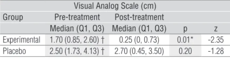
Through intragroup comparison, it can be observed that the ten applications of cathodal HVES promoted a reduc-tion in pain intensity in the EG (p=0.01), while no difference was observed in the PG (p=0.20), as shown in Table 1. In the same table, which also shows the intergroup comparison, it can be observed that, before treatment, the EG and the PG did not shown any differences in pain intensity (p=0.23), a fact that demonstrates the homogeneity of the sample.
Table 2 shows that there is no difference between the EG and the PG after the application of HVES (p=0.65). However, when examining the values of the difference between the pre- and post-treatment and the range between the first and third quartiles, it can be seen that the EG presented greater reduction in pain intensity when compared to the PG.
Discussion
The results of this study showed that ten applications of cathodal HVES reduced pain intensity in the EG. In the PG, no changes were observed. When comparing both groups post-treatment, we found no significant differences between them. However, from a clinical point of view, the results indicated that the EG had a greater reduction in pain intensity than the PG.
he results of the present study agree with the indings of Almeida19, who assessed the efect of ten applications of an-odic HVES ( frequency of 10 Hz, twin pulses lasting 20 µs with interpulse interval of 100 µs, voltage above 100 volts with stimulation at motor threshold) on women with TMD and found pain reduction, evaluated through VAS, both between sessions and at the end of treatment. Pain reduction was also observed by Rodrigues-Bigaton et al.15, who concluded that
both TENS (10 Hz modulated in 50%, 200 µs, and intensity at the motor threshold) and anodic HVES (10 Hz, 20 µs twin pulses with interpulse interval of 100 µs, voltage above 100 volts with stimulation at motor threshold) promoted the re-duction in pain intensity in women with TMD, showing that HVES is also indicated for the treatment of these patients.
A comparison of the results of the present study and those found by Almeida19 and Rodrigues-Bigaton et al.15 suggests that the analgesia can be obtained with both the anodic and cathodal HVES, which corroborates the asser-tion by Holcomb18 that both poles are suitable for analgesia. Therefore, it appears that the analgesic effect of HVES is more closely connected to the frequency and voltage of the current (motor threshold) than to the polarity.
Mohr, Akers, and Wessman22 observed that HVES pro-moted increased blood circulation in rats’ hindlimbs and suggested that this increase was more closely related to the intensity of muscular contraction than to the polar effect produced by the current. The polar effect of HVES is dis-cussed because Mendel and Fish23 believe that, in this type of stimulation, the duration of the pulse is too small to pro-mote chemical reactions under the electrodes. In contrast, Davini et al.24 reviewed the literature on HVES as a treat-ment option and concluded that, despite the controversies, open wound repair is faster when there is alternating polar-ity starting with cathodal stimulation and that the circula-tory effect is most effective when cathodal HVES is used at motor threshold. Thus, it can be stated that the polar effect of HVES with the purpose of promoting analgesia must be better investigated.
Generally, it is already known that when electrical stim-ulation is performed at the motor threshold and with low frequency (non-tetanic muscle contraction), in addition to increasing the arterial blood flow to the stimulated area, the current generates analgesia due to the stimulation of group III and IV afferent fibers causing the release of endogenous opioids from the central nervous system25. In this way, the electrical stimulation applied at motor threshold and at low frequency is effective in modulating clinical pain and experimentally-induced pain26.
In the present study, as in the work of Almeida19 and Rodrigues-Bigaton et al.15, HVES was applied using low frequency and high voltage, therefore it is believed that the analgesic effect promoted by HVES is more closely related to the frequency and voltage of the current than to its polar effect. In the intra-group comparison, it was possible to ob-serve that the current did not reduce pain intensity in the PG after the ten applications of cathodal HVES. Thus, the presence of the PG in this study was of great importance as it allowed the verification of the selected method.
Tramèr et al.27 observed that, in clinical contexts with no gold standard and in treatments with wide-ranging values, the absence of the PG results in improbable conclusions. In many cases, the recruitment of patients in clinical trials is questionable and, therefore, the absence of this group can produce unrealistic results. Also according to the author, the PG allows the estimation of the effectiveness of the treatment.
Although the comparison between the EG and the PG did not show any statistical differences, it was clinically possible to observe that the EG had a greater reduction in pain intensity compared to the PG. It has been suggested that, in order to obtain significant results in the intergroup comparison, it would be necessary to increase the sample size, which is a limitation of the present study.
The hypothesis of the study was confirmed because the results showed that cathodal HVES reduced pain intensity in women with TMD. Therefore, this resource can be incor-porated into the clinical practice of physical therapists.
Acknowledgments
To the Conselho Nacional de Desenvolvimento Cientí-fico e Tecnológico (CNPq) process no. 567190/2008 0 and to the Programa Institucional de Bolsas de Iniciação Científica (PIBIC/CNPq/UNIMEP) for the support granted for this re-search, and to Maria Imaculada de Lima Montebelo for the contribution in the data analysis.
Table 1. Comparison of pain intensity recorded in the VAS in cm before HVES (pre-treatment) and after HVES (post-treatment) in the experimental group (n=10) and the placebo group (n=10).
Visual Analog Scale (cm)
Group Pre-treatment Post-treatment
Median (Q1, Q3) Median (Q1, Q3) p z
Experimental 1.70 (0.85, 2.60) † 0.25 (0, 0.73) 0.01* -2.35
Placebo 2.50 (1.73, 4.13) † 2.70 (0.45, 3.50) 0.20 -1.28
Q1=quartile 1; Q3=quartile 3; (*) indicates significant difference (p<0.05); Wilcoxon Test; (†) indicates intragroup comparison using the Mann-Whitney Test (p=0.23 and z=1.21).
Table 2. Intergroup comparison obtained by the difference in pain intensity recorded in the VAS in cm before HVES (pre-treatment) and after HVES (post-treatment) in the experimental group (n=10) and the placebo group (n=10).
Visual Analog Scale (cm)
Group Experimental Placebo
Median (Q1, Q3) Median (Q1, Q3) p z
Pre-post 1.25 (0.13, 2.28) 1 (-0.18, 1.88) 0.65 -0.45
Q1=quartile 1; Q3=quartile 3; (*) indicates significant difference (p<0.05); Mann Whitney Test.
1. Tvrdy P. Methods of imaging in the diagnosis of temporomandibular joint disorders. Biomed Pap Med Fac Univ Palacky Olomouc Czech Repub. 2007;151(1):133-6.
2. Frisardi G, Chessa G, Sau G, F Frisardi. Trigeminal Electrophysiology: a 2×2 matrix model for differential diagnosis between temporomandibular disorders and orofacial pain. BMC Musculoskelet Disord. 2010;11:141.
3. Gonçalves DA, Dal Fabbro AL, Campos JA, Bigal ME, Speciali JG. Symptoms of temporomandibular disorders in the population: an epidemiological study. J Orofac Pain. 2010;24(3):270-8.
4. Cooper BC, Kleinberg I. Examination of a large patient population for the presence of symptoms and signs of temporomandibular disorders. Cranio. 2007;25(2):114-26.
5. Magnusson T, Egermark I, Carlsson GE. A longitudinal epidemiologic study of signs and symptoms of temporomandibular disorders from 15 to 35 years of age. J Orofac Pain. 2000;14(4):310-9.
6. Oliveira AS, Bermudez CC, Souza RA, Souza CMF, Dias EM, Castro CES, et al. Impacto da dor na vida de portadores de disfunção temporomandibular. J Appl Oral Sci. 2003;11(2):138-43.
7. Tullberg M, Alstergren PJ, Ernberg MM. Effects of low-power laser exposure on masseter muscle pain and microcirculation. Pain. 2003;105(1-2):89-96.
8. Okada K, Yamaguchi T, Minowa K, Inoue N. The influence of hot pack therapy on the blood flow in masseter muscles. J Oral Rehabil. 2005;32(7):480-6.
9. Jung A, Shin BC, Lee MS, Sim H, Ernst E. Acupuncture for treating temporomandibular joint disorders: a systematic review and meta-analysis of randomized, sham-controlled trials. J Dent. 2011;39(5):341-50.
10. Furto ES, Cleland JA, Whitman JM, Olson KA. Manual physical therapy interventions and exercise for patients with temporomandibular disorders. Cranio. 2006;24(4):283-91.
11. Biasotto-Gonzalez DA, Bérzin F. Electromyographic study of patients with masticatory muscles disorders, physiotherapeutic treatment (massage). Braz J Oral Sci. 2004;3(10):516-21.
12. La Touche R, Fernández-de-las-Peñas C, Fernández-Carnero J, Escalante K, Angulo-Díaz-Parreño S, Paris-Alemany A, et al. The effects of manual therapy and exercise directed at the cervical spine on pain and pressure pain sensitivity in patients with myofascial temporomandibular disorders. J Oral Rehabil. 2009;36(9):644-52.
13. Hotta PT, Hotta TH, Bataglion C, Bataglion SA, de Souza Coronatto EA, Siéssere S, et al. Emg analysis after laser acupuncture in patients with temporomandibular dysfunction (TMD).
References
Implications for practice. Complement Ther Clin Pract. 2010;16(3):158-60.
14. Rodrigues D, Oliveira AS, Bérzin F. Effect of conventional TENS on pain and electromyographic activity of masticatory muscles in TMD patients. Braz Oral Res. 2004;18(4):290-5.
15. Rodrigues-Bigaton D, Almeida AFN, Berni KCS, Pedroni CR, Gonçalves RN, Bérzin F. Utilização de diferentes estimulações elétricas para o tratamento da dor em mulheres com disfunção temporomandibular. Rev Bras Fisioter. 2008;12(6):476-81.
16. Nelson RM, Hayes KW, Currier DP. Eletroterapia Clínica. 3ª ed. Barueri: Manole; 2003.
17. Daeschlein G, Assadian O, Kloth LC, Meinl C, Ney F, Kramer A. Antibacterial activity of positive and negative polarity low-voltage pulsed current (LVPC) on six typical Gram-positive and Gram-negative bacterial pathogens of chronic wounds. Wound Repair Regen. 2007;15(3):399-403.
18. Holcomb WR. A practical Guide to Electrical Therapy. J Sport Rehabil. 1997;6(3):272-82.
19. Almeida AFN. Efeito do tratamento com estimulação elétrica de alta voltagem sobre a dor e a atividade eletromiográfica dos músculos mastigatórios em mulheres com DTM [dissertação]. Piracicaba: UNIMEP; 2004.
20. Schwarzenbeck A. Efeito da estimulação elétrica de alta voltagem sobre os sinais e sintomas da disfunção temporomandibular [dissertação]. Piracicaba: UNIMEP; 2009.
21. Low J, Reed A. Eletroterapia explicada. 3ª ed. São Paulo: Manole; 2001.
22. Mohr T, Akers TK, Wessman HC. Effect of high voltage stimulation on blood flow in the rat hind limb. Phys Ther. 1987;67(4):526-33.
23. Mendel FC, Fish DR. New perspectives in edema control via electrical stimulation. J Athl Train. 1993;28(1):63-74.
24. Davini R, Nunes CV, Guirro ECO, Guirro RRJ. Estimulação elétrica de alta voltagem: uma opção de tratamento. Rev Bras Fisioter. 2005;9(3):249-56.
25. Cox P, Kramer JF, Hartsell H. Effect of different TENS stimulus parameters on ulnar motor nerve conduction velocity. Am J Phys Med Rehabil. 1993;72(5):294-300.
26. Robinson AJ, Snyder ML. Eletrofisiologia clínica: eletroterapia e teste eletrofisiológico. 2ª ed. Porto Alegre: Artmed; 2001.
27. Tramèr MR, Reynolds DJ, Moore RA, McQuay HJ. When placebo controlled trials are essential and equivalence trials are inadequate. BMJ. 1998;317(7162):875-80.