Artigo
*e-mail: [email protected]
CHEMICAL CONSTITUENTS FROM THE STEM BARK OF Annona pickelii (Annonaceae)
Emmanoel Vilaça Costaa,b,*, Marília Fernanda Chaves Sampaioc, Marcos José Salvadord, Angelita Nepele and Andersson
Barisone
aDepartamento de Química, Universidade Federal de Sergipe, Avenida Vereador Olímpio Grande, S/N, Campus Prof. Alberto
Carvalho, 49500-000 Itabaiana – SE, Brasil
bDepartamento de Química, Universidade Federal do Amazonas, Avenida General Rodrigo Otavio Jordão Ramos, 6200, Campus
Sen. Arthur Virgílio Filho, 69077-000 Manaus – AM, Brasil
cDepartamento de Química, Universidade Federal de Sergipe, Av. Marechal Rondon, S/N, Cidade Universitária Prof. José Aloísio
de Campos, 49100-000 São Cristóvão – SE, Brasil
dDepartamento de Biologia Vegetal, Instituto de Biologia, Universidade Estadual de Campinas, CP 6109, 13083-970 Campinas
– SP, Brasil
eDepartamento de Química, Universidade Federal do Paraná, Centro Politécnico, CP 19081, 81531-990 Curitiba – PR, Brasil
Recebido em 23/10/2014; aceito em 05/03/2015; publicado na web em 28/04/2015
The phytochemical investigation of the stem bark of Annona pickelii yielded four steroids (β-sitostenone, β-sitosterol, stigmasterol and campesterol), three lignans (eudesmin, magnolin and yangambin), twelve alkaloids (liriodenine, lysicamine, atherospermidine, anonaine, analobine, asimilobine, discretamine, stepholidine, coclaurine, orientaline, juziphine and stepharine), and a benzenoid (2-methoxybenzoic acid). These compounds support their recent reclassification from Rollinia to Annona, and that it is a typical species of the family Annonaceae. Significant antifungal and antioxidant activities were found for the methanol crude extract as well as for the lignans eudesmin, magnolin, yangambin and the alkaloid discretamine. In addition, several items of NMR data for the alkaloids were reviewed and unequivocally described in this work.
Keywords:Annonaceae; Annona pickelii; steroids; lignans; alkaloids; NMR.
INTRODUCTION
Annona L. belongs to the family Annonaceae and comprises approximately 175 species of trees and shrubs, found predominantly in lowland tropical regions.1 Economically, this genus is the most
im-portant of the family Annonaceae due to its edible fruits and medicinal properties.2 Previous chemical and pharmacological investigations
on some species of this genus revealed the presence of important bioactive compounds, exhibiting several pharmacological activities, including cytotoxicity against tumor cell lines,3-5 antimicrobial,6-8
antioxidant,6-8 antiparasitic properties, mainly against Leishmania
sp. and Trypanosoma cruzi,5-11 and analgesic and anti-inflammatory
activities.12 These activities generally are attributed to alkaloids,
acetogenins, and terpenes.
In this context, Annona pickelii (Diels) H. Rainer [synonym: Rollinia pickelii Diels] is a small tree endemic to Brazil, popularly known as ‘araticum-do-mato’, ‘araticum-da-mata’, ‘jaquirinha-do--mato’, and ‘jussara’ found in Paraíba, Pernambuco and recently in Sergipe, Brazil,13 where this species is endemic and commonly
found in Atlantic forest remainders. Previous phytochemical and pharmacological investigations on this species described the isolation of terpenes, lignans and alkaloids.6-11,13,14
In our continuous search for bioactive natural products from Sergipe annonaceous plants, four steroids (1-4), three lignans (5-7), twelve alkaloids (8-19) and one benzenoid (20) (Figure 1) were obtai-ned according to a bio-guided systematic investigation from the stem bark of A. pickelii. Their structures were established on basis of spec-trometric data, including 1D (1H and 13C) and 2D (HSQC and HMBC)
NMR experiments, as well as 1D NOE and MS analysis. Moreover, many of these compounds were isolated a long time ago, and their 1H
and 13C NMR chemical shifts were performed only on basis of general
chemical shifts arguments. The NMR chemical shift assignments ba-sed on two-dimensional NMR experiments have not been performed. Therefore, their previous 13C assignments contain some ambiguities.
In the same way, their previous 1H NMR chemical shifts as well as the
scalar coupling constants values have not been specifically assigned and/or obtained. In this work, the complete and unequivocal assign-ments of 1H and 13C NMR chemical shifts are described for several
alkaloids. Additionally some biological activities were demonstrated for the pure compounds. This is the first report on the phytochemical investigation and biological activities of the stem bark of this plant. EXPERIMENTAL
General procedures
MS analyses were performed on a Shimadzu QP5050A GC-MS system equipped with an AOC-20i auto-injector, and an Rtx®-5Sil
MS fused capillary chromatography column (30 m x 0.25 mm x 0.25 µm film thickness) coated with 5%-diphenyl-95%-dimethylpoly-siloxane. MS analysis were taken at 70 eV with a scan interval of 0.5 s and fragments from 40-500 Da. LR-ESI-MS were obtained in the positive ion mode on an ultra-high performance Waters Acquity UHPLC-TQD LC-MS system, equipped with an ESI source. 1D and 2D NMR data were acquired at 303 K in CDCl3 or CDCl3 plus some
drops of CD3OD on a Bruker AVANCE III 400 NMR spectrometers,
operating at 9.4 Tesla, observing 1H and 13C at 400.13 and 100.61
MHz, respectively. The spectrometer was equipped with a 5-mm multinuclear direct detection probe with z-gradient. One-bond and long-range 1H-13C correlation from HSQC and HMBC NMR
expe-riments were optimized for an average coupling constant 1J H,C and LRJ
H,C of 140 and 8 Hz, respectively. All
shifts (δ) are given in ppm related to the TMS signal at 0.00 ppm as internal reference, and the coupling constants (J) in Hz. Silica gel 60 (70-230 mesh) was used for column chromatography, while silica gel 60 F254 was used for analytical (0.25 mm), and pre parative (1.00
mm) TLC. Compounds were visualized by exposure under UV254/365
light and spraying of p-anisaldehyde reagent followed by heating on a hot plate, and Dragendorff’s reagent.
Botanical material
The stem bark of Annona pickelii were collected at ‘Mata do Crasto’, in the city of Santa Luzia do Itanhy [coordinates: 11° 23’ 01” S, 37° 25’ 13” W], Sergipe, Brazil, in March 2010. The identity of the plant was confirmed by Dr. A. P. N. Prata, Department of Biology, Sergipe Federal University (UFS), Brazil, and a voucher speci men (#15442) has been deposited in the Herbarium of Sergipe Federal University (ASE/UFS).
Extraction and isolation
Dried at room temperature and powdered stem bark of A. pickelii (790 g) was succes sively extracted with n-hexane (2.5 L, five times) followed by MeOH (2.5 L, five times), yielding hexane (5.53 g) and
MeOH (129.32 g) extracts, after solvent removal under reduce pressure. An aliquot of the hexane extract (5.0 g) was initially subjected to silica gel column chromatography (CC) eluted with in creasing concentrations of CH2Cl2 in n-hexane (0 to 100, v/v), followed by EtOAc in CH2Cl2 (0
to 100, v/v), and MeOH in EtOAc (0 to 70, v/v), affording 177 fractions (25 mL each), that were pooled in 23 groups (G1 to G23), according to TLC analysis. Group G17 (446.0mg) eluted with CH2Cl2-EtOAc
(80:20, v/v), was submitted to a new silica gel CC eluted with in creasing concentrations of CH2Cl2 in n-hexane (0 to 100, v/v) followed by EtOAc
in CH2Cl2 (0 to 100, v/v), giving 201 fractions (each 20 mL) that were
pooled in 31 groups (G17.1 to G17.31), according to TLC analysis. Groups G17.6 to G17.8 were also pooled (213.0 mg) and submitted to preparative TLC eluted with n-hexane-EtOAc (80:20, v/v, two times), affording 1 (5.3 mg) and a mixture of 2, 3 and 4 (48.9 mg). Group G18 (346.0 mg) eluted with CH2Cl2-EtOAc (70:30, 60:40 and 50:50,
v/v) was submitted to a new silica gel CC eluted with the same eluent system as described for initial CC, yielding 78 fractions (25 mL each), that were subsequently pooled into 14 groups (G18.1 to G18.14). Group G18.10 (76.1 mg) was submitted to preparative TLC eluted with n-hexane-EtOAc (95:05, v/v, four times), giving 5 (4.3 mg), 6 (27.4 mg), and 7 (20.1 mg).
was initially subjected to an acid-base extraction to give alkaloid (0.68 g) and neutral (3.04 g) fractions.15 An aliquot of alkaloid
frac-tion (0.60 g) was submitted to silica gel CC previously treated with a 10% NaHCO3 solution,
15 and eluted with increasing concentrations of
CH2Cl2 in n-hexane (0 to 100, v/v), followed by EtOAc in CH2Cl2 (0
to 100, v/v), and MeOH in EtOAc (0 to 50, v/v), giving 130 fractions (15 mL each), that were pooled in 12 groups (G1 to G12), according to TLC analysis. Group G4 (195.0mg) eluted with CH2Cl2 (100%)
and CH2Cl2-EtOAc (95:05 and 90:10, v/v) was submitted to a new
silica gel CC eluted with in creasing concentrations of CH2Cl2 in n
-hexane (0 to 100, v/v) followed by MeOH in CH2Cl2 (0 to 50, v/v),
affording 27 fractions (25 mL each) that were subsequently pooled into 5 groups (G4.1 to G4.5). Group G4.2 (29.9 mg) was submit-ted to preparative TLC elusubmit-ted with CH2Cl2-MeOH (95:05, v/v, two
times) yielding a mixture of 8 and 20 (3.1 mg) and 11 (18.3 mg). Group G6 (27.3 mg) eluted with EtOAc (100%) was also submit-ted to preparative TLC elusubmit-ted with CH2Cl2-MeOH (95:05, v/v, two
times) giving 14 (4.8 mg) and 15 (9.5 mg), respectively. Group G7 (128.8 mg) eluted with EtOAc-MeOH (95:05, v/v) was submitted to a new silica gel CC eluted with in creasing concentrations CH2Cl2 in
n-hexane (0 to 100, v/v) followed by MeOH in CH2Cl2 (0 to 100, v/v)
resulting in 38 fractions (25 mL each), that were subsequently pooled into 7 groups (G7.1 to G7.7). Group G7.2 (58.8 mg) was submitted to preparative TLC eluted with CH2Cl2-MeOH (90:10, v/v, three
times) yielding a mixture of 12 and 16 (9.9 mg), and a mixture of 17 and 18 (6.1 mg). Group G8 (50.2 mg) eluted with EtOAc-MeOH (95:05 and 90:10, v/v) was subjected to a new silica gel CC eluted with in creasing concentrations of CH2Cl2 in n-hexane (0 to 100, v/v)
followed by MeOH in CH2Cl2 (0 to 100, v/v) resulting in 30
frac-tions (20 mL each) that were pooled into 7 groups (G8.1 to G8.7). Groups G8.3 and G8.4 were also pooled (21.0 mg) and submitted to preparative TLC eluted with CH2Cl2-MeOH (90:10, v/v, two times)
yielding a mixtureof 8, 9 and 10 (7.4 mg), 8 (2.7 mg), 12 (5.1 mg), and 19 (1.7 mg), respectively.
β-sitostenone (1) and Mixture of β-sitosterol (2), stigmasterol (3) and campesterol (4): white needles; identified by comparison with literature data (co-TLC, mp, 1H NMR and 13C NMR);16,17 EI-MS m/z
412, 414, 412, and 400 [M]+., respectively.
Eudesmin (5), Magnolin (6) and Yangambin (7): Yellow amor-phous powder (CHCl3); identified by comparison with literature data
(1H NMR and 13C NMR);18,19 EI-MS m/z 386, 416 and 446 [M]+.,
respectively.
Liriodenine (8), Atherospermidine (9) and lysicamine (10): Orange crystals (CH2Cl2:MeOH 3:1); identified by comparison with
literature data (1H NMR and 13C NMR).9-11
Anonaine (11): Brown amorphous powder (MeOH); 1H and 13C
NMR data, see Table 1; LR-ESI-MS [M+H]+m/z 266.5.
Asimilobine (12): Brown amorphous powder (MeOH); 1H and 13C NMR data, see Table 1. LR-ESI-MS [M+H]+m/z 268.4.
Analobine (13) and coclaurine (16): Brown amorphous powder (MeOH); 1H and 13C NMR data, see Tables 1 and 3, respectively.
Discretamine (14): Yellow amorphous powder (MeOH); 1H and 13C NMR data, see Table 2. LR-ESI-MS [M+H]+m/z 328.6.
Stepholidine (15): Yellow amorphous powder (MeOH); 1H and 13C NMR data, see Table 2; LR-ESI-MS [M+H]+m/z 328.6.
Mixture of orientaline (17) and juziphine (18): Brown amor-phous powder (MeOH); 1H and 13C NMR data, see Table 3.
Stepharine (19): Brown amorphous powder (CHCl3:MeOH 3:1); 1H and 13C NMR data, see Table 4. EI-MS [M]+.m/z 297.
Mixture of 2-Methoxybenzoic acid (20) and liriodenine (8): Mixture of yellow and white needles (CH2Cl2:MeOH 3:1); identified
by comparison with literature data (1H and 13C NMR).9-11,20-22
Antifungal and antibacterial activities (in vitro)
Crude extracts, fractions and isolated compoundswere evaluated for antifungal and antibacterial activities using the broth microdilu-tion method (96-well microtiter plates), as previously described by Salvador et al.23 in the concentration of 12 to 5000 µg mL-1. The
mini-mal inhibitory concentration (MIC) was the lowest concentrations that completely inhibit a tested strain. In these assays, chloramphenicol and ketoconazole were used as positive controls, while a DMSO solution in distilled water (5:95, v/v) was the negative control. Each assay was performed in duplicate for each microorganism evaluated and repeated three times. The strains of microorganisms utilized are shown in Table 5.
Antioxidant assay by ORAC-FL kinetic assay (in vitro)
The antioxidant capacities of the extracts, fractions and pure compounds were assessed through the Oxygen Radical Absorbance Capacity (ORAC) assay (Table 5). This technique measures the scav-enging activity against the peroxyl radical [Azobis (2-amidinopro-pane) dihydrochloride (AAPH), using fluorescein as the fluorescent probe. The ORAC assays were carried out on a Biotek Synergy 2 multidetection microplate reader system. The incubator temperature was set at 37 °C. The procedure was carried out according to the method established by Ou et al.24 with modifications Salvador et
al.25 The data are expressed as µmol of Trolox
(6-hydroxy-2,5,7,8-tetramethylchroman-2-carboxylic acid) equivalents (TE) per gram of extracts and fractions on a dry basis (µmol of TE g-1) and as the
relative Trolox equivalent for pure compounds. In these evaluations, quercetin, isoquercitrin, and caffeic acid were used as positive controls and the assays were performed in triplicate.
RESULTS AND DISCUSSION
Once a time the crude extracts of the stem bark of A. pickelii were found to have antimicrobial and antioxidant activities (Table 5), they were subjected to successive chromatographic separations as de-scribed in the Experimental leading to the isolation and identification of 20 chemical constituents, being four steroids (1-4), three lignans (5-7), six aporphinoid alkaloids (8-13), two tetrahydroprotoberberine alkaloids (14-15), three benzyltetrahydroisoquinoline alkaloids ( 16-18), one proaporphine alkaloid (19), and one benzenoid (20) (Figure 1). Compounds 2, 5-8, 10 and 13 were recently described in the leaves of this species,13 while the other compounds are described here for
the first time in this species.
All compounds were identified by comparing their spectrometric data with those reported in the literature, as well as, extensive analysis of NMR data as β-sitostenone (1),16,17 β-sitosterol (2),16,17
stigmas-terol (3),16,17 campesterol (4),16,17 eudesmin (5),13,18 magnolin (6),13,18
yangambin (7),13,19 liriodenine (8),9-11,13 atherospermidine (9),9-11
lysicamine (10),26-30 and 2-methoxybenzoic acid (20).24 Although,
as the alkaloids 11-19 have been described a long time ago, their 1H
to literature data. In this work, the complete and unequivocal 1H and 13C NMR data were revised according to 1D and 2D NMR
experi-ments (Tables 1-4).
Compound 11 displayed two spin system in the 1H NMR spectrum
which is characteristic of aporphine alkaloids, one consisting of the signals at δ2.67 and δ3.01 (1H each, m, H-4) and δ3.02 and δ3.41 (1H each, m, H-5), and the other comprising the signals at δ3.99 (1H, m, H-6a) and δ2.83 and δ2.96 (1H each, m, H-7). The singlet at δ6.57 (1H, H-3) indicated a 1,2,3,4,5-pentasubstituted benzene ring, while the other aromatic signals revealed any substitution on the D ring (Table 1). The location of the methylenedioxy at C-1 and C-2 were established on the basis of the long-range 1H-13C correlation map
from HMBC NMR experiment. In this, the hydrogens at δ6.57 (H-3) as well as the methylenedioxy hydrogens showed correlation with the carbons at δ142.5 (C-1) and δ146.8 (C-2). The overall analysis of 1D and 2D NMR data enable the complete and unequivocal 1H
and 13C NMR chemical shifts assignments (Table 1).
Compound 12 presented 1H and 13C{1H} NMR spectra very
sim-ilar to those of 11. However, in this case, the methylenedioxy signals were replaced by signals suggesting the presence of a methoxyl and hydroxyl groups at C-1 and C-2. The location of the methoxyl group at C-1 was established on the basis of the long-range 1H-13C
correla-tion map from HMBC NMR experiment. In this, the hydrogen at δ 6.68 (H-3) as well the methoxyl hydrogens at δ3.60 showed strong correlation with the carbon at δ143.5 (C-1) (Table 1).
Compound 14 showed several signals in the aliphatic region of 1H
NMR spectrum that along with 1H-1H correlation map from COSY
NMR experiment revealed three spin systems, which are typical for tetrahydroprotoberberine alkaloids (Table 2). The signals at δ3.82 and δ3.88 (3H each, s) indicated the presence of two methoxyl groups in the structure. Furthermore, the 1H NMR spectrum
exhib-ited two signals at δ 6.71 (1H, H-1) and δ6.64 (1H, H-4), indicating a 1,2,4,5-tetrasubstituted benzene ring and two doublets at δ 6.81
(1H, J 8.2 Hz, H-12) and δ6.77 (1H, J 8.2 Hz, H-11) suggesting a 1,2,3,4-tetrasubstituted benzene ring. The complete assignments of the 1H and 13C NMR chemical shifts were established on basis of
one-bond and long-range 1H-13C NMR correlation experiments and 1D
experiments (Table 2). In this, the hydrogen at δ6.64 (H-4) and the methoxyl hydrogens at δ3.88 showed long-range 1H-13C correlation
with the carbon at δ145.8 (C-2), while the hydrogen at δ6.77 (H-11) and the methoxyl hydrogens at δ3.82 showed long-range 1H-13C
correlation with the carbon at δ143.6 (C-9). The hydroxyl groups at C-3 and C-10 were established on the basis of the long-range 1H-13C
correlations of hydrogen at δ6.71 (H-1) with the carbon at δ144.4 (C-3) and the hydrogen at δ6.81 (H-12) with the carbon at δ147.2 (C-10), that showed any correlation with the methoxyl groups (Table 2). Moreover, the selective irradiation of the resonance frequency of the methoxyl hydrogens at δ3.88 caused a NOE enhancement in the signal at δ6.71 (H-1), while the selective irradiation of the reso-nance frequency of the methoxyl hydrogens at δ3.82 caused a NOE intensification in the signal at δ4.20 (H-8 pseudoequatorial) (Table 2). Compound 15 showed 1H and 13C{1H} NMR spectra very similar
to those of 14. However, in this case, the hydrogen at δ6.77 (H-1) as well as the methoxyl hydrogens at δ3.86 showed 1H-13C long-range
correlation with the same carbon at δ146.3 (C-3), while the hydrogen at δ6.61 (H-4) showed 1H-13C long-range correlation with the carbon
at δ144.6 (C-2), that showed any correlation with methoxyl groups. Thus, the methoxyl group at C-1 in 14 was replaced by a hydroxyl group in 15. In addition, the selective irradiation of the resonance frequency of the methoxyl hydrogens at δ3.86 showed a NOE in-tensification in the signal of H-4 at δ6.61 and any enhancement in the signal of H-1 at δ6.77. The overall NMR data are described in (Table 2).
Compounds 13 and 16 were obtained in a mixture once its NMR data indicated the presence of two set of signals with different ratios. The first set of signals in the 1H NMR spectrum displayed two spin
Table 1. NMR data (400 and 100 MHz for 1H and 13C, respectively) for the aporphine alkaloids 11-13
Position Anonaine (11) Asimilobine (12) Analobine (13)
δC mult.
a,c δ
H mult. (J in Hz)
a δ
C mult.
b,c δ
H mult. (J in Hz)
b δ
C mult.
b,c δ
H mult. (J in Hz) b
1 142.5, C 143.5, C 143.0, C
1a 116.1, C 125.9, C 115.7, C
2 146.8, C 148.9, C 148.3, C
3 107.9, CH 6.57 s 115.1, CH 6.68 s 107.6, CH 6.50 s
3a 126.4, C 129.2, C 127.2, C
3b 128.1, C 127.4, C 127.3, C
4pseudoeq
4pseudoax
29.0, CH2 3.01 m
2.67 m
28.3, CH2 3.01 m
2.70 m
29.4, CH2 2.67 m
2.30 m 5pseudoeq
5pseudoax
43.1, CH2 3.41 m
3.02 m
42.8, CH2 3.35 m
3.02 m
43.9, CH2 3.33 m
2.96 m
6a 53.3, CH 3.99 dd (14.2; 4.9) 53.4, CH 3.81 dd (13.8; 4.6) 54.5, CH 3.89 m
7pseudoeq
7pseudoax
36.8, CH2 2.96 dd (14.2; 4.9)
2.83 dd (14.2; 14.2)
36.9, CH2 2.86 dd (13.8; 4.6)
2.76 brd (13.8)
37.4, CH2 2.84 m
2.74 m
7a 134.9, C 135.7, C 137.6, C
8 128.1, CH 7.25 m 127.3, CH 7.31 ddd (6.7, 2.0,
0.6)
115.8, CH 6.70 d (2.7)
9 127.5, CH 7.22 m 127.6, CH 7.22 m 158.0, C
10 127.00 CH 7.31 m 128.0, CH 7.22 m 114.9, CH 6.71 dd (8.3, 2.7)
11 127.03, CH 8.07 brd (7.7) 127.6, CH 8.32 dd (8.1, 0.6) 129.5, CH 7.91 d (8.3)
11a 131.1, C 132.9, C 123.9, C
1-OCH2O-2 100.6, CH2 5.94 d (1.4)
6.09 d (1.4)
101.7, CH2 5.90 d (1.2)
6.07 d (1.2)
H3CO-1 60.2, CH3 3.60 s
The experiments were acquired at 293 K with TMS as internal reference (0.00 ppm) in CDCl3 a or CDCl
3 with some drops of CD3OD
b. cMultiplicities determined
system characteristic of aporphine alkaloids at δ2.30 and δ2.67 (H-4), δ2.96 and δ3.33 (H-5), δ3.89 (H-6a) and δ2.84 and δ2.74 (H-7) (Table 1). The singlet at δ6.50 (1H, H-3) indicated a 1,2,3,4,5-pen-tasubstituted benzene ring and the spins system at δ6.70 (1H, d, J 2.7 Hz, H-8), 6.71 (1H, dd, J 8.3 and 2.7 Hz, H-10) and 7.91 (1H, d, J 8.3 Hz, H-11), indicating a 1,2,4-trisubstituted benzene ring. The location of the hydroxyl and the methylenedioxy at C-9 and C-1 and C-2 were established on the basis of the long-range 1H-13C correlation
map from HMBC NMR experiment. In this, the hydrogens at δ6.50 (H-3) as well as the methylenedioxy hydrogens showed correlation with the carbons at δ143.0 (C-1) and δ148.3 (C-2). On the other hand, the hydrogen at 7.91 (H-11) showed 1H-13C long-range
correla-tion with the carbon at 158.0 (C-9), that showed no correlacorrela-tion with hydrogens from methoxyl group. The overall analysis of 1D and 2D NMR experiments enable the complete and unequivocal 1H and 13C
NMR chemical shifts assignments (Table 1).
The 1H NMR spectrum showed also a second NMR dataset
consisting of two spin system characteristic of a benzyltetrahidroiso-quinoline alkaloid at δ4.13 (H-1), δ2.93 and δ3.22 (H-3), δ2.78 (H-4) and δ2.84 and δ3.17 (H-9) (Table 3). The signals at δ6.65 (1H, s, H-5) and δ6.69 (1H, s, H-8) indicated a 1,2,4,5-tetrasubstituted benzene ring, while the signals at δ7.07 (2H, d, J 8.5 Hz, H-11 and H-15) and δ6.77 (2H, d, J 8.5 Hz, H-12 and H-14) indicted presence of a p-substituted benzene ring in the structure. The location of the two hydroxyl groups at C-7 and C-13 and the methoxyl group at C-6, were established on the basis of the long-range 1H-13C correlation map
from HMBC NMR experiment. In this, the hydrogens at δ6.69 (H-8) as well as the methoxyl hydrogens showed correlation with the same
carbon at δ148.0 (C-6). The hydroxyl groups at C-7 and C-13, were established through the 1H-13C long-range correlations of hydrogens at
δ6.65 (H-5) and δ7.07 (H-11 and H-15) with the carbons at δ145.7 (C-7) and δ157.2 (C-13), respectively, that showed no correlation with hydrogens from methoxyl groups. The overall analysis of 1D and 2D NMR experiments enable the complete and unequivocal 1H
and 13C NMR chemical shifts assignments (Table 3).
Compounds 17 and 18 presented NMR data indicating the pres-ence of two set of signals from two benzyltetrahydroisoquinoline alkaloids. The first set of signals in the 1H NMR spectrum displayed
two spin system at δ3.82 (H-1) and δ2.77 and δ3.07 (H-9) and δ 2.84 and δ3.22 (H-3) andδ2.73 and δ2.88 (H-4) (Table 3). The signal at 2.54 (3H, s) revealed the presence of an N-CH3 group. The
singlet at δ6.64 (1H, s, H-5) and δ6.14 (1H, s, H-8) indicated a 1,2,4,5-tetrasubstituted benzene ring and the spins system at δ 6.54 (1H, dd, J 8.2 and 2.1 Hz, H-11), δ6.80 (1H, d, J 8.2 Hz, H-12) and δ 6.63 (1H, d, J 2.1 Hz, H-15), indicating a 1,2,4-trisubstituted benzene ring. The overall analysis of 1D and 2D NMR experiments enable to locate the methoxyl and hydroxyl groups in the structure as well as to perform the complete and unequivocal 1H and 13C NMR chemical
shifts assignments (Table 3).
The second NMR dataset in the 1H NMR spectrum showed two
very similar spin system (Table 3). However, for this compound only one signal for a methoxyl group was observed at δ 3.86 (3H, s), while the signal at δ 2.43 (3H, s) revealed the presence of an N-CH3 group, as well. The main difference was the two doublets at
δ 6.61 (H-5) and δ6.83 (H-6) (1H each, J 8.4 Hz) that indicated a 1,2,3,4-tetrasubstituted benzene ring and the two doublets at δ 7.11 Table 2. NMR data (400 and 100 MHz for 1H and 13C, respectively) for tetrahydroprotoberberine alkaloids 14 and 15
Position Discretamine (14) Stepholidine (15)
δC mult.
a,b δ
H mult. (J in Hz)
a NOE δ
C mult.
a,b δ
H mult. (J in Hz)
a NOE
1 108.3, CH 6.71 s H3CO-2; H-13pseudoeq 112.0, CH 6.77 s
2 145.8, C 144.6, C
3 144.4, C 146.3, C
4 114.7, CH 6.64 s H-5pseudoeq 111.4, CH 6.61 s H3CO-3; H-5pseudoeq
4a 127.0, C 125.5, C
5pseudoeq
5pseudoax
28.4, CH2 3.08 ddd (17.0; 12.0;
5.0) 2.67 m
28.7, CH2 3.11 m
2.69 m
6pseudoeq
6pseudoax
51.6, CH2 3.19 ddd (10.9; 5.0;
1.6) 2.63 m (11.9; 10.8;
2.9)
52.0, CH2 3.21 m
2.67 m
8pseudoeq
8pseudoax
53.9, CH2 4.20 d (15.5)
3.56 d (15.5)
H-8pseudoax 54.1, CH2 4.20 d (15.6)
3.55 d (15.6)
H-8pseudoax
8a 127.8, C 127.9, C
9 143.6, C 143.8, C
10 147.2, C 147.5, C
11 115.1, CH 6.77 d (8.2) 115.4, CH 6.76 d (8.3)
12 124.7, CH 6.81 d (8.2) 124.7, CH 6.80 d (8.3)
12a 126.5, C 126.4, C
13pseudoeq
13pseudoax
35.9, CH2 3.28 dd (16.0; 3.8)
2.82 dd (16.0; 11.4)
35.8, CH2 3.28 dd (16.0; 3.9)
2.78 dd (16.0; 11.4)
13a 59.6, CH 3.59 dd (11.4; 3.8) 59.6, CH 3.57 dd (11.4; 3.9)
13b 128.7, C 129.9, C
H3CO-2 56.2, CH3 3.88 s H-1
H3CO-3 56.0, CH3 3.86 s H-4
H3CO-9 60.3, CH3 3.82 s H-8pseudoeq 60.2, CH3 3.83 s H-8pseudoeq
aThe experiments were acquired at 293 K with TMS as internal reference (0.00 ppm) in CDCl
3 + drops of CD3OD.
bMultiplicities determined by DEPT 135,
(H-11 and H-15) and δ6.70 (H-12 and H-14) (2H each, J 8.4 Hz) indicted the presence of a p-substituted benzene ring in the structure. The overall analysis of 1D and 2D NMR experiments enable to locate the methoxyl and hydroxyl groups in the structure as well as to perform the complete and unequivocal 1H and 13C NMR chemical
shifts assignments (Table 3).
Compound 19 showed a 1H NMR spectrum and HSQC correlation
mapconsistent with six typical signals for a proaporphine alkaloid at δ2.21 (H-7 pseudoaxial), δ2.41 (H-7 pseudoequatorial), δ2.79 (H-4 pseudoaxial/pseudoequatorial), δ3.14 (H-5 pseudoaxial), δ3.44 (H-5 pseudoequatorial), and δ4.30 (H-6a). Furthermore, the 1H NMR
spectrum exhibited one singlet at δ 6.66 indicated a 1,2,3,4,5-penta-substituted benzene ring, two double doublets at δ 6.30 and δ 6.42 (each 1H, dd, J 10.0 and 1.9 Hz), and two other double doublets were observed at δ 6.93 and δ 7.06 (each 1H, dd, J 10.0 and 2.9 Hz). The HSQC and HMBC correlations maps showed 14 signals, being one at δ 186.7 from a carbonyl group, ten between δ 154.3 and δ112.7, two methoxyl at δ56.6 and δ61.4, three methylenes at δ 48.2, δ 44.8 and δ26.1, one methine at δ 57.9, and a quaternary carbon at δ51.4 (Table 4). Moreover, the hydrogen at δ6.66 (H-3) as well as the methoxyl hydrogens at δ3.60 and δ 3.81 showed the long-range
1H-13C correlations with the same carbons at δ144.3 (C-1) and δ 153.4
(C-2). It showed a strong correlation with the carbon at δ 144.3 and a weak correlation with de carbon at δ 153.4 revealing 3J
H,C and 2J
H,C
correlations, respectively. The presence of a proaporphine moiety was also established on basis of long-range 1H-13C correlation of the
hydrogens at δ7.06 and δ6.93 (H-8 and H-12) with the carbon at δ186.7 (C-10). The overall analysis of 1D and 2D NMR experiments enabled its complete and unambiguous 1H and 13C NMR chemical
shift assignments (Table 4).
Among the compounds found in A. pickelli, the aporphinoids al-kaloids liriodenine (8), anonaine (11), and asimilobine (12),20,22,26-30 are
Table 4. NMR data (400 and 100 MHz for 1H and 13C, respectively) for the
proaporphine alkaloid 19
Position Stepharine (19)
δC mult.
a,b δ
H mult. (J in Hz) a
1 144.3, C
2 153.4, C
3 112.7, CH 6.66 s
3a 128.8, C
4pseudoeq
4pseudoax
26.1, CH2 2.79 m
2.79 m
5pseudoeq
5pseudoax
44.8, CH2 3.44 ddd (12.7; 6.6; 2.3)
3.14 ddd (12.7; 10.3; 6.6)
6a 57.9, CH 4.30 dd (10.8; 6.3)
6b 134.7, C
7pseudoeq
7pseudoax
48.2, CH2 2.41 dd (12.1; 6.3)
2.21 dd (12.1; 10.8)
8 150.8, CH 7.06 dd (10.0; 2.9 )
9 127.6, CH 6.30 dd (10.0; 1.9)
10 186.7, C
11 128.4, CH 6.42 dd (10.0; 1.9)
12 154.3, CH 6.93 dd (10.0; 2.9)
12a 51.4, C
12b 137.0, C
H3CO-1 61.4, CH3 3.60 s
H3CO-2 56.6, CH3 3.81 s
aThe experiments were acquired at 293 K with TMS as internal reference
(0.00 ppm) in CDCl3.
bMultiplicities determined by DEPT 135, HSQC e
HMBC experiments.
Table 3. NMR data (400 and 100 MHz for 1H and 13C, respectively) for the benzyltetrahydroisoquinoline alkaloids 16-18
Position Coclaurine (16) Orientaline (17) Juziphine (18)
δC mult.
a,b δ
H mult. (J in Hz)
a δ
C mult.
a,b δ
H mult. (J in Hz)
a δ
C mult.
a,b δ
H mult. (J in Hz) a
1 57.8, CH 4.13 dd (9.4; 4.4) 65.8, CH 3.82 m 61.9, CH 4.39 dd (8.9; 2.8)
3pseudoeq
3pseudoax
41.2, CH2 3.22 m
2.93 m
47.4, CH2 3.22 m
2.84 m
46.0, CH2 3.31 m
2.82 m 4pseudoeq
4pseudoax
28.8, CH2 2.78 m
2.78 m
25.5, CH2 2.88 m
2.73 m
24.2, CH2 2.91 m
2.63 m
4a 126.1, C 124.6, C 126.5, C
5 112.7, CH 6.65 s 112.4, CH 6.64 s 120.1, CH 6.61 d (8.4)
6 148.0, C 148.0, C 111.4, CH 6.83 d (8.4)
7 145.7, C 145.2, C 146.6, C
8 114.0, CH 6.69 s 115.5, CH 6.14 s 144.2, C
8a 129.6, C 129.3, C 124.0, C
9 41.7, CH2 3.17 dd (14.2, 4.4)
2.84 dd (14.2, 9.4)
40.9, CH2 3.07 m
2.77 m
38.8, CH2 3.08 m
2.93 m
10 128.8, C 132.9, C 132.9, C
11 131.3, CH 7.07 d (8.5) 121.8, CH 6.54 dd (8.2; 2.1) 131.2, CH 7.11 d (8.6)
12 116.6, CH 6.77 d (8.5) 112.7, CH 6.80 d (8.2) 116.0, CH 6.70 d (8.6)
13 157.2, C 147.3, C 156.6, C
14 116.6, CH 6.77 d (8.5) 147.5, C 116.0, CH 6.70 d (8.6)
15 131.3, CH 7.07 d (8.5) 117.4, CH 6.63 d (2.1) 131.2, CH 7.11 d (8.6)
H3CO-6 56.4, CH3 3.83 s 56.3, CH3 3.81 s
H3CO-7 56.6, CH3 3.86 s
H3CO-14 56.4, CH3 3.83 s
H3C-N 42.3, CH3 2.54 s 42.7, CH3 2.43 s
aThe experiments were acquired at 293 K with TMS as internal reference (0.00 ppm) in CDCl
3 + drops of CD3OD.
bMultiplicities determined by DEPT 135,
the most representative of the family Annonaceae, once they are found in most genera of this family being considered as chemotaxonomic markers.13,20,21 On the other hand, the lignans eudesmin (5), magnolin
(6), and yangambin (7) have been found only in the genus Annona. Therefore, this finding is very important from the chemotaxonomic point of view, once this species belonging to the genus Rollinia was recently reclassified as Annona.1 Thus, these lignans may be useful
as chemotaxonomic markers of the genus.
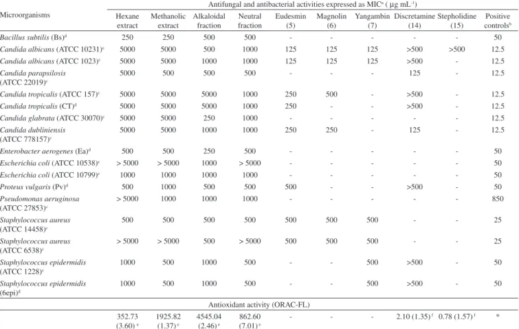
The hexane and methanol crude extracts, as well as the alkaloidal and neutral fractions from methanol extract showed antifungal and antibacterial activities (Table 5). Therefore, the major compounds eudesmin (5), magnolin (6), yangambin (7), discretamine (14), and stepholidine (15), mixture of β-sitosterol (2), stigmasterol (3) and campesterol (4), and β-sitostenone (1) were also evaluated. Among them, only the lignans and the tetrahydroprotoberberine alkaloid discretamine (14)showed significant activities (Table 5). The alka-loids liriodenine (8), anonaine (11) and asimilobine (12) has been previously investigated.6-8 The lignans were active against C.
albi-cans (ATCC 10231 and ATCC 1023), all with MIC of 125 µg mL-1.
Eudesmin (5) and magnolin (6) were also active against C. dublin-iensis (ATCC 778157), while only eudesmin (5) was active against C. tropicalis (ATCC 157 and CT), both with MIC of 250 µg mL-1
(Table 5). The tetrahydroprotoberberine alkaloid, discretamine (14) was active against C. parapsilosis (ATCC 22019) and C. dubliniensis (ATCC 778157) with MIC of 125 µg mL-1. On the other hand, all
the steroids were inactive until 500 µg mL-1. MIC values lower than
100 µg mL-1 were considered as good activity, between 100 and
1000 µg mL-1 moderate activity and small activity to MIC value
higher than 1000 µg mL-1 for extracts. On the other hand, for pure
compounds MIC value lower than 100 µg mL-1 was considered as
good activity, between 100 and 500 µg mL-1 moderate activity and
small activity to value higher than 500 µg mL-1.25
Among crude extracts evaluated, methanol shows the high anti-oxidant activity which ORAC of 1925.82 µmol of TE g-1 (Table 5).
After acid-base extraction, the antioxidant activity was revealed only in the alkaloidal fraction with ORAC value of 4545.04 µmol of TE g-1. In a previous work, the aporphinoid alkaloids liriodenine (8),
anonaine (11), and asimilobine (12) were evaluated on ORAC assay and asimilobine (12) was the most active. In this work the tetrahydro-protoberberine alkaloids discretamine (14) and stepholidine (15) were investigated, although only discretamine was active with ORAC of 2.10 trolox equivalents relative (Table 5). According to these results, the antioxidant activity of the methanol extract can be attributed in part to the alkaloids asimilobine (12) and discretamine (14). Those extracts and pure compounds that shown ORACvalues higher than 1000.00 µmol of TE g-1 and 1.00 Trolox equivalent relative were
considered to have good antioxidant capacity.5,23
CONCLUSION
This is the first phytochemical and biological investigation of the stem bark of A. pickelii. The lignans found in this species support their recent reclassification from Rollinia to Annona. In this way, the alkaloids liriodenine, anonaine, and asimilobine support that A.
Table 5. Biological activities of the crude extracts, fractions and pure compounds
Microorganisms
Antifungal and antibacterial activities expressed as MICa ( µg mL-1)
Hexane extract
Methanolic extract
Alkaloidal fraction
Neutral fraction
Eudesmin (5)
Magnolin (6)
Yangambin (7)
Discretamine (14)
Stepholidine (15)
Positive
controlsb
Bacillus subtilis (Bs)d 250 250 500 500 - - - - - 50
Candida albicans (ATCC 10231)c 5000 5000 500 1000 125 125 125 >500 >500 12.5
Candida albicans (ATCC 1023)c 5000 5000 1000 1000 125 125 125 >500 - 12.5
Candida parapsilosis
(ATCC 22019)c
5000 500 500 500 - - - 125 - 12.5
Candida tropicalis (ATCC 157)c 5000 5000 5000 1000 250 500 - >500 - 12.5
Candida tropicalis (CT)d 5000 5000 5000 1000 250 - - >500 - 12.5
Candida glabrata (ATCC 30070)c 5000 5000 250 1000 - - - - - 12.5
Candida dubliniensis
(ATCC 778157)c
5000 5000 1000 1000 250 250 - 125 - 12.5
Enterobacter aerogenes (Ea)d 500 500 250 500 - - - - - 50
Escherichia coli (ATCC 10538)c > 5000 > 5000 1000 > 5000 - - - - - 50
Escherichia coli (ATCC 10799)c 1000 1000 1000 1000 - - - - - 50
Proteus vulgaris (Pv)d 500 1000 500 500 500 - - >500 - 50
Pseudomonas aeruginosa
(ATCC 27853)c
> 5000 1000 1000 1000 - - - 850
Staphylococcus aureus
(ATCC 14458)c
500 500 500 500 500 500 500 - - 25
Staphylococcus aureus
(ATCC 6538)c
> 5000 > 5000 500 > 5000 500 500 500 - - 25
Staphylococcus epidermidis
(ATCC 1228)c
1000 500 1000 500 - - 500 >500 - 50
Staphylococcus epidermidis
(6epi)d
1000 500 1000 500 - - 500 >500 - 50
Antioxidant activity (ORAC-FL) 352.73
(3.60) e
1925.82
(1.37) e
4545.04
(2.46) e
862.60
(7.01) e
- - - 2.10 (1.35) f 0.78 (1.57) f *
aMIC (minimum inhibitory concentration with 100% of microorganism inhibition) in µg mL-1; bpositive controls (chloramphenicol for bacteria strains and
ketoconazole for yeast strains); cstandard strain; dfield strain. eORAC data expressed as µmol of TE g-1 of extract or fraction, mean (%RSD, relative standard
deviation) of triplicate assays; fORAC data expressed as Trolox equivalent relative, mean (%RSD, relative standard deviation) of triplicate assays; *Positive
pickelii is a typically species of the family Annonaceae. Significant antifungal and antioxidant activities were found to the lignans and the alkaloids from A. pickelii, and then it can be a promising source for bioactive compounds. In addition, several NMR data for the al-kaloids were reviewed and are unequivocally described in this work. SUPPLEMENTARY MATERIAL
NMR data including spectra and correlation maps for compounds 11-19 are available free of charge at http://quimicanova.sbq.org.br as PDF file.
ACKNOWLEDGMENTS
The authors are grateful to FAPITEC/SE (Editais # 07/2009 and 10/2009), CNPq, CAPES, FINEP, UFPR and FAPESP for financial support and fellowships, and Professor Dr. Ana Paula do Nascimento Prata for botanical identification.
REFERENCES
1. Chatrou, L. W.; Pirie, M. D.; Erkens, R. H. J.; Couvreur, T. L. P.; Neubig, K. M.; Abbott JR.; Mols, J. B.; Maas, J. W.; Saunders, R. M. K.; Chase, M.W.; Bot. J. Linn. Soc.2012, 169,5.
2. Corrêa, M. P.; Dicionário das plantas úteis do Brasil e das exóticas cultivadas, IBDF, Rio de Janeiro, RJ, 1984.
3. Stévigny, C.; Bailly, C.; Quetin-Leclercq, J.; Curr. Med. Chem.: Anti-Cancer Agents2005, 5, 173.
4. Alali, F. Q; Liu X.-X.; McLaughlin, J. L; J. Nat. Prod.1999, 62, 504. 5. Costa, E. V.; Dutra, L. M.; Salvador, M. J.; Ribeiro, L. H. G.; Gadelha,
F. R.; Carvalho, J. E.; Nat. Prod. Res. 2013,27, 997.
6. Costa, E. V.; Da Cruz, P. E. O.; Lourenço, C. C.; Moraes, V. R. S.; Nogueira, P. C. L.; Salvador, M. J.; Nat. Prod. Res. 2013,27, 1002. 7. Costa, E. V.; Dutra, L. M.; De Jesus, H. C. R.; Nogueira, P. C. L.;
Mo-raes, V. R. S.; Salvador, M. J.; Cavalcanti, S. C. H.; Dos Santos, R. L. C.; Prata, A. P. N.; Nat. Prod. Commun.2011, 6, 907.
8. Costa, E. V.; Pinheiro, M. L. B.; Silva, J. R. A.; Maia, B. H. L. N. S.; Duarte, M. C. T.; Amaral, A. C. F.; Machado, G. M. C.; Leon, L. L.; Quim. Nova2009, 32, 78.
9. Costa, E. V.; Pinheiro, M. L. B.; Souza, A. D. L.; Barison, A.; Campos, F.R.; Valdez, R. H.; Ueda-Nakamura, T.; Filho, B. P. D.; Nakamura, C. V.; Molecules2011, 16, 9714.
10. Siqueira, C. A. T.; Oliani, J.; Sartoratto, A.; Queiroga, C. L.; Moreno, P. R. H.; Reimão, J. Q.; Tempone, A. G.; Fischer, D. C. H.; Rev. Bras. Farmacogn.2011, 21, 33.
11. Costa, E. V.; Marques, F. A.; Pinheiro, M. L. B.; Braga, R. M.; Delar-melina, C.; Duarte, M.C.T.; Ruiz, A. L. T. G.; Carvalho, J. E.; Maia, B. H. L. N. S.; J. Braz. Chem. Soc. 2011, 22, 1111.
12. Chavan, M. J.; Wakte, P. S.; Shinde, D. B.; Inflammopharmacology 2011, 19, 111.
13. Dutra, L. M.; Costa, E. V.; Moraes, V. R. S.; Nogueira, P. C. L.; Vendramin, M. E.; Barison, A.; Prata, A. P. N.; Biochem. Syst. Ecol. 2012, 41, 115.
14. De Mesquita, L. M.; Roque, N. F.; Quintana, L. M. B.; Paulo, M. D. Q.; Barbosa Filho, J. M.; Biochem. Syst. Ecol.1988, 16, 379.
15. Costa, E. V.; Pinheiro, M. L. B.; Xavier, C. M.; Silva, J. R. A.; Amaral, A. C. F.; Souza, A. D. L.; Barison, A.; Campos, F. R.; Ferreira, A. G.; Machado, G. M. C.; Leon, L. L. P. J.; J. Nat. Prod.2006, 69, 292. 16. Facundo, V. A.; Polli, A. R.; Rodrigues, R. V.; Militão, J. S. L. T.;
Sta-belli, R. G.; Cardoso, C. T.; Acta Amazonica 2008, 38, 733.
17. Della Greca, M.; Monaco, P.; Previtera, L.; J. Nat. Prod.1990, 53, 1430. 18. Batista, A. N. L.; Batista Junior, J. M.; López, S. N.; Furlan, M.; Cava-lheiro, A. J.; Silva, D. H. S.; Bolzani, V. S.; Nunomura, S. M.; Yoshida, M.; Quim. Nova2010, 33, 321.
19. Ahmed, A. A.; Mahmoud, A. A.; Ali, E. T.; Tzakou, O.; Couladis, M.; Mabry, T.J.; Gáti, T.; Tóth, G.; Phytochemistry2002, 59, 851. 20. Xue, L.; Shi, L.; Han, Y.; Xia, C.; Huynh, H. V.; Li, F. Dalton Trans.
2011, 40, 7632.
21. Leboeuf, M.; Cavé, A.; Bhaumik, P. K.; Mukherjee, B.; Mukherjee, R.; Phytochemistry1982, 21, 2783.
22. Da Cruz, P. E. O.; Costa, E. V.; Moraes, V. R. S.; Nogueira, P. C. L.; Vendramin, M. E.; Barison, A.; Ferreira, A. G.; Prata, A. P. N.; Biochem. Syst. Ecol.2011, 39, 872.
23. Salvador, M. J.; Ferreira, E.O.; Pral, E. M. F.; Alfieri, S. C.; Albuquer-que, S.; Ito, I. Y.; Dias, D. A.; Phytomedicine2002, 9, 566.
24. Ou, B.; Hampsch-Woodill, M.; Prior, R.L.; J. Agric. Food Chem.2001, 49, 4619.
25. Salvador, M. J.; Ferreira, E. O.; Mertens-Talcott, S. U.; Castro, W. V.; Butterweck, V.; Derendorf, H.; Dias, D. A.; Z. Naturforsch., C: J. Biosci. 2006, 61, 19.