Article
0103 - 5053 $6.00+0.00
*e-mail: [email protected]
The Photochemical Reactivity of Triplet
β
-Lapachone-3-sulfonic Acid towards
Biological Substrates
José C. Netto-Ferreira,*,a,b Virginie Lhiaubet-Vallet,b Andrea R. da Silva,a
Ari M. da Silva,a Aurelio B. B. Ferreiraa and Miguel A. Mirandab
aDepartamento de Química, Universidade Federal Rural do Rio de Janeiro,
BR 465 km 7, 23970-000 Seropédica-RJ, Brazil
bInstituto de Tecnología Química UPV-CSIC, Universidad Politécnica de Valencia,
Av. de los Naranjos s/n, 46022 Valencia, Spain
A reatividade fotoquímica do ácido 3-sulfônico da β-lapachona (1) frente a amino ácidos, bases nucleicas ou nucleosídeos foi determinada empregando a técnica de fotólise por pulso de laser de nanossegundo. A excitação (λ = 355 nm) de soluções deaeradas de 1, em acetonitrila, resultou na formação do seu estado excitado triplete, o qual foi suprimido eicientemente por L-triptofano, éster metílico de L-triptofano, L-tirosina, éster metílico de L-tirosina e éster metílico de L-cisteína (kq≅ 10
9 L mol-1 s-1). Para L-triptofano, L-tirosina e seus ésteres metílicos novos transientes foram formados no proceso de supressão, os quais foram atribuídos ao par de radicais resultante de uma transferência inicial de elétron do amino ácido, ou dos seus ésteres metílicos, à quinona excitada, seguida por uma transferência de próton rápida. Não foi possível a obtenção de constantes de velocidade de supressão para timina e timidina, em acetonitrila, o que pode ser devido tanto ao caráter π π* de 1 quanto ao baixo valor para a sua energia triplete. Por outro lado, a supressão de 1 por 2’-deoxaguanosina foi tão eiciente quanto para triptofano ou tirosina (kq≅ 10
9 L mol-1 s-1). O rendimento quântico para a formação de oxigênio singlete (1O
2) a partir de 1 foi determinado empregando-se estudos de emissão resolvida no tempo na região do infravermelho próximo, tendo-se obtido um valor consideravelmente alto para este rendimento quântico (Φ∆= 0,7).
The photochemical reactivity of β-lapachone-3-sulfonic acid (1) towards amino acids, nucleobases or nucleosides has been examined employing the nanosecond laser lash photolysis technique. Excitation (λ = 355 nm) of degassed solutions of 1, in acetonitrile, resulted in the formation of its corresponding triplet excited state. This transient was eficiently quenched by L-tryptophan, L-tryptophan methyl ester, L-tyrosine, L-tyrosine methyl ester and L-cysteine methyl ester (kq≅ 10
9 L mol-1 s-1). For L-tryptophan, L-tyrosine and their methyl esters new transients were formed in the quenching process, which were assigned to the corresponding radical pairs resulting from an initial electron transfer from the amino acids, or their esters, to the excited quinone, followed by a fast proton transfer. No measurable quenching rate constants could be observed in the presence of thymine or thymidine, in acetonitrile solution, which is probably due to the π π* character of triplet 1 as well as to its low triplet energy. On the other hand, the rate constant obtained when 1 was quenched by 2’-deoxyguanosine is reasonably fast (kq≅ 109 L mol-1 s-1). The quantum eficiency of singlet oxygen (1O
2) formation from 1 was determined employing time-resolved near-IR emission studies upon laser excitation and showed a considerably high value (Φ∆= 0.7).
Keywords: laser lash photolysis,ortho-quinones, triplet excited state, coupled electron/ proton transfer
Introduction
Quinones show several biological and pharmacological
activities,1-9 with their mechanism of action being
related to redox cycling, which leads to the formation of reactive oxygen species that can damage cellular
macromolecules.10,11 The quinone cytotoxicity to many
human cancer cell lines is amply recognized,12-17 acting
through inhibition of DNA repair enzymes,18 inhibition
topoisomerase IIa-mediated DNA breaks,20 andinhibition
of poly(ADP-ribose) polymerase-1.21 More recently,
Boothman and co-workers23 have clearly demonstrated that
β-lapachone, an ortho-quinone, activates a novel apoptotic
response in a number of cell lines. It was shown that the enzyme NAD(P)H:quinone oxidoreductase (NQO1)
substantially enhances the toxicity of β-lapachone in a
number of tumor types (i.e., breast, pancreatic, colon,
prostate and lung) and during neoplastic transformation.22
The characterization and reactivity of triplet
β-lapachone-3-sulfonic acid (1) has been recently reported
by us. Upon laser excitation (266 or 355 nm), 1 leads to the
formation of its triplet excited state which shows absorption maxima at 300, 380 and 650 (broad band) nm, with a
lifetime of 5 µs. Hydrogen abstraction rate constant for the
triplet 1 towards 2-propanol or 1,4-cyclohexadiene is quite
low (105 L mol-1 s-1), which has been associated with its ππ*
character. On the other hand, 4-methoxyphenol or indole
quenches the triplet excited state of 1 with a rate constant
of 109 L mol-1 s-1. Triplet 1 also reacts with electron donors,
such as triethylamine, at an almost diffusion-controlled rate, yielding the corresponding long-lived anion radical.
1
Several mechanisms account for the photosensitization
process toward biomolecules.24-26 For excited carbonyl
groups showing nπ* character, Paternò-Büchi reaction
between triplet carbonyl and thymine can yield oxetanes.24
In case the triplet carbonyl compound is higher in energy than the triplet thymine, thymine dimerization can be
observed through a triplet-triplet energy transfer process.25
More general photosensitizing mechanisms can involve either photooxidation of nucleic acid components by the sensitizer, yielding the corresponding radical pair and ultimately leading to sensitizer-protein photobinding, or a triplet-triplet energy transfer to molecular oxygen, resulting
in formation of singlet oxygen O2 (1∆
g) and other reactive
oxygen species, such as superoxide anion, hydrogen peroxide and hydroxyl radical.
In this work we show results of the laser lash photolysis
studies on the reactivity of β-lapachone-3-sulfonic acid (1)
towards biological substrates such as amino acids and their methyl esters, nucleic bases and nucleosides, as well as its
ability to form singlet oxygen, O2 (1∆
g).
Materials and Methods
Material
The solvent acetonitrile was used as received. Lapachol, 1,2-naphthoquinone, L-tryptophan, L-tryptophan methyl ester, L-tyrosine, L-tyrosine methyl ester, L-cysteine, thymine, thymidine, 2’-deoxyguanosine and perinaphthenone, from Aldrich, were used as received
(purity> 99%).
β-Lapachone-3-sulfonic acid (1) was prepared by
drop-wise addition of concentrated H2SO4 to a suspension
of lapachol in Ac2O, at 20-30 oC. After cooling and
iltering, the orange-red crystals were washed with dry
ether and recrystallized from ethanol (mp 158-160 oC).
Its spectroscopic and spectrometric properties are in full
accord with the structure proposed.27
Laser Flash Photolysis
These experiments were carried out using either the 3rd (λ
exc = 355 nm) or the fourth harmonic (λexc = 266 nm)
of a Quantel pulsed Nd:YAG laser. The single pulses were
ca. 10 ns duration, and the energy was ca. 15 mJ pulse-1.
A Xenon lamp was employed as the detecting light source. The laser lash photolysis apparatus (Luzchem, model mLFP112) consisted of a Xe lamp, a monochromator, and a photomultiplier (PMT) system made up of side-on PMT, PMT housing, and a PMT power supply. The output signal from the Tektronix oscilloscope was transferred to a personal computer for study. Samples were contained in 10 mm × 10 mm cells made of Suprasil quartz and were deaerated for at least 20 min with oxygen-free nitrogen
prior to the experiments. Concentration for the ortho
-quinone 1 was adjusted to yield an absorbance of ca. 0.3
at the excitation wavelength. Stock solutions of quenchers were prepared so that it was only necessary to add microliter volumes to the sample cell in order to obtain appropriate concentrations of the quencher.
Quenching experiments were performed using the 3rd
harmonic of the Nd-YAG laser (λexc = 355 nm) since at
this wavelength β-lapachone-3-sulfonic acid (1) is the
only absorbing species. Rate constants for the reaction of
the triplet excited state of 1 with the different quenchers
employed in this work were obtained from Stern-Volmer
plots,28 following equation 1.
kobs = ko + kq[Q] (1)
where: ko is the triplet decay rate constant in the absence of
quencher; kq is the triplet decay rate constant in the presence
O O
O
of the quencher and [Q] is the quencher concentration in mol L-1.
Singlet oxygen measurements
Singlet oxygen generation was monitored through the characteristic phosphorescence at 1270 nm, upon laser excitation of isoabsorptive solutions (Abs = 0.3)
of β-lapachone-3-sulfonic acid (1) relative to a standard
solution of perinaphthenone (quantum yield of 1.0, in
acetonitrile).29 All samples were thoroughly oxygenated
before measurement. The quantum yield of singlet oxygen formation was determined from the slope of the linear plots
of signal intensity at zero time versus laser light intensity,
employing equation 2. In all cases acetonitrile was used as solvent. A set of neutral density ilters was employed to obtain different laser intensities.
I
sample
Φsample = –––––––––– Φperinaphthenone (2)
I
perinaphthenone
where Ιsample is the emission intensity recorded for the
sample,Ιperinaphthenoneis the perinaphthenone emission
intensity (used as standard) and Φperinaphthenone is the quantum
yield of singlet oxygen formation from perinaphthenone. The singlet oxygen luminescence (1270 nm) was detected by means of an Oriel 71614 germanium photodiode
(5 mm2), which was coupled to the laser photolysis cell in
right-angle geometry. A Xe/HCl excimer laser (LEXTRA50
Lambda Physik) was used for excitation at λ = 308 nm
(80 mJ pulse-1, 10 ns pulse-1). A 1050 nm cut-off silicon ilter
(5 mm thick, 5 cm diameter) and a 1270 nm interference ilter were placed between the diode and the cell. The output current of the photodiode was ampliied and fed into a
TDS-640A Tektronix oscilloscope via a Co-linear 150 MHz,
20 dB ampliier.The output signal from the oscilloscope was
treated and studied by means of a personal computer.
Results and Discussion
Laser lash photolysis studies
Laser excitation (266 nm) of a deoxygenated acetonitrile
solution of 1 leads to the formation of its triplet excited state
which shows absorption bands at 300, 380 and 650 nm, as
previously reported (Figure 1).23 This transient decays by irst
order kinetics and shows a lifetime of 5 µs (Figure 1, inset).
Quenching studies for triplet 1 by amino acids were
performed employing L-tyrosine, L-tryptophan, their corresponding methyl esters, and cysteine methyl ester. In
all cases, linear quenching plotsfollowing equation 1 were
obtained and the resulting quenching rate constants (kq)
are of the order of 109 L mol-1 s-1, as shown in Table 1. It
is worth noting that in the quenching experiments samples
were excited at 355 nm (3rd harmonic of a Nd-YAG laser),
since at this wavelength the only absorbing species is the
β-lapachone-3-sulfonic acid.
Transient absorption spectra recorded for samples
containing β-lapachone-3-sulfonic acid (1), in the presence
of L-tyrosine, L-tryptophan and their corresponding methyl esters, showed the disappearance of the absorption bands corresponding to its triplet excited state and the formation of new absorption bands at 330 (weak) and 370 (strong) nm (Figure 2). These absorptions were assigned
to the semiquinone radical derived from 1 (Scheme 1),
which is easily formed when quinones are irradiated in the presence of suitable hydrogen donors. At shorter timescales (400 ns), one can observe that the semiquinone
radical (λmax = 370 nm) grows-in with irst order kinetics
(Figure 2, inset). Due to the strong absorption at 380 nm
for the semiquinone radical derived from 1, we were not
able to observe the L-tyrosinyl radical (λmax = 380 nm)30,31
formed through hydrogen transfer reaction from L-tyrosine
or L-tyrosine methyl ester to triplet 1 (Figure 2). On the
Table 1. Second order rate constants for the quenching of β-lapachone-3-sulfonic acid (1) by nucleic acids constituents, in acetonitrile
Quencher kq (L mol-1 s-1)
L-Tyrosine (1.1 ± 0.1) × 109
L-Tyrosine methyl ester (7.3 ± 0.6) × 109
L-Tryptophan (3.6 ± 0.8) × 109
L-Tryptophan methyl ester (3.9 ± 0.3) × 109
L-Cysteine methyl ester (5.5 ± 1.0) × 109
Thymine a
Thymidine a
2’-Deoxyguanosine (1.3 ± 0.1) × 109 ano measurable rate constants.
other hand, when L-tryptophan or its methyl ester were employed as quenchers, the L-tryptophanyl radical, which
shows a broad absorption in the 450-550 nm region,30,31 was
easily observed (Figure 3).
The hydrogen transfer reaction from phenol and indole to triplet excited carbonyls can be usually explained by a mechanism in which, after the formation of an initial hydrogen-bonded triplet exciplex, an electron transfer process is followed by an ultra-fast proton transfer, as
exemplified in Scheme 2 for the reaction of triplet 1
with phenol. This mechanism will lead ultimately to
the semiquinone-phenoxyl radical pair.23,32-43 A similar
mechanism can be used to explain the high values for the
quenching rate constants of triplet 1 by the amino acids
L-tryptophan and L-tyrosine and their methyl esters.
Figure 3. Absorption spectra for the transient generated upon 355 nm excitation of β−lapachone-3-sulfonic acid, in acetonitrile, in the presence of 1.3x10⁻⁴ mol L-1 tryptophan methyl ester and recorded () 1.4; ()
10.0; () 35.0 and () 70.5 µs after the laser pulse.
L-Cysteine is an amino acid that can mimic the reactivity of thiol-containing enzymes such as topoisomerases. The
quenching rate constant of triplet 1 by L-cysteine methyl
ester is of the same order of magnitude as that observed
for the other amino acids: ca. (5.5 ± 1.0) × 109 L mol-1 s-1
(Table 1). However, for this amino acid, no new transients could be observed. This could be related to the fact that one of the proposed mechanisms for the cytotoxic action of quinones involves the alkylation of thiol groups on the
thiol-containing enzymes.44 This same mechanism could
be operating in the quenching of the triplet excited state of
β-lapachone-3-sulfonic acid, which would explain the lack
of observation of the semiquinone radical.
As previously reported by our group, triplet 1 has ππ*
coniguration23 and, as stated before, oxetane formation
O O
O
SO3H
OH O
O
SO3H
O OH
O
SO3H
hν
RH, ACN
OH O
O
SO3H
.
O OH
O
SO3H
.
.
.
Scheme 1. Figure 2. Absorption spectra for the transient generated upon 355 nm excitation of β−lapachone-3-sulfonic acid, in acetonitrile, in the presence of 1.3x10⁻⁴ mol L-1 tyrosine methyl ester, and recorded 0.2 (); 12.3 ();
in the quenching by thymine of triplet carbonyls is only possible for those compounds having a triplet state with
nπ* coniguration.24 Furthermore, since the triplet energy
for β-lapachones is relatively low (ET = 46 kcal mol-1 for
β-lapachone),45 triplet-triplet energy transfer from 1 to
thymine, leading to its dimerization, is not expected. Indeed, we were not able to measure quenching rate constants for
triplet 1 when employing thymine or thymidine as quenchers.
Unlike what was observed for these two nucleic acid constituents, 2’-deoxyguanosine efficiently quenches
the triplet excited state of β-lapachone-3-sulfonic acid
(kq = (1.3 ± 0.1) × 109 L mol-1 s-1) (Table 1). In this case, the
semiquinone radical derived from 1 was readily observed,
as shown in Figure 4. The broad absorption observed in the 500-600 nm range can be assigned to the 2’-deoxyguanosyl
radical,since it displays absorption bands at 315, 380
and 540 nm,46,47 with its irst two bands probably being
overlapped by the semiquinone radical absorptions, which has strong bands at the same wavelengths.
The above results indicate that the photochemical
behavior of β-lapachone-3-sulfonic acid (1) towards
biological substrates is similar to that observed for
β-lapachone and nor-β-lapachone, which was recently
Scheme 2.
O O
O
SO3H
ACN *3
+
OH
O O
O
SO3H
H O
*3
δ+
.
δ
-.
exciplex
electron transfer
O O
O
SO3H
+
OH +
.
.
-ultra-fast proton transfer OH
O
O
SO3H
+ O
.
.
O OH
O
SO3H
.
OO
O
SO3H
.
reported by us.43 Thus, it is easy to conclude that the
presence of the sulfonic acid group in 1 does not affect the
photoreactivity of the ortho-quinone chromophore.
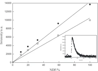
Singlet oxygen formation
Figure 5 shows plots for the singlet oxygen phosphorescence intensity versus energy dependence for
oxygenated solutions of perinaphthenone and β
-lapachone-3-sulfonic acid in acetonitrile, from which a quantum
efficiency of singlet oxygen formation Φ∆of 0.7 was
obtained. This high value is fully in accord with earlier
suggestions48-53 that a ππ* triplet is required for highly
eficient singlet oxygen formation. Similar values were
reported for other β-lapachones.43 A decay of 70 µs was
measured for singlet oxygen phosphorescence generated
by energy transfer from the lapachone 1 to oxygen (inset
of Figure 5), which is similar to that previously observed
when acetonitrile was employed as the solvent.54
Conclusions
In conclusion, it was shown that β-lapachone-3-sulphonic
acid (1) is able to act as photosensitizer for the one-electron
oxidation of L-tryptophan, L-tyrosine and their methyl esters, as well as of 2’-deoxyguanosine. Furthermore, L-cysteine
methyl ester also is an eficient quencher of triplet 1, but
may be acting by a different mechanism. Besides, eficient
singlet oxygen formation was measured for this β-lapachone
derivative (φ∆= 0.7). These results clearly demonstrate that 1
is able to photosensitize biological substrates by both type I and type II mechanisms, with its reactivity being similar
to other β-lapachones, as already reported in the literature.
Acknowledgments
Financial Support by the Spanish Government (Grant PHB2008-0104-PC) is gratefully acknowledged. We thank Prof. Julia Pérez-Prieto for the use of the Laser Flash Photolysis facilities at the Universidad de Valencia,
Spain.V.L-V thanks the Ramon y Cajal program of the
Spanish government for a research contract.AMS and
ARS thank Coordenação de Aperfeiçoamento do Pessoal do Ensino Superior (CAPES-Brazil) and Conselho Nacional de Desenvolvimento Cientíico e Tecnológico (CNPq-Brazil), respectively, for graduate fellowships. JCN-F thanks Generalitat Valenciana for a Visiting Professor fellowship.
References
1. Subramanian, S.; Ferreira, M. M. C.; Trsic, M.; Struct. Chem. 1998, 9, 47.
2. Pinto, C. N.; Dantas, A. P.; Moura, K. C. G.; Emery, F. S.; Polequevitch, P. F.; Pinto, M. D.; Castro, S. L.; Pinto, A. V.; Arzneim. Forsch. 2000, 50, 1120.
3. Dos Santos, A. F.; Ferraz, P. A. L.; Pinto, A. V.; Pinto, M. C. F. R.; Goulart, M. O. F.; Sant’Ana, A. E. G.; Int. J. Parasitol. 2000, 30, 1199.
4. Dos Santos, A. F.; Ferraz, P. A. L.; Abreu, F. C.; Chiari, E.; Goulart, M. O. F.; Sant’Ana, A. E. G.; Planta Med. 2001, 67, 92.
5. Teixeira, M. J.; De Almeida, Y. M.; Viana, J. R.; Holanda, J. G.; Rodrigues, T. P.; Prata, J. R. C.; Coelho, I. V. B.; Rao, V. S.; Pompeu, M. M. L.; Phytotherapy Res. 2001, 15, 44. 6. Rao, K. V.; Cancer Chemother. Rep. Part 3 Prog. Inf.-Suppl.
1974, 4, 11.
7. Gafner, S.; Wolfender, J.-L.; Nianga, M.; Stoeckli-Evans, H.; Hostettmann, K.; Phytochemistry 1996, 4, 1315.
8. Moura, K. C. G.; Emerya, F. S.; Neves-Pinto, C.; Pinto, M. C. F. R.; Dantas, A. P.; Salomão, K.; Castro, S. L.; Pinto, A. V.; J. Braz. Chem. Soc. 2001, 12, 325.
9. Pardee, A. B.; Li, Y. Z.; Li, C. J.; Curr.Cancer Drug Targets 2002, 2, 227.
10. Powis, G.; Pharmacol. Ther.1987, 35, 57.
11. DoCampo, R.; Cruz, F. S.; Boveris, A.; Muniz, R. P.; Esquivel, D. M.; Biochem. Pharmacol. 1979, 28, 723.
12. Bolton, J. L.; Trush, M. A.; Penning, T. M.; Dryhurst, G.; Monks, T. J.; Chem. Res. Toxicol. 2000, 13, 136.
13. Driscoll, J. S.; Hazard, G. F.; Wood, H. B.; Cancer Chemother. Rep. Part 2 Suppl. 1974, 4, 1.
14. Lopes, J. N.; Cruz, F. S.; DoCampo, R.; Ann. Trop. Med. Parasitol. 1978, 72, 523.
15. Li, C. J.; Zhang, L. J.; Dezubw, B. J.; Crumpacker, C. S.; Pardee, A. B.; Proc. Natl. Acad. Sci. USA 1993, 90, 1839.
Figure 5. Plots for singlet oxygen phosphorescence emission versus
16. Dolan, M. E.; Frydman, B.; Thompson, C. B.; Diamond, A. M.; Garbiras, B. J.; Safa, A. R.; Beck, W. T.; Marton, L.; Anti-Cancer Drugs 1998, 9, 437.
17. Li, C. J.; Averboukh, I.; Pardee, A. B.; J. Biol. Chem. 1993, 268, 22463.
18. Boothman, D. A.; Trask, D. K.; Pardee, A. B.; Cancer Res. 1989, 49, 605.
19. Planchon, S. M.; Wuerzberger, S.; Frydman, B.; Witiak, D. T.; Hutson, P.; Church, D. R.; Wilding, G.; Boothman, D. A.; Cancer Res. 1995, 55, 3706.
20. Frydman, B.; Marton, L. J.; Sun, J. S.; Neder, K.; Witiak, D. T.; Liu, A. A.; Wang, H.-M.; Mao, Y.; Wu, H.-V.; Sanders, M. M.; Liu, L. F.; Cancer Res. 1997, 57, 620.
21. Bentle, M. S.; Bey, E. A.; Dong, Y.; Reinicke, K. E.; Boothman, D. A.; J. Mol. Hist. 2006, 37, 203.
22. Pink, J. J.; Planchon, S. M.; Tagliarino, C.; Varnes, M. E.; Siegel, D.; Boothman, D. A.; J. Biol. Chem. 2000, 275, 5416. 23. Netto-Ferreira, J. C.; Bernardes, B. O.; Ferreira, A. B. B.;
Miranda, M. A.; Photochem. Photobiol. Sci. 2008, 7, 467. 24. Encinas, S.; Belmadoui, N.; Climent, M. J.; Gil, S.; Miranda,
M. A.; Chem. Res. Toxicol. 2004, 17, 857
25. Chouini-Lalanne, N.; Defais, M.; Paillous, N.; Biochem. Pharmacol. 1998, 55, 441.
26. Lhiaubet, V.; Paillous, N.; Chouini-Lalanne, N.; Photochem. Photobiol. 2001, 74, 670.
27. Fieser, L. F.; J. Am. Chem. Soc. 1948, 70, 3232. 28. Stern, O.; Volmer, M.; Phys. Z. 1919, 20, 183.
29. Nonell, S.; González, M.; Trull, F. R.; Ainidad 1993, 50, 445. 30. Merényi, G.; Lind, J.; Shen, X. H.; J. Phys. Chem. 1988, 92,
134.
31. Schuler, R. H.; Neta, P.; Zemel, H.; Fessenden, R. W.; J. Am. Chem. Soc. 1976, 98, 3825.
32. Pérez-Prieto, J.; Boscá, F.; Galian, R. E.; Lahoz, A.; Domingo, L. R.; Miranda, M. A.; J. Org. Chem. 2003, 68, 5104. 33. Das, P. K.; Encinas, M. V.; Scaiano, J. C.; J. Am. Chem. Soc.
1981, 103, 4154.
34. Turro, N. J.; Engel, R.; J. Am. Chem. Soc. 1969, 91, 7113. 35. Biczók, L.; Bérces, T.; Linschitz, H.; J. Am. Chem. Soc. 1997,
119, 11071.
36. Leigh, W. J.; Lathioor, E. C.; St Pierre, M. J.; J. Am. Chem. Soc.
1996, 118, 12339.
37. Miranda, M. A.; Lahoz, A.; Boscá, F.; Metni, M. R.; Abdelouahab, F. B.; Pérez-Prieto, J.; Chem. Commun.2000, 2257.
38. Miranda, M. A.; Lahoz, A.; Matínez-Mañez, R.; Boscá, F.; Castell, J. V.; Pérez-Prieto, J.; J. Am. Chem. Soc.. 1999, 121, 11569.
39. De Lucas, N. C.; Correa, R. J.; Albuquerque, A. C. C.; Firme, C. L.; Garden, S. J.; Bertoti, A. R.; Netto-Ferreira, J. C.; J. Phys. Chem. A 2007, 111, 1117.
40. Lathioor, E. C.; Leigh, W. J.; Photochem. Photobiol. 2006, 82, 291.
41. Netto-Ferreira, J. C.; Bernardes, B. O.; Ferreira, A. B. B.; Lhiaubet-Vallet, V.; Miranda, M. A.; Phys. Chem. Chem. Phys. 2008, 10, 6645.
42. De Lucas, N. C.; Elias, M. M.; Firme, C. L.; Corrêa, R. J.; Garden, S. J.; Nicodem, D. E.; Netto-Ferreira, J. C.; J. Photochem. Photobiol., A 2009, 201, 1.
43. Netto-Ferreira, J. C.; Bernardes, B. O.; Ferreira, A. B. B.; Lhiaubet-Vallet, V.; Miranda, M. A.; Photochem. Photobiol. 2009, 85, 153.
44. Neder, K.; Marton, L. J.; Liu, L. F.; Frydman, B.; Cell. Mol. Biol. 1998, 44, 465.
45. Ci, X. H.; Silva, R. S.; Nicodem, D. E.; Whitten, D. G.; J. Am. Chem. Soc. 1989, 111, 1337.
46. Steenken, S.; Jovanovic, S. V.; J. Am. Chem. Soc. 1997, 119, 617.
47. Misiaszeck, R.; Crean, C.; Geacintov, N. E.; Shairovich, V.; J. Am. Chem. Soc. 2005, 127, 2191.
48. Darmanyan, A. P.; Foote, C. S.; J. Phys. Chem. 1993, 97, 5032. 49. Redmond, R. W.; Braslavsky, S. E.; Chem. Phys. Lett. 1988,
148, 523.
50. Schmidt, R.; Tanielian, C.; Dunsbach, R.; Wolff, C.; J. Photochem. Photobiol. A 1994, 79, 11.
51. Oliveros, E.; Suardi-Murasecco,P.; Aminian-Saghai, T.; Braun, A. M.; Hansen, H.-J.; Helv. Chim. Acta 1991, 74, 79. 52. Nau, W. M.; Scaiano, J. C.; J. Phys. Chem. 1996, 100, 11360. 53. Hora Machado, A. E.; Miranda, J. A.; Oliveira-Campos, A. M.
F.; Severino, D.; Nicodem, D. E.; J. Photochem. Photobiol. A 2001, 146, 75.
54. Wilkinson, F.; Helman, W. P.; Ross, A. B.; J. Phys. Chem. Ref. Data 1995, 24, 663.
Received: October 5, 2009