Mg-Containing Hydroxyapatite Coatings Produced by Plasma Electrolytic
Oxidation of Titanium
César Augusto Antônioa,b*, Elidiane Cipriano Rangela, Steven Frederick Durranta, Adriana de
Oliveira Delgado-Silvac, Manfredo H. Tabacniksd, Nilson Cristino da Cruza
Received: September 18, 2016; Revised: January 17, 2017; Accepted: February 12, 2017
Plasma Electrolytic Oxidation (PEO) is promising for the processing of biomaterials because it enables the production of surfaces with adjustable composition and structure. In this work, aimed at the improvement of the bioactivity of titanium, PEO has been used to grow calcium phosphide
coatings on titanium substrates. The efects of the addition of magnesium acetate to the electrolytes
on the composition of the coatings produced during 120 s on Ti disks using bipolar voltage pulses and solutions of calcium and magnesium acetates and sodium glycerophosphate as electrolytes have been studied. Scanning electron microscopy, X-ray energy dispersive spectroscopy, Rutherford
backscattering spectroscopy, X-ray difractometry with Rietveld reinement and proilometry were used to characterize the modiied samples. Coatings composed of nearly 50 % of Mg-doped hydroxyapatite have been produced. In certain conditions up to 4% Mg can be incorporated into the coating without any observable signiicant structural modiications of the hydroxyapatite.
Keywords: Plasma Electrolytic Oxidation, Mg-doped hydroxyapatite, titanium
* e-mail: [email protected]
1. Introduction
Owing to high biocompatibility, good corrosion and wear resistance, good mechanical properties, low density and chemical inertness, titanium and its alloys are widely used in the production of dental and orthopedic implants1,2.
Although titanium is biocompatible3,4, its intrinsic
bioinertness does not stimulate spontaneous bone-implant integration5. To expand the successful applications as dental and orthopedic implants several methods are used to modify the titanium surface with the objective of improving its bioactivity. In this context, great improvement in biological properties, consequently reducing the rejection of implants6,
has been achieved with calcium phosphate coatings.
Hydroxyapatite (Ca10 (PO4)6 (OH)2) (HA) is a calcium phosphate that constitutes about 70% in weight of human
bones. HA is a natural mineral that has excellent biological properties. When it is synthetically produced on the surface of titanium, it can promote bioactivity and improve the connection with bone tissue7. Nowadays it is possible to
produce synthetic HA with similar characteristics to those of human bone, including bioactivity and biocompatibility, that can be used in biomedical applications8,9.
Over the last decade, various methods for producing HA coatings on titanium and its alloys have been developed10-25. These include plasma spray10-18, sputtering19,20, electrophoretic
deposition21,22, immersion in simulated body luid (SBF)23,
biomimetic techniques24, sol-gel procedures25,26 and laser
ablation27. There are, however, some problems related to the
hydroxyapatite coatings, such as poor control of the chemical composition and structure and poor coating adhesion to the substrate5,28-30. Therefore, there is still a demand for methods to produce HA with greater bioactivity and similarity to human bone combined with good mechanical properties31,32.
The absorption of nutrients and the growth of bone are improved by the presence of ions such as Zn+2, Mg+2, Na+, CO
3 -2
and F in non-stoichiometric compounds of low crystallinity33-35. Owing to that, improvements in the biological properties and greater similarity to human bone can be achieved by the incorporation of ionic elements into synthetic HA36,37. Magnesium is the fourth most abundant ion present in the
human body, where it helps to inhibit crystallization, to reduce crystal size, to decrease the proliferation and activities of osteoblast-like cells38-41. Therefore, magnesium deiciency can afect bone metabolism and growth, reducing osteoplastic
activity and resulting in fragile bones42-46.
Plasma Electrolytic Oxidation (PEO) combines the efects
of conventional electrolysis with micro-arc discharges that appear on the sample surface. When the voltage applied
a Laboratory of Technological Plasmas, Sorocaba Institute of Science and Technology, Paulista State
University - UNESP, Sorocaba, SP, Brazil
between two electrodes immersed in electrolytic solutions exceeds several hundred volts, the dielectric barrier of the oxide coating produced on the metal surface at the anode is
broken down mostly by impact ionizations. Consequently,
many high energy micro-arcs able to melt the oxide coating arise on the surface of the sample. In this condition, chemical elements present in the electrolytic solution can be incorporated into the coating. Also, the high thermal energy from the micro-arcs can produce ceramic coatings with complex structures. This method has been successfully used to produce ceramic coatings on light weight metals
such as Ti, Al, Nb, Ta and Mg47.
Recent studies have shown that HA can be produced by PEO of titanium and its alloys. Usually, however, long process times (>20 min) or two-step procedures are required and the HA produced is of low crystallinity48,49. In titanium
oxidation using PEO other structures such as anatase, rutile, and phosphates are also produced. Results of X-ray
difraction (XRD) studies have shown that the proportion
of HA is generally low, i.e., anatase and rutile phases are predominant50. To our best knowledge there were no studies
of the production of HA by PEO with Mg incorporation
into the coating for improved bioactivity of the synthetic HA. Thus, this study aims to produce a coating on Grade 4
titanium with a high concentration of Mg-doped HA with
very short treatment time.
2. Experimental description
The HA coatings were deposited on grade 4 titanium disks 8 mm in diameter and 2 mm thick. After machining and polishing, the substrates were ultrasonically cleaned and stored. Figure 1 shows schematically the treatment system. The treatments have been performed using a two liter stainless steel tank, enclosed in a cooling system that keeps the temperature of the solution constant. The substrate was connected to the positive terminal of a pulsed bipolar voltage supply and the tank itself served as the cathode. The electrolytic solution used for the treatment was prepared
by diluting 0.2 M of calcium acetate and 0.02 M of sodium
glycerophosphate in deionized water. The variable process
parameter was the amount CM of magnesium acetate (MgA) added in concentrations of 0.0, 0.02, 0.04, 0.06 and 0.08 M to
the electrolytic solution. The samples have been biased with positive pulses of 480 V with frequency and duty cycle of
100 Hz and 60 %, respectively. The substrates were treated
for 120 s in the potentiostatic mode. After PEO treatment, the samples were cleaned with distilled water and dried.
A previous study by the authors51 concluded that a
treatment time of 120 s was suicient to produce a coating
with a high HA content. Therefore, based on that study, all the
other process parameters were maintained; only a diferent amount of MgA in the electrolytic solution was used.
Figure 1. Schematic representation of the treatment cell.
After metallographic preparation of cross-sectioned samples, coating thickness was measured using scanning electron microscopy
(SEM). Roughness was investigated using proilometry and surface morphology was examined by SEM.
The chemical composition of the samples was analyzed
using Rutherford backscattering spectrometry (RBS). A He+
beam, from a Pelletron-tandem accelerator produced by
National Electrostatic Corporation (NEC) model 5SDH,
was directed onto the samples in a vacuum chamber at 7° incidence and 170° scattering angles. The energy of the ions
was 3.29 MeV and the beam charge was 10 µC. Chemical composition and elemental depth proiles were determined by computer simulations using RUMP52 and SIMNRA53 softwares.
Analysis of the coating crystalline structure was based
on X-ray difraction data obtained using a Panalytical X-Pert Powder difractometer in theta-2 theta geometry, which employed Cu-Kα radiation (λ = 1.54 Å). Data acquisition was performed at difraction angles of between 20 and 60°, using a step-size of 0.02° at 5 s per step. The Rietveld Reinement
technique was employed to determinate the proportion of each phase as well as hydroxyapatite cell parameters. The
reinement was performed using X’Pert HighScorePlus software with the structural model (ISCD database) listed
in Table 1. The quantity of each phase was calculated, but the cell parameters were determined only for the HA phase, the main object of study in this research. The electric current through the electrolytic cell was measured with a digital amperimeter every 2 s during the treatment. Three measurements were performed for each treatment condition.
3. Results and Discussion
3.1. Current density characteristics.
Table 1. Structural model (ISCD database) of the possible phases present in the PEO coating.
Phase Formula ICSD code
Anatase TiO2 200392
Rutile TiO2 36413
Titanium Ti 43614
Calcium Phosphate Ca5P8 74854
Hydroxyapatite Ca5(PO4)3 (HO) 203027
Magnesium Phosphide Mg3P2 24489
Figure 2. (a) Current density as a function of treatment time and
(b) average current as a function of CM.
In a general way, the addition of MgA to the electrolytic
solutions increased the current density due to the increment of ion densities in solution. The maximum value was observed
for treatments with CM = 0.06 M. The current density in these
treatment conditions was 0.99 (± 0.012) A cm-2, which is 24%
higher than the current density measured during treatments
with CM = 0.0 M.
Inspection of Figure 2 (b) reveals some variations in the current density, which were caused by instabilities generated by micro-arc discharges produced when the ion density was
increased. The amount of MgA increased linearly but the
micro-arcs occur in thermodynamic non-equilibrium and the electrolysis does not occur according to the usual electrochemical laws.
Consequently, detailed explanations of the observed variations
in the current densities are not available.
3.2. Thickness and roughness
The coating thicknesses were measured from MEV images
of sample cross-sections prepared by metallography. Averages and standard deviations determined from measurements of the thicknesses of three samples are given. Figures 3
(left and right) show the thicknesses as a functions of CM
and typical cross-sections of samples treated by PEO. The addition of magnesium acetate to the electrolytic solutions
did not change signiicantly the coating thickness. Samples treated in solutions with CM up to 0.06 M had thicknesses of around 10 µm but samples treated at a CM of 0.08 M had thicknesses of around 8.0 µm. It was also was observed
that dispersion of the thickness measurements decreases
when the CM was 0.06 M. Therefore, the results show that this condition (CM = 0.06 M) produced a more uniform
coating because the process combined the best parameters of current density, voltage, composition of the electrolytic solution and treatment time.
Figure 3. (left) SEM micrograph of a typical cross section of a
samples treated with CM = 0.06 M. (right) Thickness of the coatings
as a function of CM.
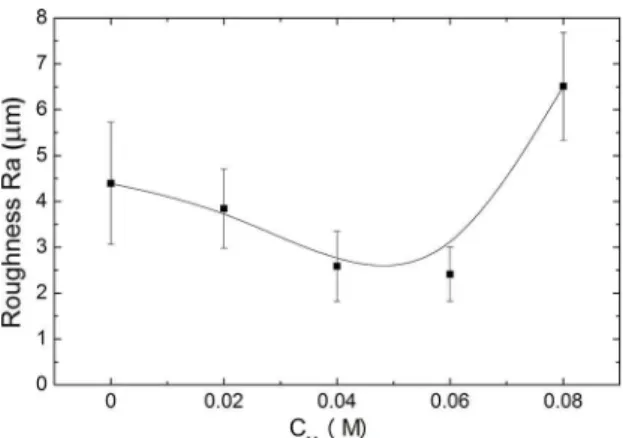
As it can be noticed in Figure 4, which shows the Ra
roughness as a function of CM, the roughness decreases
with the increasing of CM up to 0.06 M and then grows to reach 6.5 ± 1.17 µm when CM = 0.08 M. This result relects the modiication of the intensities of the micro-arc as the proportion of magnesium acetate is modiied.
Figure 4. Roughness of titanium samples treated by PEO at
diferent CM.
3.3. Coating Morphology
Figure 5 presents SEM micrographs of surfaces produced in solutions with various CM. It can be pointed out the presence of clusters of granular structures on the
surface of all the samples produced with CM < 0.08 M. As
previously reported53, such structures are mostly composed of hydroxyapatite. Therefore, the decrease of the density
of such clusters in Figure 5 suggests that the higher CM the lower the proportion of HA.
Figure 5. SEM micrographs of titanium samples treated by PEO
3.4. Chemical compositions
As previously noted the samples present porous irregular surfaces. As can be observed in Figure 6, which shows
typical simulated and experimental RBS spectra, such rough morphology makes rather diicult the theoretical adjustment of the measured RBS spectra. Therefore, to ensure a good
agreement between theoretical and experimental values, the simulations were restricted to the outermost 100 nm thick layer of all the samples. It is important to mention that the complexities of the simulations imposed by the irregular
surface introduced an estimated error of about 5%.
Figure 6. RBS spectrum (measured and simulated) of the sample
treated by PEO with CM = 0.06 M.
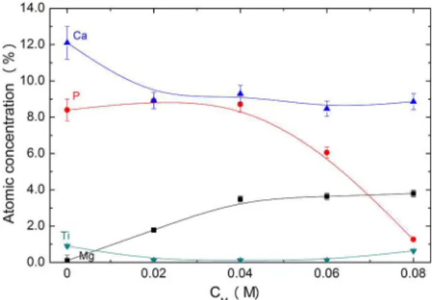
The average proportions of Ca, P, Mg, and Ti in a 100 nm thick supericial layer can be observed in Figure 7. The proportions of carbon (nearly 32%) and oxygen (approximately 47%) have been omitted since they do not provide any useful
information to the discussions that follow. According to
the results, the addition of MgA to the electrolytes caused the Ca concentration to decrease from 12.2 to 8.9 at%, independently of CM. On the other hand, the concentration
of phosphorous remained constant at 8.4 at% and decreased when MgA was added in proportions larger than 0.04 M reaching 1.2 at% when CM = 0.08 M. This strong decrease can be attributed to the low boiling point of P (553.6 K),
which can easily evaporate from sample surface. It has
been observed the intensiication of the light emitted by the
micro-arcs as magnesium acetate is added to the solution. This observation associated to the higher current observed in Figure 2 indicates the enhancement of the energy transferred
to the sample when MgA is added to the solution, which
may cause the heating of the coating. The heating near the surface of the sample makes impedes the deposition of low
melting point elements and also increases the difusion of species to inner coating regions. Both mechanisms contribute to the observed depletion of phosphorous. In addition, the Mg
concentration in the coating increased with the increasing of the proportion of magnesium acetate added to the solution,
Figure 7. Average atomic proportion of elements in samples
treated with diferent CM determined by RBS in a 100 nm thick
supericial layer.
reaching about 3.6 at% on samples treated with CM = 0.08 M. Less than 1.0% of titanium has been detected in all the
samples indicating that the electrophoretic deposition rather than the re-deposition of the quenched metal after melting by the micro-arc is the predominant mechanism of coating
growth. Accordingly to the RBS analyses, the surfaces
produced in this work are very promising for implants since
they are mostly composed by Ca, P, and Mg.
X-ray energy dispersive spectroscopy (EDS) has been
employed to evaluate the coating composition in bulk regions
deeper that those probed by RBS. As can be seen in Figure 8 the concentrations are signiicantly diferent from those
determine near the surface. In disagreement with what is
observed near the surface, the average concentrations of Ca,
P and Ti in the bulk are nearly independent of the amount
of magnesium acetate added up to CM = 0.04 M. Larger CM caused the proportions of Ca and P to decrease. When CM = 0.08 M the proportion of Ca is 35% smaller than that measured without the addition of Mg acetate while the proportion of titanium increases fourfold with the same variation of CM. It is interesting to mention that the proportions of calcium and phosphorous in all the conditions are roughly the same
as was detected by RBS on the supericial layer of the
samples produced without the addition of magnesium. This observation corroborates the supposition of the enhancement
of the difusion of species to deeper regions as a consequence
of the increase of the heating caused by larger currents when
the amount of MgA in the solution is increased. Moreover,
larger current densities can produce micro-arcs intense enough to reach the substrate beneath the coating resulting in the ejection of molten titanium towards the liquid. The quenching of the metal by the electrolyte explains the high
amount of Ti in the bulk of coatings as thick as 8 µm.
Figure 8. Atomic proportions determined by EDS in the bulk of
samples treated with diferent CM.
to observe that the treatments resulted in the formation of anatase, rutile, crystalline calcium phosphate, magnesium
phosphides, and hydroxyapatite. The difractograms also reveal the modiication of the crystalline structure of the
coatings as the proportion of magnesium acetate is increased.
It is possible to conclude that the higher CM, the smaller the peaks related to hydroxyapatite as becomes evident if one
notes that difractogram of the coating grown with CM = 0.08 M contains only the peaks produced by anatase, rutile
and metallic titanium.
Figure 9. XRD patterns of coatings produced by PEO with diferent
CM, where the letters stand for: H - Hydroxyapatite; P - Calcium
Phosphate, R - Rutile, M - Magnesium phosphide, A - Anatase,
and T - titanium.
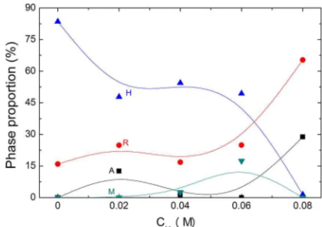
The phase composition of the samples can be better
evaluated from the results of Rietveld reinement presented
in Figure 10. The coating produced without the addition of
MgA is composed by 83.5% HA, 15,9% rutile and 0.9% calcium phosphate. Samples treated with CM = 0.02, 0.04, and 0.06 M are composed of around 50% hydroxyapatite.
The reduction in the amount of HA can be attributed to
the greater incorporation of Mg and the reduction of the
proportion of phosphorous in the coatings, as shown in Figure 8. In addition, the same amount of P was bound as
magnesium phosphide (Mg3P2).
The greatest amount of magnesium phosphide was observed in samples produced in electrolytic solutions with
Figure 10. Phases proportion calculated by Rietveld reinement
of samples treated by PEO as a function of CM. The letters stand
for: H - Hydroxyapatite, R - Rutile, M - Magnesium phosphide,
and A - Anatase
CM = 0.06 M. Therefore, this condition produced an ideal
coating for biological applications because the composition
of crystalline phases combines signiicant amounts of
hydroxyapatite, magnesium phosphide and TiO2 (rutile).
On the other hand, the samples produced with CM = 0.08 M were predominately composed by titanium (4.5%) and titanium dioxide (65.3% rutile, 28.8% anatase). No HA was formed under that condition because of the deiciency
of phosphorous.
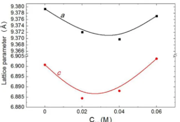
Some authors54,55 have reported that Mg2+ ions can be
incorporated into the crystalline structure of hydroxyapatite,
replacing Ca2+ ions. The Mg2+ ions have ionic radius of 0.069 nm that is smaller than radius of Ca2+ ions (0.099 nm). Thus Mg2+ can easily replace Ca2+ in the crystalline structure. This efect can also reduce the lattice parameters a and c of the hexagonal crystalline structure of the HA. In the present study, it may be observed from Figure 11 that those lattice parameters did decrease when the samples were treated with
CM = 0.02 M and 0.04 M in comparison with samples treated without addition of MgA to the electrolyte. This efect is attributed to the replacement of Ca2+ by Mg2+ ions in the
hydroxyapatite crystalline structure. The sample treated with
CM = 0.06 M presents the lattice parameters closer to the lattice parameters of samples treated without addition of MgA. The results of Rietveld reinement revealed that this coating contains 17.4% magnesium phosphide, the largest proportion
of this phase found in this study. Therefore, most of the magnesium incorporated is present as magnesium phosphide, thus explaining why the hydroxyapatite crystalline structure is similar to the crystalline structure of the hydroxyapatite
produced with CM = 0.
4. Conclusion
In the present work the eicacy of PEO to produce
microscopic observations. Journal of Biomedical Materials
Research Part A. 1993;27(6):791-800.
5. Liu F, Wang F, Shimizu T, Igarashi K, Zao L. Formation of
hydroxyapatite on Ti-6Al-4V alloy by microarc oxidation and hydrothermal treatment. Surface and Coatings Technology.
2005;199(2-3):220-224.
6. Wong M, Eulenberger J, Schenk R, Hunziker E. Efect
of surface topology on the osseointegration of implant materials in trabecular bone. Journal of Biomedical Materials
Research. 1995;29(12):1567-1575.
7. Fatehi K, Moztarzadeh F, Solati-Hashjin M, Tahriri M, Rezvannia M, Ravarian R. In vitro biomimetic deposition of apatite on alkaline and heat treated Ti6A14V alloy surface. Bulletin of Materials Science. 2008;31:101-108.
8. Halouani R, Bernache-Assolant D, Champion E, Ababou A. Microstructure and related mechanical properties of hot pressed
hydroxyapatite ceramics. Journal of Materials Science: Materials in Medicine. 1994;5(8):563-568.
9. Thomas MB, Doremus RH. Fracture strength of dense hydroxylapatite.
American Ceramic Society Bulletin. 1981;60(2):258-259. 10. Vijayaraghavan TV, Bensalem A. Electrodeposition of apatite
coating on pure titanium and titanium alloys. Journal of Materials Science Letters. 1994;13(24):1782-1785.
11. Weng W, Baptista JL. Preparation and Characterization of Hydroxyapatite Coatings on Ti6Al4V Alloy by a Sol-Gel Method. Journal of the American Ceramic Society. 1999;82(1):27-32.
12. Nishio K, Neo M, Akiyama H, Nishiguchi S, Kim HM, Kokubo T, et al. The efect of alkali- and heat-treated titanium and apatite-formed titanium on osteoblastic diferentiation
of bone marrow cells. Journal of Biomedical Materials
Research. 2000;52(4):652-661.
13. Wang CK, Lin JH, Ju CP, Ong HC, Chang RP. Structural characterization of pulsed laser-deposited hydroxyapatite ilm
on titanium substrate. Biomaterials. 1997;18(20):1331-1338.
14. Yang B, Uchida M, Kim HM, Zhang X, Kokubo T. Preparation
of bioactive titanium metal via anodic oxidation treatment.
Biomaterials. 2004;25(6):1003-1010.
15. Lim YM, Park YJ, Yun YH, Hwang KS. Functionally graded
Ti/HAP coatings on Ti-6Al-4V obtained by chemical solution deposition. Ceramics International. 2002;28(1):37-41.
16. Yang S, Man HC, Xing W, Zheng X. Adhesion strength of
plasma-sprayed hydroxyapatite coatings on laser gas-nitrided pure titanium. Surface and Coatings Technology. 2009;203(20-21):3116-3122.
17. Kozerski S, Pawlowski L, Jaworski R, Roudet F, Petit F.
Two zones microstructure of suspension plasma sprayed hydroxyapatite coatings. Surface and Coatings Technology. 2010;204(9-10):1380-1387.
18. d’Haese R, Pawlowski L, Bigan M, Jaworski R, Martel M.
Phase evolution of hydroxapatite coatings suspension plasma
sprayed using variable parameters in simulated body luid.
Surface and Coatings Technology. 2010;204(8):1236-1246.
19. Chen S, Liu W, Huang Z, Liu X, Zhang Q, Lu X. The simulation of the electrochemical cathodic Ca-P deposition process.
Materials Science and Engineering: C. 2009;29(1):108-114. Figure 11. Hydroxyapatite lattice parameters in samples produced
by PEO with various CM.
to 50% of Mg-containing HA. Results of Rietveld reinement
have shown the decrease of the lattice parameters a and c of HA crystalline structures in coatings produced in solutions
with CM = 0.02 and 0.04 M. Such characteristics suggest the replacement of Ca by Mg ions in the HA structure, conirming the production of Mg-doped HA. The treatment with the electrolyte containing 0.06 M MgA was efective
in producing HA with a crystalline structure similar to that
of HA produced with CM = 0.0 M. In that case, the coating produced contained around 17% of magnesium phosphide.
In conclusion, this work demonstrated the versatility of PEO
since the modiication of the composition of the electrolyte
allowed the adjustment of the morphology, composition, and crystalline structure of the coatings. The PEO process
has been demonstrated to produce a large amount of
Mg-doped HA chemically bonded to the substrate in only 120 s. In addition, the coating surface is porous and rough, which are important properties for biological applications.
5. Acknowledgment
The authors thank the Brazilian agencies FAPESP and CNPq for inancial support, and Conexão for providing the
titanium samples.
6. References
1. Albrektsson T, Brånemark PI, Hansson HA, Lindström J.
Osseointegrated titanium implants. Requirements for ensuring a long-lasting, direct bone-to-implant anchorage in man. Acta
Orthopaedica Scandinavica. 1981;52(2):155-170.
2. Long M, Rack HJ. Titanium alloys in total joint replacement-a
materials science perspective. Biomaterials. 1998;19(18):1621-1639.
3. Sennerby L, Thomsen P, Ericson LE. Ultrastructure of the
bone-titanium interface in rabbits. Journal of Materials Science: Materials in Medicine. 1992;3(4):262-271.
4. Stelik DE, Sisk AL, Parr GR, Gardner LK, Hanes PJ, Lake FT,
20. Blackwood DJ, Seah KHW. Electrochemical cathodic deposition
of hydroxyapatite: improvements in adhesion and crystallinity.
Materials Science and Engineering: C. 2009;29(4):1233-1238.
21. Wang J, Huang C, Wan Q, Chen Y, Chao Y. Characterization of luoridated hydroxyapatite/zirconia nano-composite coating deposited by a modiied electrocodeposition technique. Surface and Coatings Technology. 2010;204(16-17):2576-2582.
22. Yang X, Zhang B, Lu J, Chen J, Zhang X, Gu Z. Biomimetic Ca-P coating on pre-calciied Ti plates by electrodeposition
method. Applied Surface Science. 2010;256(9):2700-2704.
23. Long LH, Chen LD, Bai SQ, Chang J, Lin KL. Preparation of dense β-CaSiO3 ceramic with high mechanical strength and
HAp formation ability in simulated body luid. Journal of the European Ceramic Society. 2006;26(9):1701-1706.
24. Zhang E, Zou C, Zeng S. Preparation and characterization of
silicon-substituted hydroxyapatite coating by a biomimetic process on titanium substrate. Surface and Coatings Technology.
2009;203(8):1075-1080.
25. Cheng K, Zhang S, Weng W, Zeng X. The interfacial study of sol-gel-derived luoridated hydroxyapatite coatings. Surface and Coatings Technology. 2005;198(1-3):242-246.
26. Zhang S, Zeng X, Wang Y, Cheng K, Weng W. Adhesion strength of sol-gel derived luoridated hydroxyapatite coatings. Surface and Coatings Technology. 2006;200(22-23):6350-6354.
27. Hu J, Wang Z, Guan T, Gao Y, Lv X, Lin X, et al. In situ
synthesis and fabrication of tricalcium phosphate bioceramic coating on commercially pure titanium by laser rapid forming.
Surface and Coatings Technology. 2010;204(23):3833-3837.
28. Wang BC, Chang E, Lee TM, Yang CY. Changes in phases and
crystallinity of plasma-sprayed hydroxyapatite coatings under heat treatment: A quantitative study. Journal of Biomedical
Materials Research. 1995;29(12):1483-1492.
29. Kangasniemi IM, Verheyen CC, van der Velde EA, de Groot K. In vivo tensile testing of luorapatite and hydroxylapatite
plasma-sprayed coatings. Journal of Biomedical Materials
Research. 1994;28(5):563-572.
30. Liu F, Wang F, Shimizu T, Igarashi K, Zhao L. Hydroxyapatite formation on oxide ilms containing Ca and P by hydrothermal
treatment. Ceramics International. 2006;32(5):527-531.
31. Jahangir AA, Nunley RM, Mehta S, Sharan A; the Washington
Health Policy Fellows. Bone graft substitutes in orthopaedic surgery. AAOS Now; 2008. Available from: <http://www. aaos.org/news/aaosnow/jan08/reimbursement2.asp>. Access in: 06/03/2027.
32. Bose S, Tarafder S. Calcium phosphate ceramic systems in
growth factor and drug delivery for bone tissue engineering: a review. Acta Biomaterialia. 2012;8(4):1401-1421.
33. Elliott JC. Structure and Chemistry of the Apatite and Other Calcium Orthophosphates. In: Elliot JC, ed. Studies in Inorganic Chemistry. Amsterdam: Elsevier; 1994. p. 111-189.
34. Mayer I, Featherstone JDB. Dissolution studies of Zn-containing
carbonated hydroxyapatites. Journal of Crystal Growth. 2000;219(1-2):98-101.
35. Abdelkader SB, Khatetech I, Rey C, Jemal M. Synthese, caractérisation et thermochimie d’apatites calco-magnésiennes hydroxylées et luorées. Thermochimica Acta. 2001;376(1):25-36. 36. Narasaraju TSB, Phebe DE. Some physico-chemical aspects of
hydroxyapatite. Journal of Materials Science. 1996;31(1):1-21.
37. Gaines RV, Skinner HCV, Foord EF, Mason B, Rosenzweig A.
Dana’s New Mineralogy. Hoboken: Wiley; 1997.
38. Sun ZP, Ercan B, Evis Z, Webster TJ. Microstructural, mechanical, and osteocompatibility properties of Mg2+/F- doped nanophase
hydroxyapatite. Journal of Biomedical Materials Research. Part A. 2010;94(3):806-815.
39. Bigi A, Falini G, Foresti E, Ripamonti A, Gazzano M, Roveri N. Magnesium inluence on hydroxyapatite crystallization.
Journal of Inorganic Biochemistry. 1993;49(1):69-78.
40. TenHuisen KS, Brown PW. Efects of magnesium on the formation of calcium deicient hydroxyapatite from CaHPO4.2H2O
and Ca4(PO4)2O. Journal of Biomedical Materials Research.
1997;36(3):306-314.
41. Le Geros RZ. Calcium phosphates in oral biology and medicine.
Monographs in Oral Science. 1991;15:1-201.
42. Percival M. Bone health & osteoporosis. Applied Nutritional Science Reports. 1999;5(4):1-6.
43. Yasukawa A, Ouchi S, Kandori K, Ishikawa T. Preparation
and characterization of magnesium-calcium hydroxyapatites.
Journal of Materials Chemistry. 1996;6(8):1401-1405.
44. Okazaki M. Crystallographic behavior of luoridate hydroxyapatites containing Mg2+ and CO
2-3 Ions. Biomaterials. 1991;12(9):831-835.
45. Kim SR, Lee JH, Kim YT, Riu DH, Jung SJ, Lee YJ, et al. Synthesis of Si, Mg, substituted hydroxyapatites and their
sintering behaviors. Biomaterials. 2003;24(8):1389-1398.
46. Rude RK, Gruber HE. Magnesium deiciency and osteoporosis:
animal and human observations. TheJournal of Nutritional Biochemistry. 2004;15(12):710-716.
47. Yerokhin AL, Nie X, Leyland A, Matthews A. Characterisation of oxide ilms produced by plasma electrolytic oxidation
of a Ti-6Al-4V alloy. Surface and Coatings Technology.
2000;130(2-3):195-206.
48. Durdu S, Deniz ÖF, Kutbay I, Usta M. Characterization and
formation of hydroxyapatite on Ti6Al4V coated by plasma electrolytic oxidation. Journal of Alloys and Compounds.
2013;551:422-429.
49. Ziani S, Meski S, Khireddine H. Characterization of Magnesium-Doped Hydroxyapatite Prepared by Sol-Gel Process. International
Journal of Applied Ceramic Technology. 2014;11(1):83-91.
50. Durdu S, Usta M. The tribological properties of bioceramic
coatings produced on Ti6Al4V alloy by plasma electrolytic oxidation. Ceramics International. 2014;40(2):3627-3635.
51. Antônio CA, Cruz NC, Rangel EC, Rangel RCC, Araujo TES, Durrant SF, et al. Hydroxyapatite coating deposited on
grade 4 Titanium by Plasma Electrolytic Oxidation. Materials
Research. 2014;17(6):1427-1433.
52. Doolittle LR. Algorithms for the rapid simulation of Rutherford
Physics Research Section B: Beam Interactions with Materials and Atoms. 1985;9(3):344-351.
53. Markina E, Mayer M, Lee HT. Measurement of He and H depth proiles in tungsten using ERDA with medium heavy
ion beams. Nuclear Instruments and Methods in Physics
Research Section B: Beam Interactions with Materials and Atoms. 2001;269(24):3094-3097.
54. Okazaki M, LeGeros RZ. Crystallographic and chemical properties of Mg-containing apatites before and after suspension
in solutions. Magnesium Research. 1992;5(2):103-108.
55. Landi E, Tampieri A, Mattioli-Belmonte M, Celotti C, Sandri M, Gigante A, et al. Biomimetic Mg-and Mg,CO3-substituted
hydroxyapatites: synthesis characterization and in vitro behavior.