Identification of
Leptospira
serovars by RFLP of the RNA polymerase
beta subunit gene (
rpo
B)
Lenice Roteia Cardoso Jung
1, Maria Rosa Quaresma Bomfim
2,3, Erna Geessien Kroon
2,
Álvaro Cantini Nunes
11Departamento de Biologia Geral, Instituto de Ciências Biológicas,
Universidade Federal de Minas Gerais, Belo Horizonte, MG, Brazil. 2Departamento de Microbiologia, Instituto de Ciências Biológicas,
Universidade Federal de Minas Gerais, Belo Horizonte, MG, Brazil. 3Universidade Ceuma, Departamento de Biologia Parasitária, São Luis, MA, Brazil.
Submitted: September 03, 2012; Approved: October 29, 2014.
Abstract
Leptospires are usually classified by methods based on DNA-DNA hybridization and the conven-tional cross-agglutination absorption test, which uses polyclonal antibodies against lipopolysaccha-rides. In this study, the amplification of the rpoB gene, which encodes the beta-subunit of RNA polymerase, was used as an alternative tool to identifyLeptospira.DNA extracts from sixty-eight serovars were obtained, and the hypervariable region located between 1990 and 2500-bp in therpoB gene was amplified by polymerase chain reaction (PCR). The 600-bp amplicons of therpoB gene were digested with the restriction endonucleasesTaqI,Tru1I,Sau3AI andMslI, and the restriction fragments were separated by 6% polyacrylamide gel electrophoresis. Thirty-five fragment patters were obtained from the combined data of restriction fragment length polymorphism (PCR-RFLP) analysis and used to infer the phylogenetic relationships among theLeptospiraspecies and serovars. The species assignments obtained were in full agreement with the established taxonomic classifica-tions. Twenty-two serovars were effectively identified based on differences in their molecular pro-files. However, the other 46 serovars remained clustered in groups that included more than one serovar of different species. This study demonstrates the value of RFLP analysis of PCR-amplified rpoB as an initial method for identifyingLeptospiraspecies and serovars.
Key words:Leptospira, rpoB gene, RFLP, serovar, DNA typing.
Introduction
Leptospirosis is a zoonotic disease of global impor-tance that has emerged as a major cause of morbidity and mortality among impoverished populations (Ko et al., 2009). Based on global data, more than 500,000 new cases of leptospirosis are reported annually, with mortality rates exceeding 10% (WHO, 1999, 2006). Multiple factors, in-cluding environmental, demographic, social, and economic factors, have contributed to the emergence of this disease, which affects a broad range of mammalian hosts, including humans, wildlife, and domestic animals (Bharti and Nally, 2003; Lauet al., 2012).
The precise identification and classification of Leptospiraspp. is important for epidemiological and public health surveillance (Mohammedet al., 2011). Leptospires are usually classified by methods based on DNA-DNA hy-bridization, whereas the cross-agglutination absorption test (CAAT), which uses polyclonal antibodies against lipo-polysaccharides (LPSs), has led to the definition of sero-vars that are today considered to be the basic systematic units ofLeptospiraspp. (Cerqueira and Picardeau, 2009; Galloway and Levett, 2010). Serological methods for the characterization of Leptospira species are complex and costly, restricting their worldwide distribution and use (Ahmedet al., 2010).
DOI: http://dx.doi.org/10.1590/S1517-838246220120018
Send correspondence to Á.C. Nunes. Universidade Federal de Minas Gerais, Campus Pampulha, Av. Antônio Carlos 6627, 31270-901 Belo Horizonte, MG, Brazil. E-mail: [email protected].
Many molecular DNA techniques have been applied to identify and classify the species and serovars of Leptospira(Ahmedet al., 2012). These include restriction endonuclease analysis (REA) of chromosomal DNA (Mar-shall et al., 1981), random amplified polymorphic DNA (RAPD) fingerprinting (Ramadass et al., 1997), DNA-DNA hybridization (Yasudaet al., 1987; Brenner et al., 1999), arbitrarily primed PCR (Ramadass et al., 2002), pulsed-field gel electrophoresis (PFGE) (Galloway and Levett, 2008) and polymerase chain reaction (PCR) of spe-cific genes followed by restriction fragment length poly-morphism analysis (RFLP) (Li et al., 2009). Recently, multilocus sequence typing has been applied as an alterna-tive to immunological methods for the identification and classification of pathogenic leptospires (Ahmed et al., 2006; Pavanet al., 2008; Leonet al., 2010; Ahmedet al., 2011; Boonsilpet al., 2013). All of these techniques men-tioned above have contributed significantly to the current taxonomic classification of theLeptospiragenus (Moreyet al., 2006; Slacket al., 2009).
Quantitative DNA-DNA hybridization to measure genetic homology has been used as a reference for the clas-sification of serovars within species (Yasudaet al., 1987; Perolatet al., 1998, Brenneret al., 1999). However, this hy-bridization method is not routinely used for the identifica-tion ofLeptospiraspecies due its complex and laborious execution, which requires the use of radioactive isotopes and is therefore restricted to reference laboratories (Morey et al., 2006). It has also been observed that some serotypes are more characteristic of a single species, while others contain both pathogenic and nonpathogenic serovars (Mo-reyet al., 2006). Furthermore, little correlation has been shown between serological classification and genotypic systems because a given serogroup can often be found in several species ofLeptospira(Ahmedet al., 2012).
In addition to DNA-DNA hybridization and the other molecular methods mentioned above, specific PCR ampli-fication of the 16S rRNA gene has contributed to the mo-lecular characterization of some species of Leptospira (Ahmedet al., 2012). The advantage of this technique is that the use of a DNA template, particularly one designed based on the region that encodes the bacterially conserved 16S rRNA gene, can clearly reveal phylogenetic relation-ships among species (Moreyet al., 2006).
La Scola et al. (2006a) have designed a universal primer pair for the identification of Leptospira species based on the gene encoding thebsubunit of RNA
polymer-ase (rpoB). These primers have been used to amplify and sequence the partialrpoB gene from 16Leptospiraspecies. According to the authors, analysis of therpoB gene “may be useful as an initial screening test for the serovar identifi-cation of a new isolate ofLeptospiraand the detection or identification of Leptospira in clinical or environmental samples”.
In previous studies, the utility of therpoB gene for spirochete distinction among various bacterial species has been demonstrated (Renestoet al., 2000; Leeet al., 2000; Khamiset al., 2004; Balamuruganet al., 2013). Thus, the aim of this study was to investigate whether the PCR-amplified fragment of rpoB in conjunction with RFLP would allow for the determination ofLeptospiraserovars.
Material and Methods
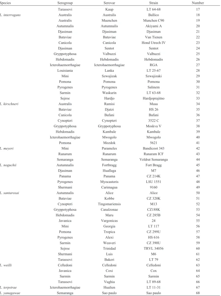
Bacterial strains, media and growth conditions For this study, sixty-eight Leptospira strains (Ta-ble 1) belonging to 11 reference species from the Pan American Institute for Food Protection and Zoonosis (INNPAZ) were used. Leptospires were grown for approxi-mately five days at 30 °C in
Ellinghausen-McCullough-Table 1- The strains, serogroups, serovars, and species of theLeptospiragenus used in this work.
Species Serogroup Serovar Strain Number
L. biflexa Andamana Andamana CH11 1
Semaranga Patoc Patoc I 2
L. borgpetersenii Autumnalis Srebarna 1409/69 3
Ballum Ballum Mus 127 4
Bataviae Moldaviae 114-2 5
Celledoni Withcombi Withcomb 6
Hebdomadis Nona Nona 7
Hebdomadis Worsfoldi Worsfoldi 8
Icterohaemorrhagiae Tonkini LT 96-68 9
Javanica Javanica Veldrat bataviae 46 10
Mini Mini Sari 11
Pyrogenes Kwale Julu 12
Sejroe Sejroe M 84 13
Tarassovi Tarassovi Perepelicin 14
L. inadai Canicola Malaya H6 15
Species Serogroup Serovar Strain Number
Tarassovi Kaup LT 64-68 17
L. interrogans Australis Australis Ballico 18
Australis Muenchen Munchen C90 19
Autumnalis Autumnalis Akiyami A 20
Djasiman Djasiman Djasiman 21
Bataviae Bataviae Van Tienen 22
Canicola Canicola Hond Utrech IV 23
Djasiman Sentot Sentot 24
Gryppotyphosa Valbuzzi Valbuzzi 25
Hebdomadis Hebdomadis Hebdomadis 26
Icterohaemorrhagiae Icterohaemorrhagiae RGA 27
Louisiania Lanka LT 25-67 28
Mini Szwajizak Szwajizaki 29
Pomona Pomona Pomona 30
Pyrogenes Pyrogenes Salinem 31
Sarmin Waskurin LT 63-68 32
Sejroe Hardjo Hardjoprajitno 33
L. kirschneri Australis Ramisi Musa 34
Bataviae Djatzi HS 26 35
Canicola Bafani Bafani 36
Cynopteri Cynopteri 3522 C 37
Gryppotyphosa Gryppotyphosa Moskva V 38
Hebdomadis Kambale Kambale 39
Icterohaemorrhagiae Mwogolo Mwogolo 40
Pomona Mozdok 5621 41
L. meyeri Mini Parameles Bandicoot 343 42
Ranarum Ranarum Ranaram ICF 43
Semaranga Semaranga Veldrat Semaranga 44
L. noguchii Autumnalis Fortbragg Fort Bragg 45
Djasiman Huallaga M7 46
Panama Panama CZ 214K 47
Pyrogenes Myocastoris LSU 1551 48
Shermani Carimagua 9160 49
L. santarosai Autumnalis Alice Alice 50
Bataviae Kobbe CZ 320K 51
Cynopteri Tingomariensis M13 52
Gryppotyphosa Canalzonae CZ188K 53
Hebdomadis Maru CZ 285B 54
Javanica Vargonicas 24 55
Mini Georgia LT 117 56
Pomona Tropica CZ 299U 57
Pyrogenes Alexi HS 616 58
Sarmin Weaveri CZ 390U 59
Sejroe Trinidad TRVL 34056 60
Shermani Luis M6 61
Tarassovi Bakeri LT 79 62
L. weilli Celledoni Celledoni Celledoni 63
Javanica Coxi Cox 64
Sarmin Sarmin Sarmin 65
Tarassovi Vughia LT 89-68 66
L. terpstrae Icterohaemorrhagiae Hualien LT 11-31 67
L. yanagawae Semaranga Sao paulo Sao paulo 68
Johnson-Harris (EMJH) culture medium (Difco) (Ellin-ghausen, 1973).
Isolation of DNA
An one-mL aliquot of eachLeptospira serovar was cultured in 5 mL EMJH medium for 7 to 10 days at 30 °C. The culture was then centrifuged at 3000 xgfor 30 min, and DNA was extracted from the bacterial pellet by adding 1 mL lysozyme solution (10 mg/mL in TE buffer (10 mM Tris and 1 mM EDTA, pH 8.0) and Wizard Genomic DNA Purification System reagents according to the manufac-turer’s instructions (Promega Co.).
PCR assays
PCR amplification of a 600-bp region of the rpoB gene was performed with the primers 1900F (5’-CCTCATGGGTTCCAACATGCA-3’) and 2500R
(5’-CGCATCCTCRAAGTTGTATTWCC-3’) as
de-scribed by La Scolaet al.(2006a). Each PCR reaction con-tained 1.5 mM MgCl2, 200mM dNTPs, 25-50 ng of DNA template, 1.5 units ofTaqDNA polymerase, and 50 pmol of each primer. The PCR amplification reactions were car-ried out in a Veriti 96-well Thermal Cycler (Applied Biosystems) under the following conditions: an initial de-naturation step of 2 min at 95 °C, 33 cycles of dede-naturation for 30 s at 94 °C, annealing at 51 °C for 30 s and extension at 72 °C for 2 min, with a final primer extension step for 10 min at 72 °C.
Restriction fragment length polymorphism (RFLP) analysis
To select enzymes for RFLP analysis, the results from in silicorestriction digestion of twenty fiverpoB sequences in GenBank®were analyzed with Webcutter 2.0 program (http://bio.lundberg.gu.se/cutter2/) to distinguish the gen-erated fragments following separation by 6% polyacryl-amide gel electrophoresis. The genomic sequences used were as follows: AE016823.1, L. interrogans serovar Copenhageni str. Fiocruz L1-130; AE010300.2, L. interrogans serovar Lai str. 56601; CP000350.1, L. borgpetersenii serovar Hardjo-bovis strain JB197; and CP000777.1, andL. biflexa serovar Patoc strain Patoc 1 (Ames). DNA sequences of therpoB gene reported by La Scolaet al.(2006a) and sequences obtained by us in this study were also used. These sequences were deposited in GenBank® under the accession numbers EU747300.1, EU747301.1, EU747302.1, EU747303.1, EU747304.1, EU747305.1, EU747306.1, EU747307.1, EU747308.1, EU747309.1, EU747310.1, EU747311.1, EU747312.1,
EU747313.1, EU747314.1, EU747317.1, and
EU747299.1, corresponding to L. interrogans serovar Bratislava, L. kirschneri serovar Grippotyphosa, L. borgpetersenii serovar Ballum, L. interrogans serovar Hardjo-prajitno, L. interrogans serovar Hebdomadis, L. borgpetersenii serovar Hardjo-bovis, L. interrogans
serovar Pomona, L. borgpeterseniiserovar Tarassovi, L. interrogansserovar Wolffi, L. biflexa serovar Andamana, L. borgpetersenii serovar Castellonis, L. borgpetersenii serovar Sejroe, L. interrogans serovar Djasiman, L. interrogansserovar Schueffneri,L. borgpeterseniiserovar Whitticombi, L. interrogans serovar Sentot, and L. interrogansserovar Canicola, respectively.
PCR products were subjected to restriction digestion withTaqI,Tru1I, Sau3AI and MslI endonucleases (Pro-mega Co.) for 3 h at the recommended temperatures. To calculate the relative molecular masses of the digested frag-ments, a 100-bp DNA Ladder was used (Promega Co.). The digestion and separation of the DNA fragments by 6% polyacrylamide gel electrophoresis were repeated at least three times for all serovars to establish the final restriction patterns.
Dendrogram construction
LabImage version 2.7.0 software (Kapelan GMBH) was used for constructing a binary matrix scored on the presence (1) or absence (2) of each fragment generated by PCR-RFLP with therpoB primers. Cluster analysis based on similarity (Nei, 1972) was performed by the unweighted pair group method (UPGMA) with the arithmetic averages clustering algorithm (Sneath and Sokal, 1973), and the ran-domization procedure implemented in Tools for Population Genetic Analysis (TFPGA) software package according to Miller (1998) was used to construct the dendrogram.
Results
In silico analysis of rpoB sequences deposited in GenBank indicated that a combination of four possible re-striction enzymes was necessary to distinguish the Leptospiraserovars as follows:TaqI,Tru1I, Sau3AI and MslI. Alone, each enzyme was able to identify only one or two different serovars.
Digestion withTaqI resulted in ten different patterns (A to J), which are schematically represented in Table 2 and had the following frequencies: A, 29.4% (20); B, 10.3% (7); C, 7.35% (5); D, 13.2% (9); E, 11.8% (8); F, 4.41% (3); G, 16.2% (11); H, 7.35% (3); I, 1.47% (1); and J, 1.47% (1). Thus,TaqI identified two serovars, Huallaga ofL. noguchii (profile I) and Alice ofL. santarosai(profile J), as shown in Figure 1. The G profile pattern was observed in almost all L. santarosaiserovars, with the exception of the serovar Alexi (profile D), and it was only identified in the Muenchen serovarL. interrogans.
The combination of both enzymes, TaqI andTru1I, generated 23 distinct patterns with some interesting results
as follows: profile A ofTaqI andTru1I (profile AA) was species-specific and was only observed forL. biflexa.
Pro-Figure 1- Polyacrylamide gel electrophoresis (6%) of the PCR products resulting from the digestion of therpoB gene with the restriction endonuclease
file AC was displayed by all serovars ofL. kirschneriand by serovar Hualien ofL. terpstrae; therefore, it is nearly species-specific. Finally, the profiles AG, FG, FE, CE, FD, BE and GE were unique to the serovars Mini, Kaup, Lanka, Szwajizak, Waskurin, Myocastoris and Maru, respectively.
Digestion with theSau3AI enzyme generated nine distinct restriction patterns, which are summarized in Table 2 with the following frequencies: A, 2.94% (2); B, 22.1% (15); C, 30.9% (21); D, 4.41% (3); E, 7.35% (5); F, 16.2% (11); G, 8.82% (6); H, 5.88% (4); and I, 1.47% (1).Sau3AI digestion identified only the serovar Ranarum to have a serovar-specific profile. However, the combination of all three enzymes generated 30 distinct profiles, including EEE, DFD, ACC, HDD, GBB and AHH, which were spe-cific for the serovars Whitcomb, Icterohaemorrhagiae, Hardjo, Ramisi, Semaranga, Vargonicas and Sarmin, re-spectively.
Finally, digestion with the enzyme MslI produced only five distinct restriction patterns, which are summa-rized in Table 2 with the following frequencies: A, 10.3% (7); B, 20.6% (14); C, 57.4% (39); D, 10.3% (7); and E, 1.47% (1). Only the serovar Saopaulo was identified by this enzyme to have a serovar-specific profile.
The combination of the four enzymes TaqI,Tru1I, Sau3AI andMslI generated 35 distinct profiles and identi-fied the serovars Parameles (EFFD) and Celledoni (HDFA). In addition, this combination helped to distin-guish the serovars Valbuzzi and Tropica, which had the profiles DFFC and GBCC, respectively (Table 3).
Out of sixty-eight serovars analyzed for RFLP poly-morphisms in the region of the coding sequence containing theb-subunit gene of RNA polymerase, 22 serovars from
nine species (32%) were identified by digestion with the enzymesTaqI,Tru1I,Sau3AI andMslI (Table 3), and the other 46 strains were clustered into 13 groups with two to seven serovars.
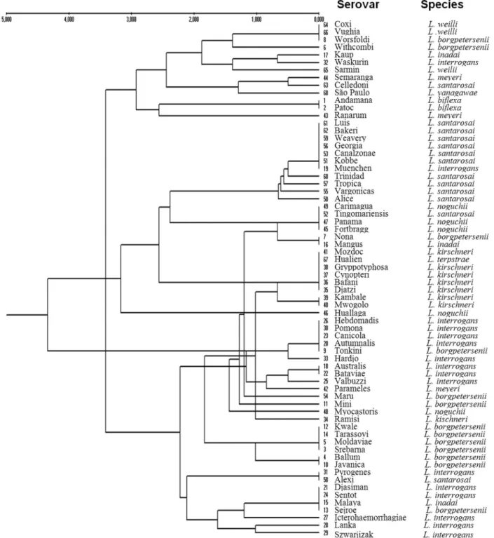
A dendrogram obtained from a matrix constructed with the results from the fragments generated by PCR-RFLP with the four restriction endonucleases (Figure 2) showed clustering of the sixty-eight reference serovars. Several of the tested strains appeared to be distant from oth-ers of the same species in relation to the current taxonomic classification. The serovar Kaup (L. inadai) was grouped with the serovar Waskurin (L. interrogans); the serovar Ranarum (L. meyeri) was similar to the nonpathogenicL. biflexa; the serovar Muenchen (L. interrogans) clustered with those of L. santarosai; the serovar Nona (L. borgpetersenii) was closer to the serovar Mangus (L. inadai); the serovar Hualien (L. terpstrae) grouped with the those of L. kirschneri; the Huallaga and Myocastoris serovars (L. noguchii) were located in different branches; the serovar Tonkini (L. borgpetersenii) grouped with the majority of those ofL. interrogans; the serovar Ramisi (L. kirschneri) was closer to those ofL. borgpetersenii; and the serovar Alexi (L. santarosai) was grouped with those of Djasiman, Pyrogenes and Sentot (L. interrogans), Malaya (L. inadai) and Sejroe (L. borgpetersenii).
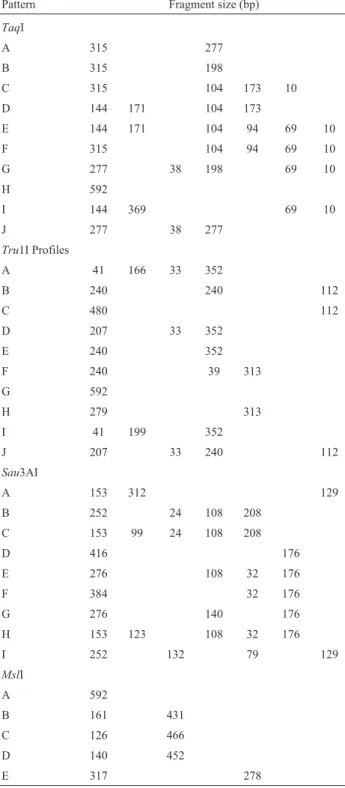
Table 2- Restriction patterns of the 600-bp fragment of therpoB gene of
Leptospira following digestion with TaqI, Tru1I, Sau3AI, and MslI
endonucleases.
Pattern Fragment size (bp)
TaqI
A 315 277
B 315 198
C 315 104 173 10
D 144 171 104 173
E 144 171 104 94 69 10
F 315 104 94 69 10
G 277 38 198 69 10
H 592
I 144 369 69 10
J 277 38 277
Tru1I Profiles
A 41 166 33 352
B 240 240 112
C 480 112
D 207 33 352
E 240 352
F 240 39 313
G 592
H 279 313
I 41 199 352
J 207 33 240 112
Sau3AI
A 153 312 129
B 252 24 108 208
C 153 99 24 108 208
D 416 176
E 276 108 32 176
F 384 32 176
G 276 140 176
H 153 123 108 32 176
I 252 132 79 129
MslI
A 592
B 161 431
C 126 466
D 140 452
Discussion
The correlation between the serological and geno-typic classifications of leptospires is low, and identification is complicated because the same serovar can be distributed among different species (Ahmedet al., 2012; Balamurugan et al., 2013). It is assumed that this lack of correlation between species and serovars is the result of horizontal transference between species of the genes that determine
serotypes, but the basis of this transference, which is re-sponsible for exchanging genetic determinants, is still unknown (Cerqueira and Picardeau, 2009). A single base difference differentiated many strains ofL. interrogansand L. kirschneri; therefore, phylogenetic representation may be less meaningful than sequence identities at variable po-sitions (Cerqueira and Picardeau, 2009).
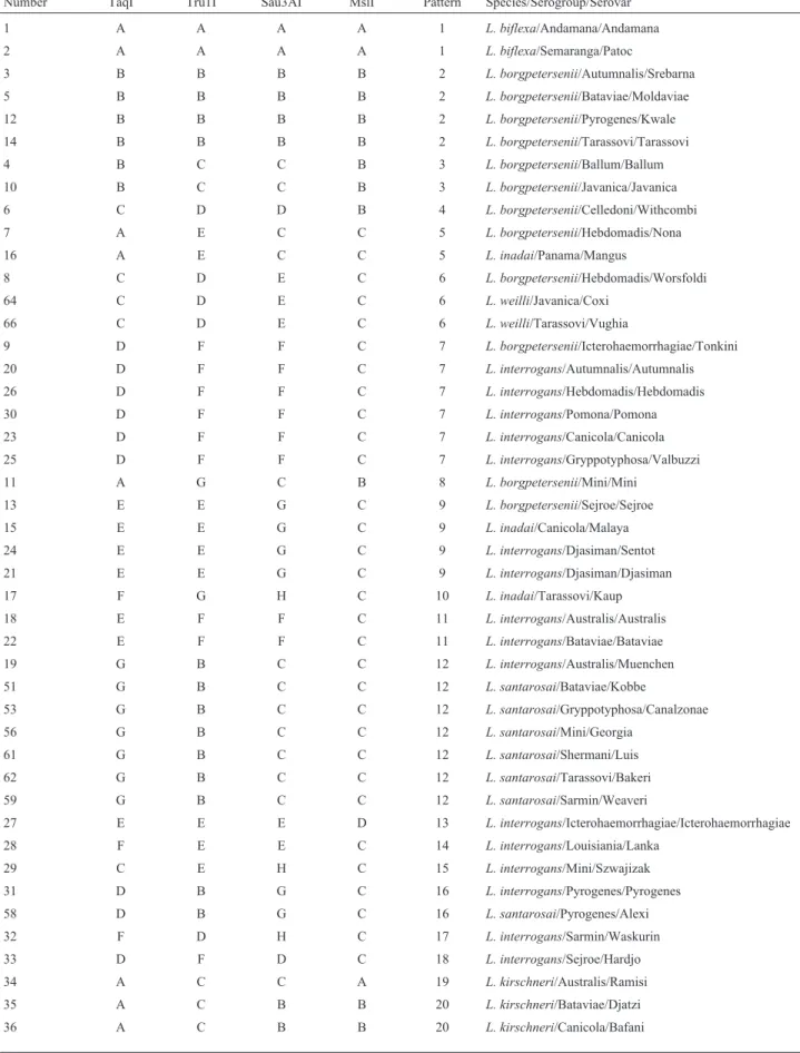
Table 3- Grouping of the serovars, serogroups and species of theLeptospiragenus based on the restriction patterns generated with the four endonucleases.
Number TaqI Tru1I Sau3AI MslI Pattern Species/Serogroup/Serovar
1 A A A A 1 L. biflexa/Andamana/Andamana
2 A A A A 1 L. biflexa/Semaranga/Patoc
3 B B B B 2 L. borgpetersenii/Autumnalis/Srebarna
5 B B B B 2 L. borgpetersenii/Bataviae/Moldaviae 12 B B B B 2 L. borgpetersenii/Pyrogenes/Kwale 14 B B B B 2 L. borgpetersenii/Tarassovi/Tarassovi 4 B C C B 3 L. borgpetersenii/Ballum/Ballum 10 B C C B 3 L. borgpetersenii/Javanica/Javanica
6 C D D B 4 L. borgpetersenii/Celledoni/Withcombi 7 A E C C 5 L. borgpetersenii/Hebdomadis/Nona
16 A E C C 5 L. inadai/Panama/Mangus
8 C D E C 6 L. borgpetersenii/Hebdomadis/Worsfoldi
64 C D E C 6 L. weilli/Javanica/Coxi
66 C D E C 6 L. weilli/Tarassovi/Vughia
9 D F F C 7 L. borgpetersenii/Icterohaemorrhagiae/Tonkini 20 D F F C 7 L. interrogans/Autumnalis/Autumnalis
26 D F F C 7 L. interrogans/Hebdomadis/Hebdomadis 30 D F F C 7 L. interrogans/Pomona/Pomona 23 D F F C 7 L. interrogans/Canicola/Canicola 25 D F F C 7 L. interrogans/Gryppotyphosa/Valbuzzi 11 A G C B 8 L. borgpetersenii/Mini/Mini
13 E E G C 9 L. borgpetersenii/Sejroe/Sejroe 15 E E G C 9 L. inadai/Canicola/Malaya 24 E E G C 9 L. interrogans/Djasiman/Sentot 21 E E G C 9 L. interrogans/Djasiman/Djasiman 17 F G H C 10 L. inadai/Tarassovi/Kaup
18 E F F C 11 L. interrogans/Australis/Australis 22 E F F C 11 L. interrogans/Bataviae/Bataviae 19 G B C C 12 L. interrogans/Australis/Muenchen 51 G B C C 12 L. santarosai/Bataviae/Kobbe
53 G B C C 12 L. santarosai/Gryppotyphosa/Canalzonae
56 G B C C 12 L. santarosai/Mini/Georgia 61 G B C C 12 L. santarosai/Shermani/Luis 62 G B C C 12 L. santarosai/Tarassovi/Bakeri 59 G B C C 12 L. santarosai/Sarmin/Weaveri
27 E E E D 13 L. interrogans/Icterohaemorrhagiae/Icterohaemorrhagiae
28 F E E C 14 L. interrogans/Louisiania/Lanka 29 C E H C 15 L. interrogans/Mini/Szwajizak 31 D B G C 16 L. interrogans/Pyrogenes/Pyrogenes 58 D B G C 16 L. santarosai/Pyrogenes/Alexi 32 F D H C 17 L. interrogans/Sarmin/Waskurin 33 D F D C 18 L. interrogans/Sejroe/Hardjo 34 A C C A 19 L. kirschneri/Australis/Ramisi 35 A C B B 20 L. kirschneri/Bataviae/Djatzi
amplify a 600-bp fragment of the coding sequence of theb
subunit of the RNA polymerase gene. TherpoB gene has been widely studied in other organisms and is considered by many researchers to be more useful than the 16S ribo-somal RNA gene for the differentiation of bacterial species (La Scolaet al., 2006a; Ahmedet al., 2006; Macheraset al., 2011; Ahmed et al., 2012). In addition, twenty-five sequences of the rpoB gene ofLeptospiraare already avail-able in databases, thereby facilitating access and minimiz-ing project costs.
In a previous report, La Scolaet al. (2006a) have compared similarities in therrsandrpoB genes between differentLeptospiraserovars. Using therpoB gene, they were able to effectively distinguish 11 of 19 serovars tested, differentiating them from other species and showing grea-ter numbers of polymorphisms in both genes, leading to the conclusion that the rpoB gene could distinguish species with a higher number of differences between base pairs.
In this study, 68Leptospiraserovars were analyzed for polymorphisms in a specific region of therpoB gene us-ing the endonucleasesTaqI,Tru1I,Sau3AI andMslI. These enzymes were selected afterin silicorestriction digestion of therpoB sequences deposited in GenBank. We were able to identify 22 strains from nine species at the serovar level
(32%). The rpoB gene has been widely used as an alterna-tive tool in the phylogeny and identification of different species of bacteria, such asCoxiella burnetii(Molletet al., 1998),Afipia(Khamiset al., 2003),Mycoplasma(Kimet al., 2003), Corynebacterium (Khamis et al., 2004), Acinetobacter (La Scola et al., 2006b), Mycobacterium (Adekambiet al., 2006; Benet al., 2008),Halobacterium (Minegishiet al., 2010) andCyanobacteria(Gagetet al., 2011).
In a recent study, the rpoB gene has been successfully used to identify or detectLeptospiraspecies in animals and humans in India (Balamuruganet al., 2013). Because each Leptospiraserovar is associated with specific host symp-toms, their identification is essential for the development of epidemiological studies (Cerqueira and Picardeau, 2009, Li et al., 2009).
Clustering analysis of the results of this study cor-rectly grouped 22 serovars by species. Considerable simi-larities in the analyzed genomic region were observed among all serovars. Analysis of dendrograms constructed from the results of each restriction enzyme and from the collective results for all of the enzymes showed the forma-tion of clusters, for which serovars of various species had identical profiles. The groups formed by therpoB gene pro-Number TaqI Tru1I Sau3AI MslI Pattern Species/Serogroup/Serovar
37 A C B B 20 L. kirschneri/Cynopteri/Cynopteri
38 A C B B 20 L. kirschneri/Gryppotyphosa/Gryppotyphosa 41 A C B B 20 L. kirschneri/Pomona/Mozdok
67 A C B B 20 L. terpstrae/Icterohaemorrhagiae/Hualien 39 A C B C 21 L. kirschneri/Hebdomadis/Kambale 40 A C B C 21 L. kirschneri/Icterohaemorrhagiae/Mwogolo
42 E F F D 22 L. meyeri/Mini/Parameles
43 A H I C 23 L. meyeri/Ranarum/Ranarum
44 H D D A 24 L. meyeri/Semaranga/Semaranga 45 A E C D 25 L. noguchii/Autumnalis/Fortbragg 47 A E C D 25 L. noguchii/Panama/Panama
49 A E C D 25 L. noguchii/Shermani/Carimagua 52 A E C D 25 L. santarosai/Cynopteri/Tingomariensis 46 I I B A 26 L. noguchii/Djasiman/Huallaga 48 B E B D 27 L. noguchii/Pyrogenes/Myocastoris 50 J B C C 28 L. santarosai/Autumnalis/Alice
54 G E C A 29 L. santarosai/Hebdomadis/Maru 55 G B B C 30 L. santarosai/Javanica/Vargonicas 57 G B C C 31 L. santarosai/Pomona/Tropica 60 G J C C 32 L. santarosai/Sejroe/Trinidad 63 H D F A 33 L. weilli/Celledoni/Celledoni
65 A H H C 34 L. weilli/Sarmin/Sarmin
files showed varying degrees of similarity and clade forma-tion. Based on this, similar banding patterns were observed among the serovars Mangus, Nona, Alexi, Pyrogenes, Sen-tot, Malaya and Sejroe, despite the fact that they belonged to different species. These findings are in accordance with similar dendrogram analyses reported previously (Perolat et al., 1998; Moreyet al., 2006; Cerqueira and Picardeau, 2009; Balamuruganet al., 2013), showing similar cluster formations and variations in serovar-species grouping.
The addition of new enzymes for the production of additional profiles should clarify the positions of other serovars. Still, these results suggest that the use of this tech-nique to assess gene sequences may reveal a precisesensu strictoclassification of these serovars.
Molecular techniques have been used for the charac-terization ofLeptospiraisolates; however, most can only make identifications to the species level (Galloway and Levett, 2010), such as 16S rRNA sequence analysis (Morey et al., 2006), RFLP (Liet al., 2009) and MLST (Boonsilpet al., 2013). PFGE has demonstrated the reliable and repro-ducible identification of Leptospira at the serovar level (Galloway and Levett, 2010). These approaches have grea-tly contributed to a revolution in bothLeptospiradetection and characterization (Ahmedet al., 2012). On the other hand, the molecular tools described so far for the character-ization ofLeptospira suffer from significant drawbacks. For example, PFGE, RFLP, and REA require large quanti-ties of purified DNA, have low levels of discrimination, produce data that is difficult to interpret, suffer from a lack of reproducibility and require specialized equipment (Ahmedet al., 2006).
Notably, the 16S rRNA gene has been considered the gold standard in molecular surveys of bacterial and archaeal diversity, but it has several disadvantages as fol-lows: it is often present in multiple copies, has little resolu-tion below the species level and cannot be readily inter-preted in an evolutionary framework (Voset al., 2012).
The main advantages of the use of therpoBgene over the 16S rRNA gene are as follows: (i) it is universally pres-ent in all prokaryotes; (ii) it typically occurs in a sin-gle-copy, essential protein-encoding gene, and sequence errors can be readily identified and removed if they intro-duce disruptions in the reading frame; (iii) it possesses both slowly and quickly evolving regions, enabling the design of probes and primers of differing specificities; (iv) it has a housekeeping function, making it less susceptible to some forms of lateral gene transfer; and (v) it is large enough in size to contain phylogenetic information, even after the re-moval of regions that are difficult to align (Case et al., 2007; Voset al., 2012).
Our findings “in vitro” indicate that the PCR-RFLP technique is a powerful and reproducible test that may be used as a complement or alternative tool to assess the distri-bution ofLeptospirastrains within species. Additionally, we recommend the use of PCR-RFLP within silico
diges-tion of the polymorphic sequences of other conserved genes already deposited in GenBank as a promising tech-nique for the genomic classification of theLeptospira ge-nus.
Conclusion
This study demonstrated that PCR-RFLP is practical and efficient, enabling the differentiation of species and serovars with good discriminatory power, reproducibility and easily interpretable results. In addition, this method is cost-effective for most research laboratories. This tech-nique has also been shown to be suitable for phylogenetic studies and the classifications of species, serovars and strains. The selected 600-bp polymorphic sequence of the rpoB gene produced restriction profiles that allowed for the accurate and timely identification of 32% of the 68 tested strains. We demonstrated that this approach achieves the stated purpose and that serological typing is unreliable for the classification of pathogenicLeptospira. However, addi-tional studies should be undertaken to reclassify these serovars within the species with which they have greater genotypic affinities based on analysis of hypervariable re-gions of multiple housekeeping genes and especially to investigate whether the clinical leptospirosis symptoms in-duced by these serovars are presented according to the spe-cies with which they are most phylogenetically related.
Acknowledgments
This study was supported by the Fundação de Ampa-ro à Pesquisa de Minas Gerais (FAPEMIG), the Conselho Nacional de Desenvolvimento Científico e Tecnológico (CNPq), the Fundação de Amparo à Pesquisa e Desen-volvimento Científico do Maranhão (FAPEMA) and the PRPq/UFMG. We thank Dr. Élvio C. Moreira from the De-partment of Preventive Medicine of the Veterinary School of UFMG for providing theLeptospirastrains and Dr. Re-gina M. Nardi Drummond and Dr. Vera Lúcia dos Santos for their assistance at the laboratory facilities with the growing and maintenance of theLeptospirastrains.
References
Adekambi T, Berger P, Raoult D et al.(2006) rpoB gene
se-quence-based characterization of emerging non-tuberculous mycobacteria with descriptions ofMycobacterium bolletii
sp. nov.,Mycobacterium phocaicumsp. nov. and Mycobac-terium aubagnense sp.nov. Int J Syst Evol Microbiol
56:133-143.
Ahmed N, Devi SM, Valverde MAet al.(2006) Multilocus se-quence typing method for identification and genotypic clas-sification of pathogenic Leptospira species. Ann Clin
Microbiol Antimicrob 5:28.
Ahmed A, Anthony RM, Hartskeerl RA (2010) A simple and rapid molecular method forLeptospiraspecies
Ahmed A, Thaipadungpanit J, Boonsilp Set al.(2011)
Compari-son of two multilocus sequence based genotyping schemes forLeptospiraspecies. PLoS Negl Trop Dis 5:e1374. Ahmed A, Grobusch MP, Klatser PRet al.(2012) Molecular
Ap-proaches in the Detection and Characterization of
Leptospira. J Bacteriol Parasitol 3:2.
Balamurugan V, Gangadhar NL, Mohandoss Net al.(2013) Char-acterization of Leptospiraisolates from animals and hu-mans: phylogenetic analysis identifies the prevalence of in-termediate species in India. Springer Plus 2:362.
Ben Salah I, Adekambi T, Raoult Det al.(2008)rpoB
sequence-based identification ofMycobacterium aviumcomplex
spe-cies. Microbiol 154:3715-3723.
Bharti AR, Nally JE (2003) Leptospirosis: a zoonotic disease of global importance. Lancet Infect Dis 3:757-771.
Brenner DJ, Kaufmann AF, Sulzer KRet al.(1999) Further
deter-mination of DNA relatedness between serogroups and sero-vars in the family Leptospiraceae with a proposal for
Leptospira alexanderi sp. nov. and four new Leptospira
genomospecies. Int J Syst Bacteriol 49 Pt2:839-858. Boonsilp S, Thaipadungpanit J, Amornchai Pet al.(2013) A
Sin-gle Multilocus Sequence Typing (MLST) Scheme for Seven PathogenicLeptospiraSpecies. PLoS Negl Trop Dis 9:e824.
Case RJ, Boucher Y, Dahllöf Iet al.(2007) Use of 16S rRNA and rpoB genes as molecular markers for microbial ecology
studies. Appl Environ Microbiol 1:278-288.
Cerqueira G, Picardeau M (2009) A century ofLeptospirastrain
typing. Infect Genet Evol 9:760-768.
Ellinghausen HC (1973) Virulence, nutrition and antigenicity of
Leptospira interrogansserotype Pomona in supplemented and nutrient deleted bovine albumin medium. Ann Micro-biol (Paris) 124:477-493.
Gaget V, Gribaldo S, Tandeau de Marsac N (2011) ArpoB signa-ture sequence provides unique resolution for the molecular typing ofcyanobacteria. Int J Syst Evol Microbiol
61:170-183.
Galloway RL, Levett PN (2008) Evaluation of a modified pul-sed-field gel electrophoresis approach for the identification ofLeptospiraserovars. Am J Trop Med Hyg 78:628-632. Galloway RL, Levett PN (2010) Application and Validation of
PFGE for Serovar Identification ofLeptospiraClinical Iso-lates. PLoS Negl Trop Dis 14:e824.
Khamis A, Colson P, Raoult Det al.(2003) Usefulness ofrpoB
gene sequencing for identification ofAfipiaandBosea spe-cies, including a strategy for choosing discriminative partial sequences. Appl Environ Microbiol 69:6740-6749. Khamis A, Raoult D, La Scola B (2004)rpoB gene sequencing for
identification ofCorynebacteriumspecies. J Clin Microbiol
42:3925-3931.
Kim KS, Ko KS, Chang MWet al.(2003) Use ofrpoBsequences
for phylogenetic study of Mycoplasma species. FEMS Microbiol Lett 226:299-305.
Ko AI, Goarant C, Picardeau M (2009)Leptospira: The Dawn of the Molecular Genetics Era for an Emerging Zoonotic Pathogen. Nat Rev Microbiol 10:736-747.
La Scola B, Bui LTM, Baranton Get al.(2006a) PartialrpoB gene sequencing for identification ofLeptospiraspecies. FEMS Microbiol Lett 263:142-147.
La Scola B, Gundi VA, Khamis Aet al.(2006b) Sequencing of the
rpoBgene and flanking spacers for molecular identification
ofAcinetobacterspecies. J Clin Microbiol 44:827-832.
Lau C, Clements A, Skelly Cet al.(2012) Leptospirosis in
Ameri-can Samoa-Estimating and mapping risk using environmen-tal data.PLoS Negl Trop Dis 6:e1669.
Lee SH, Kim BJ, Kim JHet al.(2000) Differentiation ofBorrelia burgdorferisensu lato on the basis of RNA polymerase gene (rpoB) sequences. J Clin Microbiol 38:2557-2562. Leon A, Pronost S, Fortier Get al.(2010) Multilocus Sequence
Analysis for TypingLeptospira interrogansandLeptospira kirschneri. J Clin Microbiol 48:581-585.
Li W, Raoult D, Fournier PE (2009) Bacterial strain typing in the genomic era. FEMS Microbiol Rev 33:892-916.
Macheras E, Roux AL, Bastian Set al.(2011) Multilocus se-quence analysis and rpoB sequencing of Mycobacterium abscessus(sensu lato) strains. J Clin Microbiol 49:491-499.
Marshall RB, Wilton BE, Robinson AJ (1981) Identification of
Leptospiraserovars by restriction-endonuclease analysis. J
Med Microbiol 14:163-166.
Miller MP (1998) TFPGA: Tools for Population Genetic Analy-ses for Windows. Arizona State University, USA.
Minegishi H, Kamekura M, Itoh Tet al.(2010) Further
refine-ment of the phylogeny of theHalobacteriaceaebased on the
full-length RNA polymerase subunit B9 (rpoB9) gene. Int J Syst Evol Microbiol 60:2398-2408.
Mohammed H, Nozha C, Hakim Ket al.(2011)Leptospira: Mor-phology, Classification and Pathogenesis. Bacteriol Para-sitol 2:6.
Mollet C, Drancourt M, Raoult D (1998) Determination of
Coxiellaburnetii rpoBsequence and its use for phylogenetic analysis. Gene 207:97-103.
Morey RE, Galloway RL, Bragg SLet al.(2006) Species-specific identification of Leptospiraceae by 16S rRNA gene
se-quencing. J Clin Microbiol 44:3510-3516.
Nei M (1972) Genetic distance between populations. Am Nat 106:283-292.
Pavan ME, Cairo F, Brihuega Bet al.(2008) Multiple-locus
vari-able-number tandem repeat analysis MLVA ofLeptospira interrogans serovar Pomona from Argentina reveals four new genotypes. Comp Immunol Microbiol Infec Dis 31:37-45.
Perolat P, Chappel RJ, Adler Bet al.(1998)Leptospira faineisp. nov., isolated from pigs in Australia. Int J Syst Bacteriol 48:851-858.
Ramadass P, Meerarani S, Venkatesha MDet al.(1997) Charac-terization of Leptospiral Serovars by Randomly Amplified Polymorphic DNA Fingerprinting. Int J Syst Bacteriol 47:575-576.
Ramadass P, Latha D, Senthilkumar Aet al.(2002) Arbitrarily
primed PCR- a rapid and simple method for typing of leptospiral serovars. Indian J Med Microbiol 20:25-28. Renesto P, Lorvellec-Guillon K, Drancourt Met al.(2000)rpoB
gene analysis as a novel strategy for identification of spiro-chetes from the generaBorrelia,TreponemaandLeptospira. J Clin Microbiol 38:2200-2203.
Slack AT, Khairani-Bejo S, Symonds MLet al.(2009)Leptospira kmetyisp. nov., isolated from an environmental source in
Malaysia. Int J Syst Evol Microbiol 59:705-708.
Sneath PHA, Sokal RR (1973) Numerical Taxonomy. Freeman, San Francisco, C.A.
World Health Organization. Leptospirosis worldwide. (1999). Weekly Epidemiol Rec 74:237-242.
World Health Organization (2006) Informal Consultation on Glo-bal Burden of Leptospirosis: Methods of Assessment. Avail-able at: http://www.who.int/en-tity/foodsafety/zoonoses/InformalConsultationOn
BoDLeptospirosis.pdf.
Yasuda PH, Steigerwalt AG, Sulzer KRet al.(1987)
Deoxyribo-nucleic acid relatedness between serogroups and serovars in the family Leptospiraceae with proposals for seven new
Leptospiraspecies. Int J Syst Bacteriol 37:407-415.
Associate Editor: Elizabeth de Andrade Marques