Insert here an image
Sónia das Neves Nicolau Nunes Leitão
Epidemiological studies of
Streptococcus pneumoniae
carriage in the post-vaccination era among two risk
groups: children and the elderly.
Insert here an image
with rounded corners
Dissertation presented to obtain the Ph.D. degree
in Biology/Molecular Biology
Instituto de Tecnologia Química e Biológica | Universidade Nova de Lisboa
Oeiras,
Sónia das Neves Nicolau Nunes Leitão
Dissertation presented to obtain the Ph.D. degree
in Biology/ Molecular Biology
Epidemiological studies of
Streptococcus pneumoniae
carriage in the post-vaccination
era among two risk groups:
children and the elderly.
in Biology/ Molecular Biology
Instituto de Tecnologia Química e Biológica | Universidade Nova de Lisboa
iv
Financial support from Fundação para a Ciência e a Tecnologia, Portugal through grant SFRH/BD/40706/2007 awarded to Sónia Nunes.
First edition, September 2012 Second edition, November 2012 Cover design by Gonçalo Nunes © Sónia Nunes
Supervisors:
Raquel Sá-Leão Hermínia de Lencastre
Examiners:
Ron Dagan Carmen Muñoz-Almagro
Mário Ramirez Isabel Couto
ACKNOWLEDGMENTS
First of all, I would like to thank my supervisor, Dr. Raquel Sá-Leão, Head of the Laboratory of Molecular Microbiology of Human Pathogens at Instituto de Tecnologia Química e Biológica, for her support and supervision during my PhD. I am intensely grateful for encouraging me to pursue PhD. She is not only a supervisor, she is also a friend. Thank you for the confidence, guidance and critical sense.
I would like to thank Dr Hermínia de Lencastre, my co-supervisor, for the opportunity to start this PhD in the Laboratory of Molecular Genetics. Thank you for trusting me enough and making me believe in myself demystifying the bad “wolf” for me - the English language to “finally” start my PhD. Thank you for critical sense and helpful commentaries.
I would like to thank my PhD thesis committee, Dr. Josefina Liñares and Dr. Ron Dagan, for their availability, critical sense and interesting discussions. I learned a lot with them.
All my friends and colleagues at the Laboratory of Molecular Genetics/Laboratory of Molecular Microbiology of Human Pathogens at the Instituto de Tecnologia Química e Biológica for their encouragement, sometimes help in the bench, critical sense and for providing a good work environment.
To Hospital Pediátrico de Coimbra, mainly to Dr. Fernanda Rodrigues, for the collaboration in a study that resulted in a publication.
viii
I would like to thank Dr. Ilda Santos-Sanches my first supervisor in research training.
I would like to thank also old colleagues and friends that shared with me my first adventure in research, Rosario Mato, Marta Aires de Sousa, Susana Gardete, Carla Alves, and Natacha Sousa.
À Sra. D. Manuela Nogueira, Sra. D. Maria Cândida e Sra. D. Isilda pela sua amizade e ajuda desde o primeiro instante.
I would like to thank the Instituto de Tecnologia Química e Biológica that accepted me as PhD student and in particularly the Laboratory of Molecular Genetics and more recently the Laboratory of Molecular Microbiology of Human Pathogens, which provided excellent conditions without which it would have been impossible to perform this work.
It is also important to mention the financial support of Fundação para a Ciência e Tecnologia (SFRH/BD/40706/2007, my PhD grant, and PEst-OE/EQB/LA0004/2011 to Associate Laboratory of Oeiras).
Aos meus amigos, que por várias vezes me apoiaram, tanto ficando com os meus filhos quando tinha que me deslocar a congressos ou necessitava de ficar a trabalhar até mais tarde, como dando força para continuar.
Aos meus pais e ao meu irmão, que sempre me apoiaram.
ABSTRACT
Streptococcus pneumoniae is a global cause of disease including pneumonia,
otitis media, conjunctivitis, sepsis, and bacterial meningitis. These infections are not essential to the transmission or long-term survival of the bacterium; indeed,
S. pneumoniae depends on asymptomatic colonization of the human
nasopharynx for its dissemination to additional hosts. Considering this, colonization studies are a good way to monitor changes in the pneumococcal epidemiology that may result from the use of antibiotics and vaccines. The molecular characterization of pneumococci is crucial to assess these changes which highlight the need for the development and validation of easier and faster methods of molecular typing.
Since 1996 our group has been monitoring the pneumococcal population colonizing children attending day care centers. However, for several years these studies have been confined to the Lisbon area. In this PhD we have addressed this situation by including other regions of Portugal in our study. In addition, we have started to study pneumococcal colonization in the elderly, the other age group where the incidence of pneumococcal infections is high.
This thesis summarizes five studies conducted during this PhD. The first four studies were focused on the pneumococcal epidemiology among the two age groups where the rates of pneumococcal disease are highest: children up to six years old and adults older than 60 years. The fifth and last study describes the evaluation and validation of a new genotyping strategy for pneumococci.
x
sequence typing (MLST), and antibiotyping, we observed that this serotype had been circulating in Portugal since at least 1996, it is genetically diverse, and often antibiotic resistant.
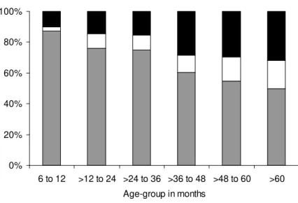
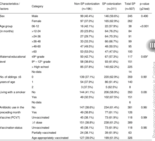
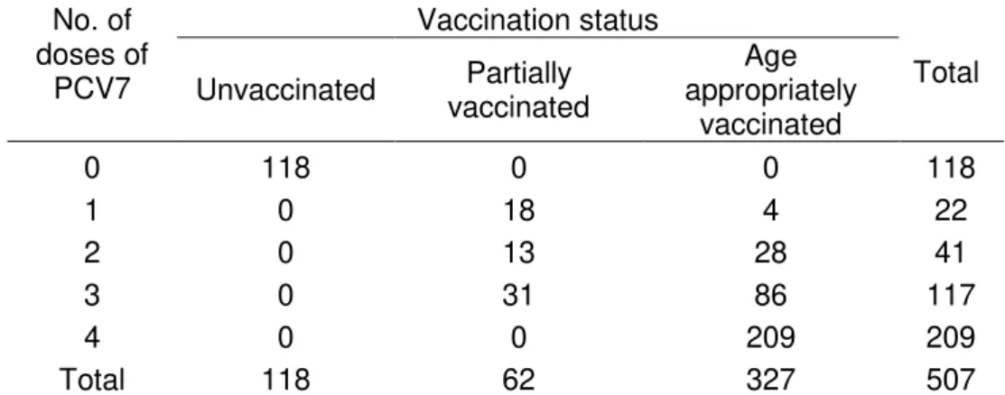
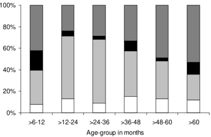
The second study was conducted in Coimbra and aimed to determine the prevalence of pneumococcal carriage and to characterize the pneumococcal strains colonizing children attending day care centers in this region in the era of pneumococcal vaccines. Between January and February 2007, nasopharyngeal swabs were obtained from 507 children (76.7% had received at least one dose of PCV7) and 61.3% were pneumococcal carriers. All 311 pneumococcal samples were antibiotyped and serotyped and the pneumococcal strains that were resistant to at least one antibiotic were also typed by PFGE. We have found in Coimbra similar rates of colonization and antimicrobial resistance patterns and similar genotypes to those previously described in the Lisbon area.
pneumococcal conjugate vaccines in Portugal, antibiotic consumption remains a main driving force for the maintenance of antimicrobial resistant pneumococci in the community.
To gain insights into the pneumococcal carriage patterns among the elderly, the fourth study was performed in two distinct areas: Oeiras and Montemor-o-Novo. In this study, 1,298 nasopharyngeal samples were collected. All pneumococci were antibiotyped, serotyped and characterized by MLST. Association between pneumococcal carriage, socio-demographic and clinical factors was evaluated using a logistic regression. The rates of colonization in this age group were low (2.2%), but there were high variability of colonizing serotypes and genetic backgrounds. Out of the 30 pneumococcal isolated, sixteen showed antimicrobial resistance. Smoking was a risk factor for pneumococcal colonization and living in the rural area seemed to increase the rate of pneumococcal colonization in this population. This study provided an important baseline to monitor the impact of pneumococcal vaccines on the patterns of colonization among the elderly.
xii
Altogether, these studies have contributed to improve our knowledge on pneumococcal colonization in Portugal in two age groups, children and the elderly.
The main conclusions of this thesis were:
i) the recently described serotype 6C is frequently carried by healthy young children in Portugal, is genetically diverse, and has been circulating in our country at least since 1996;
ii) the patterns of pneumococcal colonization among healthy children living in Coimbra are similar to those living in the Lisbon area;
iii) antibiotic consumption remains a main cause for the maintenance of antimicrobial resistance, in the era of widespread use of pneumococcal conjugate vaccines;
iv) the rates of pneumococcal colonization in the elderly are low and the serotype and genotype diversity are high;
RESUMO
Streptococcus pneumoniae, ou pneumococos, é responsável por várias
doenças em todo o mundo nas quais se incluem as infeções do trato respiratório, do ouvido médio, conjuntivite, meningite e sepsis. No entanto, é a elevada prevalência na nasogaringe em portadores assintomáticos que contribui para a sua disseminação na população. Assim, os estudos de colonização por pneumococos são importantes pois permitem monitorizar a flora da nasofaringe em vários grupos etários, e estudar a influência da utilização de vacinas e do uso de antibióticos. Igualmente importante é a possibilidade de otimização das vacinas de acordo com as características da população alvo. A caracterização molecular de pneumococos é crucial para a avaliação dessas alterações, o que alerta para a necessidade do desenvolvimento e validação de métodos de tipagem molecular fáceis e rápidos.
Desde 1996 que o nosso grupo tem vindo a estudar a população pneumocócica em crianças saudáveis que frequentam creches e jardins de infância. No entanto, esses estudos estiveram desde sempre confinados à área da grande Lisboa. Com o intuito de obter uma amostra populacional mais abrangente e variada, esta tese inclui estudos de outras regiões de Portugal não só em crianças mas também na população idosa, uma vez que este último constitui também um grupo etário de risco, com elevada incidência de doença pneumocócica.
xiv
último estudo descreve e faz-se a validação de uma nova estratégia de tipagem molecular para pneumococos.
O primeiro trabalho é um estudo retrospectivo que descreve a epidemiologia de um serotipo recentemente descoberto, 6C. Este estudo utilizou amostras que foram recolhidas da nasofaringe de crianças saudáveis que frequentavam creches e jardins de infância entre 1996 e 2007. Nesse estudo, utilizou-se a serotipagem por PCR complementada com a reacção de Quellung, electroforese em campo pulsado (PFGE), “multilocus sequence typing” (MLST) e antibiograma, para caracterizar os isolados. Concluiu-se que este serotipo já circulava em Portugal pelo menos desde 1996, que é geneticamente diverso, e que frequentemente apresenta resistência a antibióticos.
O segundo estudo foi realizado em Coimbra e teve como objectivos determinar a prevalência de portadores de pneumococos e caracterizar as estirpes de pneumococos que colonizam crianças que frequentam creches e jardins de infância nesta região, na era das vacinas pneumocócicas. Entre Janeiro e Fevereiro de 2007 foram pesquisadas 507 crianças (76.7% tinham recebido pelo menos uma dose de vacina pneumocócica conjugada 7-valente) das quais 61.3% eram portadores de pneumococos. Todos os 311 pneumococos isolados foram serotipados e foi realizado o antibiograma. Às estirpes de pneumococcus que apresentavam resistência a pelo menos um agente antimicrobiano foi também realizada a tipagem por PFGE. Em Coimbra as taxas de colonização, os padrões de resistência a antibióticos e os genotipos encontrados foram semelhantes aos descritos previamente na área metropolitana de Lisboa.
respectivamente. Os pneumococos foram caracterizados por serotipagem e antibiograma. As taxas de colonização encontradas foram semelhantes nas duas regiões (c.a. 61%). No entanto, na área urbana o nível de resistência a agentes antimicrobianos foi mais elevado (32.4% vs 21.6%, p<0.001), o mesmo acontecendo relativamente às taxas de consumo de antibióticos um mês antes da amostragem (16.7% vs 11.6%, p=0.004). Foi realizada uma análise estatística multivariada para identificar os fatores associados às diferenças encontradas nas duas regiões. O consumo de antibióticos durante o mês antecedente à recolha da amostra, estar colonizado com o serotipo 19A ou com uma estirpe não-tipável, e frequentar creche ou jardim de infância em Oeiras foram considerados fatores de risco para a colonização por pneumococos resistentes a antibióticos. Este estudo alerta para o facto do consumo de antibióticos continuar a ser a principal causa para a manutenção dos níveis de pneumococos resistentes a antibióticos encontrados na comunidade apesar da grande utilização de vacinas pneumocócicas conjugadas na comunidade infantil em Portugal.
xvi
referência para monitorizar o impacto das vacinas pneumocócicas nos padrões de colonização nos idosos.
Na última parte desta tese foi descrita e validada uma nova estratégia de “multiple-locus variable tandem repeat analysis” (MLVA) aplicada a pneumococos que permite de uma forma fácil e rápida, fazer uma análise genotípica deste microrganismo. Este método foi comparado com os métodos mais utilizados atualmente MLST e PFGE. Os três métodos mostraram índices de diversidade de Simpson de 98.5% ou superiores. O coeficiente de Wallace para MLVA e MLST foi de 0.874, o que significa que se duas estirpes tiverem o mesmo tipo de MLVA tem 88% de probabilidade de terem o mesmo tipo de MLST. O MLVA foi mais discriminativo para alguns isolados do mesmo complexo clonal (por MLST), gerando grupos de acordo com o serotipo ou serogrupo. Este estudo mostrou que o MLVA é um método promissor para genotipar S. pneumoniae.
Em suma, os trabalhos apresentados contribuíram para melhorar o nosso conhecimento em relação à colonização por pneumococos em Portugal em duas faixas etárias, crianças e idosos. As principais conclusões desta tese foram:
i) o serotipo 6C, recentemente descrito, é frequentemente encontrado em crianças saudáveis em Portugal, é geneticamente diverso e tem estado em circulação no nosso país pelo menos desde 1996;
ii) os padrões de colonização por pneumococos em crianças saudáveis que vivem em Coimbra são semelhantes aos encontrados nas crianças que vivem na área de Lisboa;
iv) as taxas de colonização por pneumococos nos idosos são baixas mas a diversidade de serotipos e de genotipos é elevada;
xviii
THESIS OUTLINE
The studies presented in this thesis provide an overview of pneumococcal colonization in Portugal in two age groups: children and the elderly.
Chapter I provides an outline of Streptococcus pneumoniae as a bacterium that
frequently colonizes the nasopharynx but can be also responsible for serious diseases. Topics such as historical highlights, epidemiology, identification, typing methods and the effect of pneumococcal vaccines are addressed.
Chapter II describes the temporal trends and molecular epidemiology of the
recently described serotype 6C of Streptococcus pneumoniae. The study
analyses the evolution of this serotype from 1996 to 2007, by antibiotyping, serotyping and also using PFGE and MLST.
Chapter III describes the pneumococcal population colonizing the nasopharynx
of children attending day care centers in Coimbra, a city in the central region of Portugal.
Chapter IV compares the patterns of pneumococcal colonization of young
children attending day care centers in two distinct areas of Portugal: one rural, Montemor-o-Novo, and other urban, Oeiras.
Chapter V is a study of pneumococcal colonization in the elderly in Portugal
that was also performed in an urban area, Oeiras, and a rural area, Montemor-o-Novo.
Chapter VI describes and validates a novel genotyping strategy applied to
Chapter VII presents general conclusions and futures perspectives.
Chapters II to VI are reproductions of the publications indicated below. They
can be read independently.
Chapter II
Nunes S.*, C. Valente*, R. Sá-Leão, and H. de Lencastre. 2009.
Temporal trends and molecular epidemiology of recently described serotype 6C of Streptococcus pneumoniae. J Clin Microbiol. 47:472-4.
*These authors contributed equally.
Chapter III
Rodrigues F.*, S. Nunes *, R. Sá-Leão, G. Gonçalves, L. Lemos, H.
de Lencastre. 2009. Streptococcus pneumoniae nasopharyngeal
carriage in children attending day-care centers in the central region of Portugal, in the era of 7-valent pneumococcal conjugate vaccine. Microb Drug Resist. 15:269-77. *These authors contributed equally.
Chapter IV
Nunes S., C. Valente, A. S. Simões, A. C. Paulo, A. Brito-Avô, H. de
Lencastre and R. Sá-Leão. 2012. Antibiotic consumption remains a
main driving force of antimicrobial resistance in the era of pneumococcal conjugate vaccines. In preparation.
Chapter V
Nunes S., S. Almeida, A. C. Paulo, I. Valadares, S. Martins, F. Breia,
A. Brito-Avô, A. Morais, H. de Lencastre, and R. Sá-Leão. 2012. Low
xx
Chapter VI
Elberse KE*, S. Nunes *, R. Sá-Leão, H.G. van der Heide, and L.M.
Schouls. 2011. Multiple-locus variable number tandem repeat analysis
for Streptococcus pneumoniae: comparison with PFGE and MLST. PLoS
TABLE OF CONTENTS
ACKNOWLEDGMENTS vii
ABSTRACT ix
RESUMO xiii
THESIS OUTLINE xviii
TABLE OF CONTENTS xxi
CHAPTER I
General Introduction 1
Introduction 3
Historical highlights 3
Epidemiology of Pneumococci 4
Pneumococcal carriage 5
Pneumococcal disease 7
Properties of Streptococcus pneumoniae 8
Distribution of serotypes 9
Use of antibiotic and pneumococcal vaccines 11 Antimicrobial resistance 12
Pneumococcal vaccines 13
Effects of pneumococcal vaccination 15
Decrease of vaccine serotypes 15
Herd immunity 16
Serotype replacement 16
Antimicrobial resistance 18 Invasive pneumococcal disease in Portugal 19 7-valent pneumococcal conjugate vaccine and carriage 19
xxii
Antibiotyping 24
Pneumococcal serotyping 24
PFGE and MLST 26
MLVA 27
Whole genome sequencing 28
Aim of the work 29
References 30
CHAPTER II
Temporal trends and molecular epidemiology of the recently
described serotype 6C of Streptococcus pneumoniae
53
Abstract 55
Introduction 55
Materials and Methods 56
Results and discussion 58
Conclusion 62
Acknowledgments 62
References 63
CHAPTER III
Streptococcus pneumoniae nasopharyngeal carriage in children attending day-care centers in the central region of Portugal, in the era of 7-valent pneumococcal
conjugate vaccine 67
Abstract 69
Introduction 70
Materials and Methods 71
Results 75
Discussion 85
References 88
CHAPTER IV
Antibiotic consumption remains a main driving force of antimicrobial resistance in the era of
pneumococcal conjugate vaccines 95
Abstract 97
Introduction 97
Materials and Methods 98
Results 101
Discussion 107
Acknowledgments 109
References 109
CHAPTER V
Low pneumococcal carriage and high serotype diversity among
elderly living in Portugal
115
Abstract 117
Introduction 118
Materials and Methods 119
Results 121
Discussion 126
Acknowledgments 127
Funding 128
References 128
CHAPTER VI
Multiple-locus variable number tandem repeat analysis for
Streptococcus pneumoniae: comparison with
xxiv
Abstract 135
Introduction 136
Materials and Methods 139
Results 142
Discussion 150
References 153
Supplementary tables 160
CHAPTER VII
INTRODUCTION
Streptococcus pneumoniae, or pneumococcus, is a Gram-positive bacterium
responsible for high rates of mortality and morbidity worldwide. It is a leading cause of disease particularly among young children, the elderly, and the immuno-compromised of all ages. Although it colonizes asymptomatically the nasopharynx,
S. pneumoniae can cause otitis media, sinusitis, pneumonia and more severe
diseases such as bacteremia and meningitis. Pneumococcal disease remains a great concern worldwide despite great advances aiming to decrease it, such as the use of antibiotic therapy and prevention through vaccination.
Since colonization precedes disease, epidemiological studies monitoring the nasopharynx flora are valuable tools to anticipate the emergence of serotypes that may became responsible for disease in the future.
HISTORICAL HIGHLIGHTS
In 1875 Edwin Klebs was the first to recognize Streptococcus pneumoniae in lung tissue and in infected sputum (reviewed in (54)), but only six years later this bacterium was isolated by two researchers working in France and the USA. In January 1881, Louis Pasteur in France reported for the first time the isolation of S. pneumoniae, after finding bacteria in the saliva of a youngster with rabies. George Miller Sternberg was in New Orleans studying malaria fever when he sampled his own saliva (as a control for his experiments) and injected it subcutaneously in a rabbit, which quickly died. This observation was in September 1880, but did not publish his report until April 1881(reviewed in (54)).
More than 90 years went by before this bacterium was named as Streptococcus
G
en
er
al
in
tr
od
uct
io
4
Sternberg called his “Micrococcus pasteuri”. In 1883 Mátray applied the term
“pneumoniekokken” and Albert Fraenkel in 1886, after the first complete description of the bacterium, gave us the familiar name “pneumokokkus”. In the same year, Anton Weichselbaum suggested the name Diplococcus pneumoniae,
which was used until the reclassification, on the basis of its growth in chains in liquid medium, as Streptococcus pneumoniae, in1974 (reviewed in (54)).
After its isolation and identification, S. pneumoniae was the model organism used
in important discoveries, including Gram staining in 1884 and the putative use of polysaccharide antigens as vaccines (reviewed in (54)). The identification of DNA as the source of genetic information was done using pneumococcus. Griffith in 1928 showed that avirulent strains could be transformed into virulent strains. Avery, MacLeod and McCarty demonstrated in 1944 that DNA was responsible for the transformation and thus the carrier of genetic information (reviewed in (54)). The therapeutic efficacy of penicillin, the role of bacterial capsule in resistance to phagocytosis (reviewed in (54)), and the first quorum sensing factor (149) were also described for the first time in pneumococci.
EPIDEMIOLOGY OF PNEUMOCOCCI
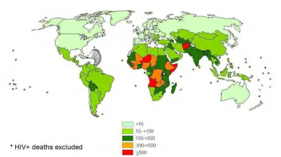
In the USA and Europe S. pneumoniae is the most common cause of pneumonia
in adults. The annual incidence of invasive pneumococcal disease (IPD) in these regions ranges from 10 to 100 cases per 100,000 (WHO, www.who.int/ith/disease/pneumococcal).
Figure 1. – Rates of Streptococcus pneumoniae death (per 100,000 children under age 5).
Data from WHO August, 2009.
Pneumococcal carriage
The pneumococcus is a normal component of the nasopharyngeal mucous membrane microflora. Acquisition is the first stage towards carriage and possible infection with pneumococcus. Carriage is usually asymptomatic. Transmission occurs by airborne droplets or direct contact with respiratory secretions (102). Recently, it has been suggested that environmental surfaces may also serve as sources of pneumococcal infection as pneumococci were found to be able to survive long-term desiccation (157).
G
en
er
al
in
tr
od
uct
io
6
Children are the major carriers and colonization occurs soon after birth (6). The peak in incidence is more or less at three years of age (15) and then declines with increasing age, until 10 years of age, remaining low in adulthood (15). Few studies have studied nasopharyngeal colonization in the elderly, but the carriage rates are typically low, ranging between 2 and 5% (2, 15, 68, 121).
Since it is in young children where the highest frequency of pneumococcal colonization and the highest crowding index are found, this group is considered the most important vector for horizontal dissemination of pneumococci in the community (88). Consequently, part of the strategy to prevent pneumococcal disease focuses on prevention of nasopharyngeal colonization.
In Portugal, data of pneumococcal carriage in healthy children attending day care centers, have been published by our group since 1996 and rates have been generally close to 60-65% (42, 43, 95, 104, 125, 128, 137). Concerning colonization in the elderly in Portugal, no studies had been conducted until recently. This topic was addressed for the first time in the pioneer study described in Chapter V of this thesis.
Most studies on pneumococcal colonization studies are based on the characterization of a single isolate of each individual. However, more than one pneumococcal serotype or clone can coexist in the nasopharynx of an individual (50, 53, 108, 129, 151). For this reason, studies on co-colonization are very important to identify changes at the individual level, such as de novo acquisition,
clearance and unmasking of pneumococcal strains (which can include serotypes or clones) (37, 90), particularly in the era of multivalent pneumococcal conjugate vaccines.
Pneumococcal disease
S. pneumoniae may spread from the nasopharynx into the respiratory tract or the
bloodstream to cause infections. These infections can be divided in two groups: invasive (isolated from sterile body sites) and invasive (isolated from non-sterile body sites). Otitis media and sinusitis are non-invasive infections. The nasopharynx is connected with the middle ear cavity via the Eustachian tube, which is a pressure regulator and prevents the entrance of the bacteria in the middle ear cavity. However, if the Eustachian tube is blocked, the bacteria may be trapped in the middle ear and cause otitis media. Sinusitis is caused when the bacteria spread locally into the sinuses and cause an accumulation of fluids in the sinuses due to an obstruction. The pneumococcus can also be responsible for more severe infections such as pneumonia, bacteremia and meningitis. From the nasopharynx, the bacteria may spread into the lungs or directly to the bloodstream. If bacteria spread into the lungs the immune system reacts and the accumulation of fluids and bacteria in the alveoli decreases oxygen transport and causes pneumonia (74). In adults, pneumonia without bacteremia is the most common non-invasive pneumococcal infection, however if the pneumococcus spreads into bloodstream, the infection becomes invasive. Pneumonia with bacteremia and meningitis are the most frequently recognized invasive disease. Pneumococcal meningitis occurs when the bacteria spread directly to bloodstream
G
en
er
al
in
tr
od
uct
io
8
cord). The meninges are filled with a liquid called cerebrospinal fluid where the bacteria can multiply freely, causing inflammation and swelling of the meninges and the brain tissue. Other invasive diseases are sepsis, peritonitis and arthritis.
Risk factors for pneumococcal infection are numerous and include, among many others, extremes of age (under 5 years of age and over 60 years old), immunosuppression, presence of underlying medical conditions, defects in host immune responses, low socioeconomic status, malnutrition, cigarette smoking and alcoholism (29, 91, 106, 107).
In Portugal, the report of pneumococcal infectious disease is not mandatory and, to my knowledge, no study has been carried out to calculate the incidence of invasive pneumococcal disease (IPD) over time. However, epidemiological studies on pneumococcal infections using data from several hospitals in different areas of Portugal have been conducted (3, 5, 63, 134, 135).
PROPERTIES OF STREPTOCOCCUS PNEUMONIAE
Streptococcus pneumoniae is a lancet-shaped Gram-positive bacterium, whose
diameter ranges between 0.5 and 1.25 µm for an individual cell. Pneumococci are frequently seen as pairs of cocci (diplococci), but they can also be in single cell and in short chains. It is a fastidious organism, growing best in media containing blood and in an enriched atmosphere with 5% CO2. On blood agar, colonies characteristically produce alpha hemolysis, form an inhibition halo ≥14 mm around
result in one or more of the assays described above (4, 32, 105, 111, 117, 127, 138).
The capsule is the main virulence factor of the pneumococcus and, with the exception of serotype 3, is covalently linked to the peptidoglycan of the cell wall (146). The capsule is 200 to 400nm thick (140), protects against opsonophagocytosis, and plays an important role in the interaction between the bacteria and the epithelium in colonization of the upper respiratory tract (103, 158). In addition, the pneumococcus may shift from transparent-phase (capsule less dense and thicker cell wall) to opaque-phase and vice-versa. This allows for attachment to the epithelial cells in carriage. The pneumococci in opaque-phase have denser capsules, providing better protection against opsonization and killing in invasive disease (159).
Based on the reaction of the capsular polysaccharide with polyclonal factor sera and more recently with the use of monoclonal antibodies, 94 serotypes are recognized up to now (16, 21, 22, 61, 115).
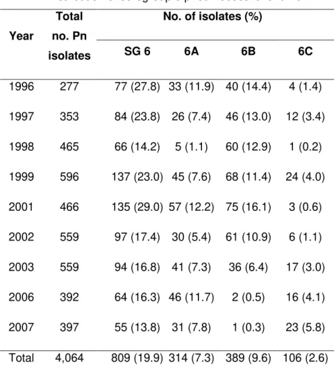
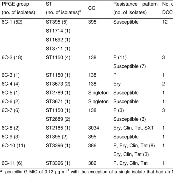
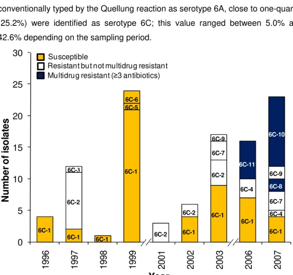
The first “novel” serotype recognized using monoclonal antibodies was 6C (115). As it was previously classified as 6A by the Quellung reaction, the real prevalence of this serotype and its characteristics were not known. In order to obtain insights on its epidemiology in colonization in Portugal, a retrospective study was performed and is described in this thesis in Chapter II.
Distribution of serotypes
Although 94 distinct pneumococcal serotypes, varying in capsular polysaccharide structure, have been described, not all seem to have the same capacity to cause disease (18, 60, 126, 136). Some serotypes can be carried in the nasopharynx asymptomatically and cause disease in a small proportion of infected individuals only. Others are rarely identified in the nasopharynx but are frequently associated
G
en
er
al
in
tr
od
uct
io
10
detected in colonization, but are responsible for a high proportion of invasive disease and are frequently associated with outbreaks (8, 13, 38, 96, 123). Before the introduction of pneumococcal conjugate vaccines 10 to 12 serogroups were responsible for the majority of pneumococcal invasive disease worldwide, including 1, 3, 4, 6A, 6B, 7F, 8, 9V, 14, 18C, 19F, and 23F (62, 112).
The prevalence of pneumococcal serotypes/serogroups causing IPD depends on several factors, such as geographic location, disease manifestation, and age of the host (15, 18, 19, 60). In young children the serogroups/serotypes 6, 14, 18, 19 and 23F are predominant due to the lower immunogenicity of these serotypes (20).
Several authors have been looking at the distribution of serotypes in colonization and disease to estimate the invasive potential (18, 19, 55, 131, 141). According to Brueggeman et al. (18) and Sleeman et al. (141) serotypes 1, 4, 5, 7F, 9V, 14,
18C, and 19A are classified as highly invasive serotypes. In a study reported by Greenberg et al. (55) the authors estimated the disease potential in pediatric
In Portugal, a study was published (126) where the invasive disease potential of serotypes and clones circulating in Portugal before the introduction of PCV7 was calculated. In this study serotypes 1, 3, 4, 5, 7F, 8, 9N, 9L, 12B, 14, 18C and 20 were found to have a high propensity to cause invasive disease, whilst serotypes 6A, 6B, 11A, 15B/C, 16F, 19F, 23F, 34, 35F, and 37 were associated with carriage. Additionally, significant differences in invasiveness were found between clones that shared the same serotype, namely among serotypes 3, 6A, 6B, 11A, 14, 19A, 19F, 22F, 23F, 34 and NT, which highlights the importance of the genetic background when analyzing the invasive disease potential of certain serotypes.
The distribution of serotypes is also affected by temporal changes, inherent of secular trends of specific serotypes, and this has been reported in several studies (57, 59, 71, 120).
Serotype distribution can be altered by antibiotic consumption and vaccination patterns. Following the introduction of the first pneumococcal conjugate vaccine, the increase of non-vaccine serotypes has been reported in several countries, in disease and also in colonization (more details are presented in the next topic).
Use of antibiotics and pneumococcal vaccines
Nowadays, the main tool to control pneumococcal disease, besides antibiotic therapy, is vaccination. Efforts to develop effective pneumococcal vaccines began in 1911, when the first clinical trial on the effectiveness of a whole-cell pneumococcal vaccine was conducted by Wright et al. in South-Africa (reviewed in
(93)). Several decades were needed before the effectiveness of pneumococcal vaccines was demonstrated. Only at the end of World War II in 1945 (92), the first capsular polysaccharide vaccine trials were undertaken.
G
en
er
al
in
tr
od
uct
io
12
Antimicrobial resistance
Before penicillin became available for medical treatment in the early 1940s, no cure for pneumonia existed and many people died with pneumococcal infection. The introduction and use of antimicrobial drugs changed this scenario and led to the withdrawn from the market of a pneumococcal vaccine that was licensed and in usage. However, it did not take long before it became clear that antimicrobials
per se had not eliminated pneumococcal disease.
In 1964, Austrian and Gold reported that one in four patients admitted with pneumococcal bacteremia died even with antimicrobial therapy (12). In 1967, the first pneumococcal isolate with intermediate resistance to penicillin was described by Hansman et al. (58) in Australia. Within a decade, similar strains were found in
South Africa, Europe and North America (77). The first multiresistant pneumococcal isolates were reported by Jacobs et al. in 1978 (70) and since then
we have been observing a widespread of multidrug resistance has been observed (33, 97).
penicillin. Resistance to erythromycin was very prevalent, 72.7%, and multidrug resistance was observed in 59.3% of the isolates (76) (reviewed in (144)).
In Portugal, among invasive disease isolates in adults, the latest data (2006-2008) showed rates of penicillin non-susceptibility around 17% and the resistance to macrolides was 18% (63). In children (including children until 17 years) during the same period, around 19% of the isolates expressed non-susceptibility to penicillin and resistance to erythromycin was found in 22.9% of the isolates. Additionally 11.6% of the isolates showed simultaneously resistance to both antimicrobial agents. In young healthy children data from 2006/2007 showed rates of macrolides resistance of 25.3% and 26.6% of the strains showed decreased susceptibility to penicillin (137).
Pneumococcal vaccines
In 1977, Austrian et al. reinforced the interest of pneumococcal prevention and a
pneumococcal polysaccharide vaccine including 14 serotypes was licensed (11). This vaccine was extended to 23 serotypes and licensed in 1983 (Pneumovax23, Merck&Co, PPV23).
In children, responses induced by polysaccharide vaccines are low (44). Conjugation of the polysaccharides with a carrier protein induced better antibody responses in infants due to the T-cell dependent immune pathway. In 2000, the first pneumococcal conjugate vaccine was licensed (Prevnar™; Wyeth-Lederle, PCV7). It included the seven serotypes most common in pneumococcal disease in children under 2 years old in the United States (4, 6B, 9V, 14, 18C, 19F and 23F). In PCV7 the polysaccharide of each serotype is individually conjugated to the protein carrier, CRM197. In Portugal, this vaccine was neither introduced in the National Vaccination Plan nor subsidized by the state. However, the majority of pediatricians recommended this vaccine what explained high intake of the vaccine;
G
en
er
al
in
tr
od
uct
io
14
months. Data obtained from Pfizer, Portugal, and our data from colonization studies in the Lisbon area, until 2007 (137), around 70% of the children under six years old were vaccinated suggested that at least one vaccine dose.
In 2009, a new pneumococcal conjugate vaccine was introduced in the market (Synflorix®, GlaxoSmithKline Inc., PCV10). It included the same serotypes in PCV7 plus three additional serotypes (1, 5, 7F; serotypes 18C and 19F are conjugated to tetanus and diphtheria toxoids, respectively, and the remaining 8 serotypes are conjugated to the non-typeable Haemophilus influenzae protein D).
In 2010, PCV7 was replaced by PCV13 (Prevenar®; Pfizer). This vaccine included serotypes 1, 3, 4, 5, 6A, 6B, 7F, 9V, 14, 18C, 19A, 19F and 23F. These vaccines have not been introduced in the National Vaccination Plan, and are not subsidized by the state in Portugal.
According to the Global Alliance for Vaccines and Immunisation (GAVI), in July of 2012, 18 countries in the developing world started the introduction of pneumococcal conjugate vaccine and 36 GAVI eligible countries have been approved for GAVI support to introduce pneumococcal conjugate vaccine into the national immunization programmes. GAVI and partners hope that by 2015, 90 million children can be immunized with pneumococcal vaccines and the pneumococcal vaccine rollout has been achieved in 58 countries (http://www.gavialliance.org).
1,24 cases per 100,000 were reported, with higher rates in adults aged more than 50 years.
Considering the high serotype diversity within S. pneumoniae, the high cost of
these vaccines, and the fact that serotype coverage varies between geographic regions, there is the need of developing vaccines that confer broader coverage, preferentially independent of the capsular polysaccharide and with a lower cost.
Currently a 15-valent pneumococcal conjugate vaccine (PCV13 serotypes plus serotypes 22F and 33F) is in evaluation in an infant-rhesus monkey model (139) and a new approach based on whole-cell is also in development (100).
A vaccine which goal was protection against pneumococcal nasopharyngeal colonization was also designed. This vaccine was based in specific antigens that activated CD4+ T cells to secrete the cytokine interleukin-17A that mediate resistance to mucosal colonization. Recently, was reported that this vaccine was tested in a mouse model and showed protection against pneumococcal infection (99, 101).
EFFECTS OF PNEUMOCOCCAL VACCINATION
Decrease of vaccine serotypes
In the USA the introduction of PCV7 in the vaccination schedule began shortly after the vaccine was licensed, resulting in a decrease of 77% of IPD in vaccinated children aged up to 5 years old by 2005 (26). Two years later, Pilishvili et al.
analyzed IPD cases, identified between January of 1998 and December of 2007, in eight participating ABCs in the USA. They reported a decline in IPD incidence of 76% in children under 5 years old, with a decline around 100% of PCV7 types (118).
G
en
er
al
in
tr
od
uct
io
16
In Europe, Isaacman et al. observed the impact of PCV7 use on the incidence of
IPD in children from eight countries. The results were variable across studies, with the decrease of IPD incidences ranging from 28% to 68%, depending on the country, vaccine uptake and the type of disease manifestation (69).
Herd immunity
The extensive vaccination with PCV7 has also resulted in an indirect protection of the non-vaccinated population in all age groups, an effect known as herd immunity (26). This was shown for example in a surveillance study conducted in eight areas of the USA with adults older than 50 years old, from which Lexau et al. concluded
that the use of conjugate vaccine in children also benefited the adults included in the study, with a decline of 28% in IPD in this age group (89). Herd immunity was also reported in newborns, which are too young to receive the vaccine, as described by Poehling et al. (119). Also in other countries such as England and
Wales (98) and Australia (73), herd immunity was detected in the population.
Serotype replacement
Another effect of pneumococcal vaccination was serotype replacement that results from the ability of non-PCV7 serotype strains to fill the niche left vacant by the PCV7 serotypes. In several countries, although there was a reduction in IPD cases caused by PCV7 serotypes, cases associated to non-PCV7 serotypes increased post-PCV7 introduction (3, 5, 63, 64, 69, 86, 118).
with the substantial decrease of PCV7 types. In this study the increase in IPD caused by non-PCV7 types was more pronounced in meningitis and invasive pneumonia, whereas in primary bacteremia there was no change (118).
Isaacman and co-authors showed also serotype replacement in Europe in a review that describes the trends of IPD in European children between 1990 and 2008. Before the use of PCV7 the serotypes most common recovered in IPD were 6B, 14, 19F and 23F, although with some variation among countries. After the widespread use of PCV, the most common IPD serotypes in Europe were 1, 19A, 3, 6A and 7F (69).
Recently, a study from Alberta, Canada between 1998 and 2010 encompassing PCV7 introduction in the routine vaccination plan, reported that PCV7 types were eradicated from IPD in children under two years old and almost eliminated in all other age groups (86). On the other hand, they observed an increase in non-PCV7 serotype IPD, mainly 19A in children and serotypes 5 and 19A in adults (86). The authors justify this increase in serotype 5 incidence with the occurrence of an outbreak in 2007 (154). Nevertheless, after exclusion of epidemiological data from the year 2007, the increase in the IPD cases associated with serotype 5 was still significant, compared to the pre-PCV7 era (0.03 per 100,000 versus 0.51 per 100,000; p<0.001) (86).
However, decrease in IPD incidence and serotype replacement were not observed in particular populations. As an example, among the White Mountain Apache in Arizona, despite the use of PCV7, high rates of IPD continue to be seen (83). One explanation could be the low rates of coverage of this vaccine, only 56.2%, due to the fact that the serotypes that were more prevalent in that population are not included in PCV7 (83). Interestingly, in this population the rate of 19A has been stable or has decreased after vaccination.
G
en
er
al
in
tr
od
uct
io
18
These observations highlight that replacement of pneumococcal serotypes may be related to other factors apart from the introduction of a vaccine. For example, in Israel and South Korea, an increase of 19A in IPD was noted before the introduction of PCV7 in the market (30, 36). Also, in the study referred to above, in Canada, the authors mention that a significant increase of serotype 19A in all ages was noticed before the widespread use of PCV7 (86).
The use of PCV7 had also an effect in otitis media, and a global review has been recently published by Rodgers et al. (122). In the USA after the introduction of
PCV7 it has been observed a decrease in the number of PCV7 cases and an increase in the number of infections produced by non-PCV7 serotypes, such as 19A, 3, 6A (122). Another important aspect reported in some studies from countries where PCVs were introduced was the decrease in the number of medical visits mainly due to otitis and also the number of antibiotic prescriptions (14, 114, 161).
In a study conducted in Israel, with children attending day care centers, children vaccinated with PCV9 needed significantly fewer days of antibiotic uptake for treating respiratory illnesses and otitis than the control group, vaccinated with meningococcal group C vaccine (48).
Antimicrobial resistance
Antimicrobial resistance rates also suffered some alterations with the introduction of PCV7, since the serotypes included in PCV7 tended to have high rates of resistance (39). But, as described before, the decreased of PCV7 serotypes varied according to the geographic location and vaccine uptake (39, 64, 82).
disease, the rates of IPD in children less than two years decreased as well as the rates of pneumococcal antimicrobial resistance (130, 155).
Invasive pneumococcal disease in Portugal
In Portugal, serotype replacement in pediatric IPD was also reported (3, 5). However, this replacement was not as pronounced as in the USA, where PCV7 was introduced in the national vaccination program unlike what happens in Portugal. Another important aspect was the low coverage of PCV7 in IPD in Portugal, mainly due to the high prevalence of serotype 1 compared to the USA. The fact that PCV7 is not included in the national vaccination plan and the low coverage of this vaccine in IPD in Portugal affect also herd immunity: despite a marked reduction in the proportion of IPD in adults caused by PCV7 serotypes, they still accounted for 18% of the cases between 2006 and 2008 (63). In the same study a decline of serotypes 4, 8 and 14, and an increase of serotypes 1, 7F and 19A were observed in adult IPD in the post-PCV7 period. Similar with adults, in pediatric IPD, serotypes 1, 19A, 7F, 14 and 3 were the serotypes more prevalent in 2006-2008 (3).
Concerning antimicrobial resistance, it remained stable after a small decrease of penicillin non-susceptibility in IPD in children under five years old in the first post-PCV7 years (5). Penicillin non-susceptibility was observed in 18.7% of the isolates and resistance to erythromycin was found in 22.9% of the isolates, from which 18% were NVT (3). On the other hand, in IPD in adults, serotypes 19A and 14 accounted for the majority of erythromycin and penicillin non-susceptible isolates in 2008. Although penicillin non-susceptibility remained stable (17%), resistance to erythromycin has been increasing in the post-PCV7 years (63).
7-valent pneumococcal vaccine and carriage
The first report to show that conjugate pneumococcal vaccines could decrease
G
en
er
al
in
tr
od
uct
io
20
1996. In that study carried out by Dagan et al. the effect of a seven valent
pneumococcal conjugate vaccine (which included the serotypes: 4, 6B, 9V, 14, 18C, 19F and 23F conjugated to the outer-membrane protein complex of Neisseria meningitis group B) was evaluated in children 12-18 months of age (40). The
authors observed a decrease of the serotypes included in the vaccine among the vaccinated while no changes occurred in the control group. One year after vaccination, carriage of antibiotic resistant vaccine-types was still lower in vaccinated children than in the control group. After that, a second study conducted by the same group was published and both studies showed clearly that conjugate vaccine could reduce the carriage and the resistance of pneumococcal serotypes included in conjugate vaccines (41).
Later, Obaro et al., studied the effect of a pentavalent pneumococcal vaccine
(including serotypes 6B, 14, 18C, 19F and 23F conjugated to CRM197) in infants in Gambia and observed the maintenance of high pneumococcal carriage of serotypes included in vaccine in control group and a decreased of these serotypes in vaccinees. However, an increased of serotypes not included in the vaccine was observed in vaccinated children and this effect was not observed in the control group (110). This phenomenon of replacement was also observed in a study conducted in toddlers attending day-care centers in Southern Israel with 9-valent pneumococcal vaccine conjugate with CRM197 (35).
communities serotypes 19F and 6B were the PCV7 types most frequently isolated post-PCV7 vaccination, whereas serotypes 4, 9V, 14 and 18C were rarely recovered. In Norway, after vaccine introduction, the pneumococcal carriage remained high, around 80%, but a decrease in PCV7 serotypes was observed (156). In Calgary, Canada a study conducted in seven community health centers where routinely PCV7 vaccination began in 2002, a decrease of PCV7 serotypes and an increased of non-PCV7 was also observed. In this study the pneumococcal carriage rate was low, 20%, and the largest increased of non-PCV7 type was noticed in serotypes 6A, 15C, and 11A (75).
Serotype replacement has also been observed in pneumococcal colonization in Portugal. The first evidence was in a controlled study conducted before the introduction of PCV7 in the market, which included 236 vaccinated children and 354 control children that attended the same day care center (49). Additionally, a reduction on pneumococcal resistance PCV7 serotypes was replaced by an increase of resistance in non-PCV7 serotypes (49). Surveillance studies conducted after the availability of PCV7 in Portugal also described a marked serotype replacement effect in the population (125). Despite serotype replacement, maintenance of the antimicrobial resistance rates was observed, due to expansion of pre-existent non-PCV7 resistant clones (125, 137). In 2006, five years after the introduction of PCV7 in the Portuguese market, non-vaccine types 1, 6C, 7F, 15A, 16F, 21, 23A, 29, and non-typeable strains (NT) increased significantly. A non-significant increase of serotype 19A was also noticed (125). In 2006 and 2007, the major serotypes were 6A, 6C, 14, 15A, 19A, 19F, 23F and non-typeable strains. A significant decrease in clonal diversity was observed for serotypes 14 and 19F, whereas a significant increase in clonal diversity was observed for serotype 6C and non-typeable strains. For serotypes 6A and 19A no significant changes in clonal diversity occurred with introduction of PCV7 (137).
G
en
er
al
in
tr
od
uct
io
22
All the above mentioned studies were conducted in the Lisbon area; in order to evaluate if this data would be representative of the country, surveillance initiatives were expanded to other areas of Portugal and the results are in Chapters III and IV of this thesis.
Recently, a study published by Valente et al. suggested a novel potential benefit of
conjugate vaccines. In the study the effect of PCV7 on pneumococcal co-colonization was evaluated. Lower co-co-colonization rates were observed among fully vaccinated children when compared with unvaccinated children. Since a decrease of co-colonization could translate in lower opportunities for horizontal gene transfer, this effect may function as a “brake” for capsular switch or acquisition of resistant genes (151).
PERSPECTIVES FOR PCV10 AND PCV13
The introduction of PCV10 and PCV13 is expected to have an impact on pneumococcal disease and carriage. In 2010, a study conducted in the USA predicted the effectiveness of PCV13 from observed PCV7 data. According to their model PCV13 would prevent 106,000 invasive pneumococcal disease cases and 2.9 million pneumonia cases over a 10-year period (124). Recently, another study examined public-health and economic impacts of PCV pediatric national immunization programs in Germany, Greece and The Netherlands and estimated that PCV13 would eliminate 31.7%, 46.4%, and 33.8% of IPD in Germany, Greece, and The Netherlands, respectively.PCV13 was found to be cost-effective or cost saving when compared to PCV7 and PCV10 (147).
The use of the polysaccharide vaccine (PPV23) remains controversial, as described by Huss et al. in a recent meta-analysis (67). In this meta-analysis 22
control. The vaccine efficacy on clinical outcomes and the quality of the methodology used in the trials were evaluated. For the authors in all trials analyzed there was little evidence of protection of this vaccine against the elderly or in adults with chronic disease. This vaccine seems not to be effective for prevention of pneumonia, even in countries where it is currently recommended. Recently, PCV13 has been approved for prevention of pneumonia and invasive disease caused by PCV13 serotypes among adults aged 50 years and older (27). However, the efficacy of this vaccine in this age group has not been established yet. A trial is in progress in The Netherlands involving 85,000 persons aged 65 years and older (56). Nevertheless, the full impact of PCV13 in children and in adults is not known yet.
PNEUMOCOCCAL TYPING METHODS
In the era of pneumococcal vaccines, with several and rapid changes in the pneumococcal population is important and essential to have good and adequate typing methods.
The choice of a typing method will depend upon the needs, skill level, and the resources of the laboratory. An optimal typing method should show high typeability, adequate stability, high reproducibility and high discriminative power. Additionally, ease of use and interpretation, quick, and low cost are also convenient criteria.
Typing pneumococcus has been useful for understanding the evolution of the species, in epidemiological studies and also in outbreak cases (153).
Characteristics expressed by the microorganism permit to classify them by phenotyping methods. Methods that involve direct DNA-based analysis of
G
en
er
al
in
tr
od
uct
io
24
genotyping methods. In general, genotyping methods have higher discriminative power and higher typeability than phenotyping methods and constitute the best approach for bacterial comparison.
Antibiotyping
Antibiotyping consists of an antimicrobial susceptibility testing where the isolates are tested by diffusion or dilution methods against a panel of antimicrobial agents. The results are interpreted according to international guidelines. This technique is easy to perform, gives rapid results, it is cheap and readily available in the routine microbiology laboratories. However, the major disadvantage is the poor discriminative power for typing proposes. Antibiotic resistance patterns are, to some extent, influenced by the local environment and the antibiotic pressure (81).
Pneumococcal serotyping
This method consists of a reaction of the capsule polysaccharides with polyclonal factor sera (145) and recently, also with the use of monoclonal antibodies (21). Classical serotyping is performed using the Quellung or Neufeld reaction (10). With this assay the swelling of the capsule of the pneumococcus is observed using a contrast phase microscope after mixing the bacteria with serotype specific antiserum. The assay includes one chessboard sequential testing of antisera of a pool that gives a serogroup, and then a specific factor serum that gives the serotype. The bacteria will frequently also agglutinate. The subjectivity in
interpretation, the high cost of antisera, the need for a complete set of control strains, and technical expertise requirements associated with these serologic methods have resulted in development of PCR based serotyping systems (17).
to Quellung reaction. Latex agglutination tests also have been developed since at least 1988 (84). However, only in 2004 Slotved et al. (142) developed a simple,
rapid latex agglutination test (Pneumotest-Latex), which is used nowadays in several laboratories.
More recently, a PCR scheme using a sequential series of multiplex PCRs to assess serotypes or related sets of serotypes has been developed by the CDC (113) (www.cdc.org). The use of this approach to assess the serotype is convenient because of the relatively low cost compared to the classic method and the time consumed.
Recently, Elberse et al., described a new genotyping method, capsular sequence
typing (CST), based on the determination of the partial sequence of the capsular
wzh gene, using a single PCR reaction with multiple primer sets (46). Another
promising methodology published for serotyping pneumococci is based on three multiplex PCR combined with fragment analysis and automated fluorescent capillary electrophoresis (FAF-mPCR) which detects a great number of serotypes/serogroups. Although it does not differentiate some groups of serotypes, this automatic method allows the analysis of 30 samples in a few hours and at low cost (133).
Additionally, some isolates do not react with any commercially available antiserum. These atypical isolates are called non-typeable S. pneumoniae (NTPn). These
isolates are difficult to identify as their differentiation from closely related species such as Streptococcus pseudopneumoniae and other streptococcus of the mitis
group is not always straightforward. Recently, Simões et al. (138) developed a low
cost and easy assay to detect NTPn. The strategy is based on a multiplex PCR targeting lytA, cpsA, aliB-like ORF2 and 16S rDNA genes, plus a RFLP assay to
differentiate typical from atypical lytA.
G
en
er
al
in
tr
od
uct
io
26
PFGE and MLST
Currently, the gold standard genotyping methods for pneumococci are pulsed-field gel electrophoresis (PFGE) (132) and multilocus sequence typing (MLST) (47).
Pulsed-field gel electrophoresis (PFGE) was developed in 1984 by Schwartz and Cantor (132) and is based on the digestion of the total chromosomal DNA with a restriction endonuclease that cleaves the DNA infrequently. The macro-restriction fragments are separated by gel electrophoresis according to molecular weight in an apparatus which voltage is periodically switched among three directions. The different orientation of the electric pulses during electrophoresis allows the separation of large DNA fragments. Normally the total chromosomal DNA of S. pneumoniae is digested with SmaI and 10 to 19 bands of 20 to 300 kb are
obtained (87). This technique is inexpensive, shows a good typeability, reproducibility, and a good discriminative power. However, it is time consuming and standardization of the method and good quality of the gels are pre-requisites to compare results between laboratories. Despite established interpretation criteria by visual comparison (148) and the standardization of the method (31) the comparison of results remains sometimes difficult. As an alternative to visual classification, automatic methods that use band-based similarity coefficient to classify by type/subtype the microbial isolates analyzed by PFGE have been developed. Carriço et al. demonstrated a good correspondence between visual
and automatic classification for S. pneumoniae (24).
MLST for pneumococci is a nucleotide sequence based approach that includes PCR amplification and sequencing of ~450-bp internal fragments of seven housekeeping genes: aroE - shikimate dehydrogenase, gdh -
glucose-6-phosphate dehydrogenase, gki – glucose kinase, recP - transketolase, spi – signal
peptidase, xpt – xanthine phosphoribosyltransferase, and ddl –
assignedwith a number. The combination of the seven numbers defines the allelic profile or sequence type (ST). The major advantage of this technique is the unambiguous ability to compare the results obtained in different laboratories, using the online database. However, this method is time consuming if need to analyze a big collection and expensive for laboratories without a sequencer. MLST has a good discriminative power and is a good tool for local and global epidemiology.
MLVA
Multiple-Locus Variable number tandem repeat Analysis (MLVA) is based in repetitive DNA localized in multiple loci of all bacterial genome. The number of repeat sequences and the sizes of these units, named BOX elements, may vary for different isolates of the same species. These BOX elements were identified in the genome of the pneumococcus in 1992 (94). In the genome of prototype strains R6 and TIGR4 115 and 127 BOX elements, have been found, respectively. The function and origin of BOX elements are unknown; however, it is thought that they may be involved in regulating the expression of virulence-associated genes when they are located in the promoter region of genes (78, 94). Van Belkum and colleagues were the first to show the usefulness of these repeats for genotyping S. pneumoniae using PCR-BOX. However the banding profiles were difficult to
interpret and often lacked reproducibility (152). The first MLVA scheme based on BOX typing applied to pneumococcus was developed by Koeck et al. in 2005 (79).
In this scheme 16 BOX loci are analyzed after amplification in a single PCR reaction and the product are visualized by agarose gel electrophoresis. This technique has high congruence with MLST and shows high discriminative power, providing a good alternative to MLST. Although it allows for the comparison of data between laboratories using a website (www.mlva.eu), this scheme and the use of agarose gel electrophoresis makes this method very fastidious. A new scheme and easier protocol was needed and is described in chapter VI of this thesis.
G
en
er
al
in
tr
od
uct
io
28
Whole genome sequencing
The sequencing of whole genomes (WGS) represents a great advance for the study of bacterial populations because current approaches such as MLST are based on the analysis of only seven housekeeping gene fragments amounting only less than 0,2% of the total length of the genome. Horizontal gene transfer (HGT) is a fundamental process in bacterial genome evolution where a great portion of DNA can be incorporated from another species genome. Since S. pneumoniae is naturally transformable these events occur frequently. Small-scale
mutations as substitutions, deletions or insertions could also occur. With this technique all these events can be identified by comparing the sequences with a reference genome. These differences in the core genome allow the construction of phylogenetic trees to show the evolutionary relationships among the isolates of the same species. WGS can also be useful in epidemiological studies, namely to follow the spread of a clone which isolates seem to be identical in several countries. One example of this was demonstrated with the genome sequencing of several isolates of the international multiresistant Spain23F-1 clone, which showed that the genome sequence of each isolate differs slightly and these differences represent an evolution of the specie, which was impossible to evaluate without WGS (34).
In addition WGS has the capacity to simultaneously elucidate serotype (if the capsule is expressed), sequence type and details of genetic information, which provide a good approach to monitor the impact of pneumococcal vaccines.
Recently, Golubchick et al. reported that using whole-genome resequencing
episode of recombination multiple and large additional DNA fragments can be imported (52).
Although very promising the price of WGS remains high and the bioinformatic tools for data analysis need further development for routine use by hospitals and by scientific community in general like as mention by Török et al. (150).
AIM OF THE WORK
The aims of this thesis were to gain insights into: (i) the epidemiology of the recently discovered serotype 6C, (ii) the pneumococcal colonization patterns in different regions of Portugal among healthy children and (iii) the pneumococcal colonization patterns in the elderly. Additionally, the need for the development and validation of easier and faster methods of molecular typing was also addressed.