Morphological diversification
through the evolution of
developmental hierarchies
Leila Teruko Shirai
Dissertation presented to obtain the Ph.D degree in Evolutionary
Biology
Instituto de Tecnologia Química e Biológica | Universidade Nova de Lisboa
Oeiras,
November, 2014
Morphological diversification
through the evolution of
developmental hierarchies
Leila Teruko Shirai
Dissertation presented to obtain the Ph.D degree in Evolutionary
Biology
Instituto de Tecnologia Química e Biológica | Universidade Nova de Lisboa
Research work coordinated by:
Oeiras,
developmental hierarchies
PhD thesis, Instituto Gulbenkian de Ciência, Universidade Nova de Lisboa, 2014
In English, with summary in Portuguese
This thesis has been scanned for plagiarism on Oct 15th 2014 and there was no conflict with published works.
Cover image by the author © 2014
The wing of Bicyclus anynana (Satyrinae, Nymphalidae) butterfly was dissected at the end of pupal life, air-dried, sputter-coated with gold, and imaged under Scanning Electron Microscopy. With this method, we are able to see the maturation of cells in the center and external ring of the eyespot. Scale bar: 100μm
Supervisor
Dr. Patrícia Beldade (Instituto Gulbenkian de Ciência, Portugal)
Jury members
Dr. Abderrahman Khila (Institut de Génomique Fonctionnelle de Lyon, France) Dr. Casper Breuker (Oxford Brookes University, UK)
Dr. Élio Sucena (Instituto Gulbenkian de Ciência, Portugal)
Dr. Filipa Alves (Instituto Gulbenkian de Ciência, Portugal)
The research presented in this thesis was supported by: Fundação para a Ciência e a Tecnologia (FCT) fellowships SFRH/BI/33842/2009 and SFRH/BD/51180/2010, Instituto Gulbenkian de Ciência, research grants to P. Beldade (PTDC/BIA-BEC/ 099808/2008 and PTDC/BIA-EVF/2170/2012), the Society for the Study of Evolution (Rosemary Grant Award and a travel award to attend the Joint Evolution Meeting at Snowbird, USA in 2013), and Fundação de Amparo à Pesquisa do Estado de São Paulo (travel award to attend the São Paulo School of Advanced Science-evolution in 2012). This thesis was printed with support from Instituto Gulbenkian de Ciência.
This thesis is dedicated to
Noribumi Shirai and Radu Coroamă
for anchoring my left and right feet in the ground,
There is a pleasure in the pathless woods, There is a rapture on the lonely shore, There is society, where none intrudes, By the deep sea, and music in its roar: I love not man the less, but Nature more, From these our interviews, in which I steal From all I may be, or have been before, To mingle with the Universe, and feel What I can ne'er express, yet cannot all conceal.
Acknowledgements
This thesis goes for the hopeful entertainment of the curious who have the luck of researching life as a need, or as a job. I’d like to thank the following people who had made the work of the last five years easier, clearer, better, more accurate, and more fun.
First and foremost, I would like to thank Vlad Coroama, who has always kept me in track about what is important in life. You are an incredible kind man, and I wish there were more leaders like you. It was my luck to encounter, and disencounter you, to experience your love with all its idiosyncrasies. In the same hierarchical layer, thanks to my family for the solid supporting love, for giving me freedom and opportunity, for being family, and for the Marukai franchise. And welcome, Umi, Izu, Luisa, and Helena!
My greatest gratitude to the dear people who willingly or not followed this process. To people from before, whom I hope will stay forever around: Daniel Damineli, Rogério Hirata, André Belluco, Tiago Pexe Hermenegildo, Gabriel Marroig, Carol Fernandes, José Natali, Jéssica Camargo, Bruno Velutini, Amalia Hellwig, Gepeto Müller, Migue Mitne, Leonardo Borges, Vitor Hugo Rodrigues, Harley Sebastião, and Renata Pardini. Also to those who were crucial and generous with the first of the past five years, Ana Paula Assis, Roberta Paresque, Denise Selivon, Daniel Lahr, and Chris Klingenberg. To the troop who makes working the most awesome thing there is: Gabriel Mar-roig, Arthur Porto, Felipe Fino Oliveira. People from the present, who made
Jess Thompson, Paeter (Sander), Pat, and Marie Bonnet, Klito and Yan-nis, João Dias, Ana Sofia Leitão, and PIBS fellows, including Vitor Faria; and the evil-teca troop Daniel Damineli, Maitê Portes, Scott Rennie, Yoan Dickmann, and Edu George.
To Patrícia Beldade for having always given us freedom to go after our inter-ests and saying "by all means" so many times, for never letting us be sloppy even if drilling holes in our brains was required, for supporting my incursions into the evolution/development/butterfly communities, and for allowing me to learn me so much. Your rigor in doing science is remarkable, reflected in the consistency of your critical, attentive, and meticulous feedbacks. This is the lesson I hope to carry back with me, and get better at. In particular, thank you for the patience in peer-reviewing my pubertarian experimental practices, and the great support in this last year.
Roberto Keller, Ana Rita Mateus, Vassilis Douris, Nelson Martins, Barbara Vreede, Diogo Manoel, Kohtaro Tanaka, Marta Alba, Maria Carvalho, and Takashi Koyama for sharing a (hotter than) warm office with many laughs and discussions. The first three also for the support and for sharing their different and diverse interests. For doing the dirty job and backing me up when needed, Pedro Castanheira, Filipa Marta (also for the music) and Maria Adelina Jerónimo (also for guidance at the beginning). Marta Mari-alva and Elvira Lafuente for doing cool stuff, NOT with butterflies. Manuel Marques-Pita for being there when I was in pieces, and introducing me to climb!
barbecue, and Manuela Cordeiro for handling the boring stuff. Élio Sucena for some really good points here and there. Christen Mirth and José Pereira Leal for guidance and discussion as my thesis committee. All members of the Evo-Devo labmeeting for teaching me all I know about development. Élio Sucena, Thiago Carvalho, Christen Mirth, Alekos Athanasiadis, Lounes Chikhi, Jorge Carneiro, Luís Rocha, Henrique Heotônio, Miguel Godinho, Ana Teresa Avelar, and Moisés Mallo for sharing your interests and thoughts. Isabel Gordo for bringing many cool people to the institute. Each and ev-ery gardener who cared for our space; Zé, Luis Monteiro, Jorge Costa, Tat Almeida, all pretty-haired ladies from the washing room, Teresa Sousa, and all security men (especially of the night shift) who also cared for boring stuff; Sofia and her crew who cared for our food.
Suzanne Saenko and Nicolien Pul for being great to work with. Ana Sofia Leitão, Maria João Verdasca, Adriana Gauvea, and Sérgio Barrientos for rays of sun during my periodic cycles of darkness. Soledad Esteban and Chris Klingenberg for refreshing my morphometrics vein. André Freitas for refreshing my systematics and biogeographical arteries. Melanie Gibbs and Casper Breuker for introducing me to butterfly people (that includes Lorna!), and to London. Antónia Monteiro for hosting me at Yale; Romanita Fuca, Dan Shirai and Maria Costa for hosting me in NY; and Gepeto Müller for hosting me in London. Daniel Lahr and Antônio Marques for providing locals and gringos great days in the beautiful island, and for bringing God. For the sweet support in the writing marathon, thanks again to Charlotte Renard, Eric DeWitt, Tom Akam; Marta Marialva, Elvira Lafuente, Claudia Mendes, Adelina Jerónimo (MECA); Daniel Damineli, Maria Angela Santa Cruz, Scott Rennie, Caetano Souto Maior, Carlos Fagulha, Francisca Vas-concelos, Lu Moraes, my mama, my sissa, Jess Thompson, Clara Pereira, Manuela Cordeiro, and André Freitas. And, most importantly, to Patrícia Beldade, Vlad Coroama, and Élio Sucena.
Contents
1 Introduction 11
1.1 Summary . . . 12
1.2 The interplay of evolution and development in the study of variation . . . 13
1.3 The hierarchical development . . . 17
1.4 Butterfly wing color patterns as the model . . . 21
1.4.1 Eyespot evolution and development. . . 24
1.5 Variation of eyespot traits relate to specific developmental stages . . . 28
1.6 Aims and thesis synopsis . . . 34
1.7 Acknowledgements . . . 39
2 Origin and diversification of recruited circuitries for orga-nizer and color ring establishment 41 2.1 Summary . . . 42
2.2 Introduction . . . 44
2.3 Material and Methods . . . 47
2.3.1 Biological material . . . 47
2.3.3 Ancestral character reconstruction and correlation of protein recruitment history . . . 48
2.4 Results and Discussion . . . 49
2.4.1 Taxonomically wide sampling of genes expressed in the developing eyespot field . . . 50
2.4.2 Ancestral reconstruction of gene recruitment. . . 55
2.4.3 Evolutionary history of gene co-recruitment . . . 58
2.4.4 Variation in gene expression and in adult phenotype . 60
2.5 Conclusion. . . 63
2.6 Annex: comparative expression patterns for the ring estab-lishment stage. . . 64
2.7 Acknowledgements . . . 69
3 Tools for the study of gene function during butterfly wing
pattern development 71
3.1 Summary . . . 72
3.2 Introduction . . . 74
3.2.1 Is Wingless necessary and sufficient to induce eyespot formation? . . . 75
3.2.2 Are melanin synthesis enzymes necessary for pigment deposition? . . . 78
3.3 Material and Methods . . . 80
3.3.1 Biological material . . . 80
3.3.2 Bead application of drugs targeting the Wg pathway . 80
3.3.3 Wings in culture with drugs against melanin pathway 86
3.4 Results and Discussion . . . 87
3
3.4.2 Wings in culture with drugs against melanin pathway 98
3.5 Conclusion. . . 107
3.6 Acknowledgements . . . 107
4 Timing of differentiation in the development of different species and morphologies 109 4.1 Summary . . . 110
4.2 Introduction . . . 112
4.2.1 Conservation of the time of differentiation across species and in different phenotypes within a species . . . 114
4.2.2 Cell identity, cell location, and time of cell differentiation118 4.3 Material and Methods . . . 120
4.3.1 Biological material . . . 120
4.3.2 Timing of differentiation . . . 121
4.3.3 Duration of color deposition . . . 122
4.3.4 Analysis of cell maturation state . . . 123
4.3.5 Left-right symmetry . . . 123
4.3.6 Pigment identity of yellow scales of mutants and wild-type . . . 124
4.3.7 Penetrance analysis. . . 125
4.4 Results and Discussion . . . 126
4.4.1 Is timing of differentiation conserved across species? . 126 4.4.2 Is timing of differentiation similar for different mor-phologies within a species? . . . 131
4.4.3 Hormonal regulation of melanogenesis . . . 137
4.4.4 Cell identity, cell location, and time of cell differentiation140 4.5 Conclusion. . . 145
5 Temporal dynamics of gene expression in butterfly wing
color development 149
5.1 Summary . . . 150
5.2 Introduction . . . 152
5.3 Material and Methods . . . 156
5.3.1 Biological material . . . 156
5.3.2 Microarrays and quality controls . . . 157
5.3.3 Gene detection and gene enrichment analysis . . . 159
5.3.4 Differential expression and temporal dynamics. . . 161
5.3.5 Candidate genes . . . 162
5.4 Results and Discussion . . . 162
5.4.1 Temporal similarities and specificities of detectable gene objects . . . 162
5.4.2 Patterns of temporal dynamics of differentially expressed gene objects . . . 172
5.4.3 Candidate gene dynamics . . . 181
5.5 Conclusion. . . 187
5.6 Acknowledgements . . . 189
6 Conclusions 191 6.1 Contributions . . . 192
6.2 Perspectives . . . 195
Bibliography 196
A A day in the life of a butterfly lab 231
5
C Chapter 3: Exploratory analyses for Wg pathway functional
tests and phenotypic variation in B. anynana wings 239
C.1 Wound-induced color patterns of butterflies and sensitive time
of treatment effect . . . 239
C.2 Agonist and antagonist drugs targeting Wg pathway . . . 242
C.3 Direct drug application in pupal wings . . . 246
C.4 Bead application of focal extracts . . . 248
C.5 PTU beads . . . 249
C.6 Quantitative responses of antagonist beads. . . 252
D Chapter 4: Timing of differentiation of B. anynana morphs and symmetry analyses 257 D.1 Proportion tests for B. anynana seasonal morphs . . . 257
D.2 Proportion tests for B. anynana mutants. . . 259
D.3 Left and right symmetry of color distribution . . . 262
E Chapter 5: Microarray exploratory analyses and quality control 267 E.1 Normalization and Detection . . . 268
E.2 Quality control results . . . 272
E.3 Differential expression . . . 275
E.4 Candidate genes . . . 282
Abstract
Changes in development impact the final form of organisms and compose the natural variation that is the raw material for evolution. Development is hierarchically structured in progressive series of cell fate determination and differentiation. How does variation in different stages of development contribute to morphological diversification? We explored the relationship of developmental hierarchies with phenotypic variation using multiple ap-proaches in a morphologically diverse, ecologically relevant, and develop-mentally tractable system: butterfly wing patterns. We focused on a par-ticular pattern element, the eyespot, because there is knowledge about its development and where particular stages of development can be linked to different aspects of the phenotype. Embryonic development is known to be characterized by a phase of reduced variation (phylotypic stage). Similarly, are there particular stages of reduced variation (developmental milestones) in post-embryonic development? Comparative expression patterns for early stages of eyespot development showed remarkable variation in the combina-tion of genes implicated in the establishment of cell identities. Different gene combinations can specify similar phenotypes, but the same set of genes can also be associated with different morphologies (developmental systems drift, Chapter 2). Establishing functional links between genes and phenotypes is central to connect variation at these two levels, and we developed a func-tional assay based on a pharmacological approach of late pupal wings in
ture demonstrating the necessity of two melanogenesis enzymes for the dif-ferentiation of pigment patterns (Chapter 3). Shifts in time (heterochrony) and in space (heterotopy) of development are important developmental evo-lutionary mechanisms. Specific colors appear in a stereotypical order during the differentiation of butterflies, which was conserved in three species with similar phenotypes, as was the time when deposition of each color occurs,
Sumário
Alterações no desenvolvimento influenciam a forma final dos organismos e compõem variação natural encontrada em populações, matéria-prima para evolução biológica. O desenvolvimento é estruturado hierarquicamente em séries progressivas de determinação e de diferenciação celular. O objetivo desta tese foi contribuir para o conhecimento de como variação em dife-rentes estágios de desenvolvimento se associam à diversificação morfológi-ca. Nós exploramos a relação entre a hierarquia do desenvolvimento e vari-ação fenotípica em um sistema morfologicamente diverso, ecologicamente relevante, e experimentalmente manipulável: padrões de cor em asas de borboleta. Este trabalho focou em um padrão específico, ocelos, cuja vari-ação em determinados estágios do desenvolvimento, já bastante explorado em outros estudos, pode ser associada a diferentes aspectos fenotípicos. O desenvolvimento embrionário é caracterizado por uma fase de reduzida vari-ação (estágio filotípico). O mesmo é observado durante o desenvolvimento pós-embrionário? Padrões de expressão gênica para estágios iniciais do de-senvolvimento de ocelos mostraram inesperada variação no conjunto de genes envolvidos no estabelecimento de identidades celulares de diferentes espé-cies. Diferentes combinações de genes podem especificar fenótipos semelhan-tes, mas o mesmo conjunto de genes também pode se associar a diferentes morfologias (developmental systems drift, Capítulo 2). Estabelecer relações funcionais entre genes e fenótipos é central para conectar variação nestes
dois níveis. Nós otimizamos um protocolo baseado em uma abordagem far-macológica para asas de pupa tardia em cultura, demonstrando a necessi-dade de duas enzimas envolvidas em melanogênese para a diferenciação de padrões de cor alares (Capítulo 3). Mudanças no tempo (heterocronia) e no espaço (heterotopia) de processos ontogenéticos são importantes mecanis-mos evolutivos do desenvolvimento. Diferentes cores se diferenciam em uma ordem estereotípica em asas de borboleta. A ordem, bem como o tempo de deposição de cada cor mostraram-se conservados em três espécies com fenótipos semelhantes, assim como em variantes genéticas dentro de uma espécie. Tal conservação no tempo de diferenciação foi discutida em termos de regulação pelo hormônio ecdisona. Ao utilizar um mutante heterotópico com alterações locais de identidade celular, demonstrou-se que o tempo de diferenciação celular depende da identidade da célula, e não da sua nova localização. Instruções adquiridas por informação posicional durante o es-tabelecimento de identidade celular provavelmente regulam a sensibilidade de cada cor aos níveis de ecdisona (Capítulo 4). Por fim, exploramos se diferentes dinâmicas temporais se associam a fases de determinação versus
Chapter 1
Introduction
1.1
Summary
13
"Characteristically, the more facts that have become known, the more
obscure the explanation of the whole problem has become".
Rupert Riedl,1977p. 356
1.2
The interplay of evolution and development in
the study of variation
Biological evolution is a process that can only be fully understood with knowledge on development. Developmental mechanisms translate genotypes into phenotypes, under the influence of the environment, and produce mor-phological variation that is the raw material for evolution by natural se-lection (Gould 1977,Klingenberg 1998,Beldade and Brakefield 2002). The origin and evolution of phenotypic variation are central themes in modern Biology, but are grounded on studies of historically independent fields (Fig.
1.1,Laubichler and Maienschein 2007).
Evolutionary Biology was the prime area to acknowledge the biological im-portance of variation, within and between taxonomic levels. It has developed methods to observe, classify, and interpret variable patterns since the natu-ralist fields of Taxonomy and Systematics,e.g. Willi Hennig’s "Phylogenetic Systematics" in 1950 (see de Queiroz and Gauthier 1992). The first evolu-tionary mechanism was proposed in 1859 by C.R. Darwin’s "On the origin of species by means of natural selection". In the early 1900s, this mecha-nism was united with Mendel’s discoveries (brought back to a large extent by W. Bateson, Newman 2007) to recognize genes as the material basis of inheritance.
R.A. Fisher, S.G. Wright, and J.B.S. Haldane. These theories, however, regarded genes as abstract entities and only considered their effect (e.g. additive, dominant, epistatic) in the composition of populational variation (seeStern and Orgogozo 2009).
Developmental Biology, on the other hand, searched for general principles of development, privileging invariant patterns such as for the design of fate maps (Gilbert 2007), and commonly disregarding that model species have their idiosyncrasies (Raff 1992). For developmental geneticists, genes have features: composition (intron, exon, intergenic region, etc.), size, number, position in the chromosome, function, and so on. But gene effect was fre-quently studied by assessing the direct impact of perturbations or poly-morphisms on a particular phenotype, not necessarily regarding pleiotropic effects and other intricacies of the process that gave rise to the altered phe-notype (Davidson et al. 2002). Even when done comparatively, the focus lied on consistent similarities or discrepancies rather than on examination of mechanisms behind possible outcomes of phenotypic variation.
The epistemological difference between Evolutionary and Developmental Bi-ology introduced a gap in the way we link variation at the genetic and morphological levels; this link is known as the genotype-phenotype map. Genotype-phenotype maps are, to date, fairly incomplete, albeit being of essence for basic and applied Biology1. The need to fill this fundamental
gap was a main driver for the recent emergence of the field of Evolutionary Developmental Biology, or Evo-Devo.
1
15
Figure 1.1: History of Evolutionary Developmental Biology. The influence of selected researchers in their respective fields for the conceptual body embraced by Evo-Devo. Decades shown on the left are approximated times of a key publication on that field, based onLaubichler and Maienschein 2007andWourms 2007(Tables 8.3 and 8.9). Image modified from Fig. 8.2 (op. cit.).
Evo-Devo "seeks as a discipline to identify those developmental mechanisms that bring about evolutionary changes in the phenotypes of organisms" (Hall 2003p. 491, also reviewed inMüller 2007). The field focuses on development because it is what connects heritable genetic variation - shaped during an in-dividual’s ontogeny by the environment and leading to phenotypic variation - to diversifying morphologies.
a challenge in Evolutionary Biology. In fact, the extrapolation of processes that happen in parts to those affecting wholes is a recurrent issue in the field (Rieppel and Grande 1994), and there is, or there can be2, no consensus so
far.
To have a phenotypic effect, genetic variation has to, directly or indirectly, lead to variation in the structure of gene products (mRNA or protein) or gene expression that interferes in the development of a trait. This alteration is integrated to systemic events of the organism to lead to the final mor-phology, and these adjustments are not necessarily done in the same way in different taxa. The complexity of developmental networks, and its some-times cryptic mechanisms impacting on morphologies, are study objects of a developmental biologist, whose primary interests need not be evolutionary. But, from an evolutionary point of view, the target of selection is the organ-ism, as opposed to its genes, and if selection is ever referred to a level below the organism, it assumes that selection acted through its phenotypic expres-sion at themorphological level (Mayr 1997). For heritable genetic variation to have evolutionary relevance, the effect must impact the organismal level. Frequently, such genes are those of major effect, biasing our understanding of the developmental basis of morphological diversification to genes of such effect.
The dissection of "evolutionary developmental mechanisms" (Hall 2003,2007) relies on knowing genotype-phenotype maps well enough to be able to predict how the maps themselves evolve. To reach a good notion of a generalized genotype-phenotype map from the many existing genetic architectures and their interactions to changing environments, we rely on associating genetic and phenotypic variation by tracking variation throughout development
(in-2
17
cluding pleiotropic effects), and assessing whether it is heritable in a case-by-case manner.
Despite its importance having been recognized since the early 20th century,
under the concept of phaenogenetics by F.C.V. Haecker (Sinnott et al. 1950, p. 405), accessible tools to explore basic aspects of this challenging task, such as detection and manipulation of gene expression levels, were only developed a few decades ago. Recently, another way to tackle the developmental basis of evolution was opened by -omics data, which is unbiased to single candidate genes.
Both approaches, united by a formal alliance of knowledge and tools from Developmental and Comparative Biology (including those within Evolution-ary Biology, e.g. Systematics), allow us to assess generalities about which aspects of organismal development influence phenotypic evolution (see Bel-dade and Brakefield 2002, Davidson and Erwin 2006, Arendt 2008). For example, comparative studies of anatomy and development revealed that the diversity in animal form is not matched by equivalent levels of diversity in underlying building blocks and mechanisms (Carroll 2005). The discov-ery of shared developmental pathways for making vdiscov-ery disparate phenotypes changed the once believed paradigm of one gene - one protein - one pheno-type.
In this thesis we aimed at contributing to the growing knowledge of the developmental basis of morphological diversity, with special regard to the contribution of changes at different stages of development to changes in different aspects of a phenotype.
1.3
The hierarchical development
regu-latory gene expression (Davidson et al. 2002,Salazar-Ciudad et al. 2003, Er-win and Davidson 2009). Stages in the development of a structure are iden-tified given singularities of genetic, biochemical, physiological, or anatomical properties, occurring at definable time intervals. At the same time that we acknowledge the uniqueness of each stage, we know that changes occurring in one stage influence the next. That is, we know that stages are inter-dependent, even if we cannot precisely measure the degree of dependence (but seeDavidson et al. 2002,Davidson and Erwin 2006). Development is determinate but also a historically (and environmentally) contingent process, and time is the principal axis connecting ontogenetic events.
The influence of previous cellular states during the formation of a structure establishes an intricate temporal hierarchy in development. This hierarchy can be classified according to the sequences of steps that lead to the adult phenotype. For instance, initial stages of development provide positional information (involving signaling molecules) that are followed by intermediate processes of spatial subdivision or the formation of future morphological patterns (involving patterning genes that respond to signaling molecules). These instructions lead to final detailed functions of cell differentiation and morphogenesis (involving effector or structural genes regulated by patterning genes,Garcia-Bellido 1975,Carroll 1998,Davidson and Erwin 2006) later in development.
Because different stages involve different genetic pathways and might impact different aspects of trait morphologies (e.g. sizeversus shape, if growth and patterning do not occur simultaneously;Klingenberg 1998,Nijhout 2011; see also Stern and Orgogozo 2009), the mechanisms underlying morphological variation and diversity may be related to specificities of hierarchical stages. For example, changes occurring at earlier ontogenetic stages tend to have amplified and potentially catastrophic effects in the organism3 (Fig. 1.2).
3
19
Thus, the underlying mechanisms occurring at these stages are expected to be particularly conserved in evolution. This prediction is supported by the conservation both in sequence and in function of homeotic genes and signaling pathways for the definition of body plans across Metazoa (e.g.
Ryan and Baxevanis 2007,Pang et al. 2010,Adamska et al. 2011).
Figure 1.2: Genetic or developmental variation acting at different times in ontogeny have different impacts. Schematic representation of genetic or developmental variation (red arrows) occurring at different times of development (t0 to t15), and distinct repercussions it may have. Each square is a cell that proliferates until t10, when growth in the structure or organism ceases.
similarity between vertebrate embryos by E. Haeckel exemplifies a period of reduced variation during embryonic development, suggesting developmental constraint and evolutionary conservation of this stage. This period, called the phylotypic period, has been reassessed by transcriptome data in insects, expanding the potential generality of such fact (Kalinka et al. 2010). It has been referred as the constriction of an hourglass, where reduced variation at an intermediate stage of embryogenesis occurs.
Reduced variation in transcriptomes also happens at other stages than the phylotypic period. These periods, named developmental milestones (Levin et al. 2012), described different phases of molecular activity in the ontogeny ofCaenorhabditisspecies that were conserved regardless of when in ontogeny they occur (Levin et al. 2012, see also Oliveira et al. 2014). Stages of re-duced molecular variation do not, however, relate with specific changes in adult morphologies and we were interested in associating variation of devel-opmental stages with different aspects of phenotypic variation.
21
within and between stages).
Put together, however, these pieces of evidence provide us with a notion of the logic of development and how developmental stages influence the genera-tion of morphological variagenera-tion and diversity. Models to study such interplay require that the system 1. is experimentally tractable so that its develop-ment can be easily dissected, 2. has morphological variation with adaptive significance and ecological relevance, and 3. has a robust phylogenetic hy-pothesis to understand the patterns of diversity. Such is the case of butterfly wing color patterns.
1.4
Butterfly wing color patterns as the model
Color in nature comes from two different sources: structural colors, which rely on how light is reflected from periodic structural arrangements; or or-ganic colors, which rely on pigments (Vukusic 2006). Pigments are present in a diverse array of cell types,e.g. chlorophyll in green photosynthetic cells, haemoglobin in red blood cells, and melanin in dark insect exoskeletons. Integumental pigments have, in particular, been the focus of many studies since they are involved in visually oriented processes such as camouflage, ad-vertisement, and mate recognition, but also function against radiation and for insulation, as well as mechanical protection and chemical defense ( Need-ham 1974). Given these roles in intra- and interspecific communication and in structural protection of the epidermis, pigments have adaptive value and are striking examples of genes in the environment. The straight forward phenotypic read-out for the human eye characterizes integumental pigmen-tation as an attractive and well-studied model morphology (Needham 1974,
Nijhout 1991,Hoekstra 2006,Wittkopp et al. 2009).
have mostly diversified (Needham 1974,Nijhout 1991).
Butterflies, in particular, explored most different pigment types in the animal kingdom, including two that were discovered in the group, papiliochromes -exclusive of butterfly family Papilionidae - and pteridines (Needham 1974). Also, there is ample morphological variation of pigment patterns found in their wings, formed by pattern formation mechanisms (e.g. reaction-diffusion, see below). With proposed phylogenies for the group (Wahlberg et al. 2005,2009,Heikkilä et al. 2012), we are able to track the evolutionary history of the diversification of these patterns. These aspects, united with their tractability for developmental studies, appoint butterfly wing patterns as a suitable model for dissecting the genetic and developmental basis of morphological diversification (Beldade and Brakefield 2002,McMillan et al. 2002,Joron et al. 2006,Nijhout 2010; see Appendix A).
In butterfly wings, cell projections, called scales, carry only one pigment. These monochromatic cells are juxtaposed in perfectly parallel rows in a two-dimensional layer of the wing tissue, as tiles in a roof (Fig. 1.3A;Nijhout 1980b, 1991, 2010, Koch and Kaufmann 1995). Wing patterns are formed by the arrangement of colored scales in pattern elements such as bands, chevrons, and concentric rings.
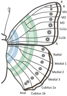
Exhaustive comparative works on butterfly wings by B.N. Schwantwitsch and F. Suffert in the 1920’s led to independent proposals of a generalized scheme of wing color patterns in nymphalids, later known as the Nymphalid Groundplan (Fig. 1.4;Schwanwitsch 1929,Nijhout 1991,Beldade and Brake-field 2002). This scheme summarized the morphological diversity of a rich family, with approximately 6,000 species (Wahlberg et al. 2009), and until today it serves as a basis of comparison for butterfly wing pattern elements. Based on morphology and on position, pattern elements are organized in three symmetry systems: basal, central, and border symmetry systems (Fig.
23
Figure 1.3: Eyespot and its traits. (A) Butterfly wing color patterns are formed by juxtaposition of monochromatic scales in patterns elements such as the eyespot. (B) The diversity of this pattern element can vary within and between species in terms of: number and shape (Junonia coenia), number of rings and position (Lasiommata megera), size (Bicyclus anynana), and color (Melanargia lachesis).
Each repeated pattern element in each symmetry system is assumed to be (serially) homologous, despite considerable variation within their particu-lar form (Nijhout 1991, 2001). At the same time, wing patterns of other non-nymphalid butterfly families have similar morphologies at correspond-ing positions (Martin and Reed 2010) and can be hypothesized as homol-ogous, even though developmental similarities have not yet been assessed (discussed inShirai et al. 2012and Chapter 2). We explored one particular pattern element, the eyespot (Fig. 1.3), homologous across nymphalids ( Ni-jhout 1990,1991,Brunetti et al. 2001,Nijhout 2001,Reed and Serfas 2004,
Monteiro et al. 2006, Martin and Reed 2010,Oliver et al. 2012), for which there is considerable knowledge on its development and evolution.
1.4.1 Eyespot evolution and development
Eyespots, called border ocelli in the Nymphalid Groundplan, are serially re-peated structures at the distal region of the wing, being part of the border symmetry system. They are circular patterns composed of rings with differ-ent colors (Fig. 1.3A), although ring shape and composition can range from single spots to semicircles to rings of one to four different colors (Nijhout 1991). Phenotypic variation of eyespots is found within and between species, and is expressed by several traits such as presence/absence (eyespot number in the wing), number of rings, position, size, shape, and color (Fig. 1.3B). Variation in many of these traits can co-occur within the same species, and it frequently does so in different wings of the same individual, in different surfaces of the same wing, and in different wing regions of the same surface (Nijhout 2010).
25
recognition (Robertson and Monteiro 2005, Costanzo and Monteiro 2007). Accordingly, their diversification is shaped by natural and sexual selection (seeOliver et al. 2009).
Similar to other repeated structures in an organism, such as teeth, petals, and body segments, despite shared developmental instructions and pheno-typic correlation, these individual repeats are able to evolve independently (Beldade et al. 2002b,Beldade and Brakefield 2003, but seeAllen et al. 2008). Furthermore, eyespots are evolutionary novelties, that is, they exist only in this group. Understanding how novel traits originate from pre-existing genetic architectures, and how do they impact species diversification, ex-emplified by adaptive radiations of angiosperms (flowers as the innovation), vertebrates (limbs), and birds (feathers), are long-standing questions of Evo-lutionary Biology.
The rings of different colors are presumably specified by a reaction-diffusion4
process. This mechanism is involved in cell fate establishment and pattern formation (Turing 1952,Murray 1981,Meinhardt and Klingler 1987,Kondo and Miura 2010), and is deployed in a wide range of structures of several species such as the Hensen’s node or Spemann organizer (prospective no-tochord in birds and amphibians, respectively), the apical ectodermal ridge (prospective tetrapod limb), and pigmentation patterns in stripes and cir-cles found in mammals (zebras, giraffes, felids), fish, birds, mollusks (shells, octopus), and arthropods (beetles, butterflies)5. The principle of this
mech-anism relies on the production and secretion of diffusible signaling molecules from an organizing center, or organizer, creating a gradient to which
neigh-4
In fact a diffusion-reaction process.
5
boring cells respond to according to the morphogen level they receive, with a threshold "function" determining different fates (Kerszberg and Wolpert 2007). Multiple factors can vary, potentially leading to phenotypic varia-tion: time of diffusion, diffusion rate, diffusion mode (in pulses, continuous), degradation rate, amount or concentration of morphogen, morphogen dif-fusibility, or tissue threshold function (tissue sensitivity).
Schwantwitsch (1929) noticed that border ocelli influence the morphology of surrounding elements (e.g. Umbra, Circuli, even the medial band), and spec-ulated on inductive properties of this pattern element. Later it was proposed that a concentration dependent signal-response would form the prospective rings of different colors, with cell fate being established by the distance to the source (Nijhout 1990,Monteiro et al. 2001, but seeOtaki 2011). It was validated by intra-specific transplants of competent cells demonstrating or-ganizers’ induction of eyespot formation at regions that usually do not bear them (Nijhout 1980a, French and Brakefield 1995), and cauteries of these cells leading to ablation or reduction of eyespots (Nijhout 1980a,Brakefield and French 1995). Artificial selection experiments for size (Monteiro et al. 1994) and color composition (Monteiro et al. 1997a) showed that these two aspects of eyespot phenotypes responded mostly by altering the signaling and response phases of ring establishment, respectively (further detailed in the next subsection).
27
rings of different colors correlate with rings of expression of transcription factors such as Engrailed,Spalt, and Distal-less (Fig. 1.5; Brakefield et al. 1996,Brunetti et al. 2001,Monteiro et al. 2006).
These developmental instructions regulate the action of effector genes during differentiation. At about 80% of pupal development (Nijhout 1980b, Koch et al. 1998, and Chapter 4), pigment synthesis pathways are activated (Fig.
1.5) and, curiously, in a stereotypic temporal fashion. Previous studies ana-lyzing the sequence of pigment deposition in different species have found an invariable time course starting with white (presumably pteridines), followed by yellow, orange, and red (presumably ommochromes), and lastly black, grey, and brown (melanins,Nijhout 1980b,Koch and Kaufmann 1995).
Figure 1.5: Eyespot development. Organizer establishment stage is illustrated by Antp in larval wings (protein detection described in Chapter 2). Ring establishment stage in early pupal wings presents rings of expression of En (green) and Sal (pink), that prefigures prospective rings of different colors in the adult (from Brunetti et al. 2001). Pigment synthesis stage is shown for the external golden ring, observed in late pupal wings.
diffuse from the organizer and, by a concentration-dependent process, pat-tern the fate of neighboring cells in early pupal wings; and 3) activation of pigment synthesis pathways, when pigments are deposited in single scales forming colored rings in late pupal wings (Fig. 1.5). Other classifications into four steps have been proposed, detailing the first stage into pre-pattern and organizer establishment (Brakefield et al. 1996), or the second stage into signal and response (Brunetti et al. 2001).
Developmental studies of genetic variants, derived from spontaneous mu-tations of large effect or artificially selected lines, done in the last decades have generated important information for linking genetic and phenotypic variation. Specifically, different aspects of eyespot phenotypes can be asso-ciated to different stages of the developmental hierarchy, by different lines of evidence involved in particular stages of development.
1.5
Variation of eyespot traits relate to specific
de-velopmental stages
Spontaneous mutations and artificial selection experiments in B. anynana
generated eyespot phenotypic variants in number, shape, position, size, color composition, and overall color (Fig. 1.6; reviewed inBrakefield 1998, Brake-field and French 1999,Brakefield 2001,Beldade and Brakefield 2002, McMil-lan et al. 2002,Beldade and Saenko 2010,Nijhout 2010).
Each of these aspects of eyespot morphology were associated with changes occurring at particular stages of eyespot development (summarized in Table
29
color relates with the pigment synthesis stage. Support for the association of particular stages determining particular traits comes from two lines of ev-idence: a) genetic variation affecting one aspect of eyespot morphology does not interfere with other traits; and b) markers of each developmental stage (e.g. expression patterns of implicated genes) are disrupted and prefigure altered phenotypes (Fig. 1.6A).
Artificial selection experiments onB. anynanawing patterns usually respond fastly (e.g. in less than 10 generations for selection on eyespot size) and with high heritabilities (h2, ranging from 0.60-0.70;Beldade et al. 2002a,b)6.
Selection on size (Monteiro et al. 1994) and color composition (Monteiro et al. 1997a) aiming at a single eyespot produced correlated responses in other eyespots, especially for those on the same wing surface. The consider-able additive genetic variation and correlated responses observed reveal the potential for evolutionary change of each selected trait, which is followed by its developmental homologues (Brakefield and French 1993,Brakefield 1998,
Brakefield and French 1999). However, selection in a trait did not affect other aspects of eyespot morphology. For instance, if selection was on color composition of an eyespot, the color scheme of other eyespots was altered, but not their position, shape, size, and so on7.
6
These studies targeted both forewing eyespots, h2
for size selection targeting one eyespot out of the two = 0.47-0.67 (Monteiro et al. 1994).
7
Figure 1.6: Evidence of differential developmental contribution for traits inB. anynana
31
A similar observation comes from mutant phenotypes that typically affect single aspects of eyespot morphology (Table 1.1). The same is true for double mutants, each with a mutation affecting a particular trait. In hybrid mutants, the additive effect of each mutation highlights their distinct genetic bases and, in all known cases, other aspects of eyespot morphology and other pattern elements are not disrupted (Table 1.2).
The association between stages determining different traits is further sup-ported by developmental markers representative of processes occurring in each stage (Table1.1). Disruptions are localized at a particular stage, with-out cascading in previous or following stages. Another important observa-tion is that within each stage the whole set of implicated genes change in the same way, suggesting that markers within each stage are linked. For exam-ple, the mutant Spotty, that bears extra eyespots in the forewing, presents expression patterns of all genes related with the first stage at a new location (Table1.1).
In this mutant, the organizer establishment stage is altered. The entire set of downstream processes following this stage are recruited to make an eyespot at a new location. Marker genes involved in the ring establishment stage and the inductive ability to form ectopic eyespots when the mutant foci are transplanted to another animal are the same as in wild-type. Pigment deposition of each colored ring at the new location is synchronous with the appearance of same colors in wild-type eyespots (Table 1.1).
Table 1.1: Complete catalogue of eyespot developmental markers associated with genetic variants produced by spontaneous mutations or artificially selected lines. In Mutant/Line: capitalized names represent dominant inheritance and normal case, recessive inheritance; segregation of each mutant (*homozygote lethal) determined in given reference, and those without: X-ray induced mutation (3+4) or selection line. In Organizer establishment and Ring establishment gene expression: "Y" (yes) for cases when expression of given genes is disrupted compared to wild-type, prefiguring the mutant phenotype; "N" for no difference. In Ring establishment, the signal (focal induction as in the donor, assessed by transplants) and response (epidermal response to transplants or wounds as in the host) are further detailed. In PigSyn (Pigment synthesis): as there is no expression pattern of candidate genes for this stage, timing of pigment deposition was used as evidence for disruption of this stage.
Mutant [ref] Morphology Wing Eyespot Other references eys: eyespots
A Spotty [1, 3, 16] extra eys FW 3+4 6, 9, 25, 30, 31, 33, 34, 35, 36, 37 B Missing [16] reduced or lost eys HW 3+4 31, 33, 34, 35, 36 C 3+4 lost (AA) or HW 3+4 28, 31, 33, 34, 36, 37, 38
reduced (Aa) eys
D comet [6, 13] comet shape both all 25, 33, 35, 37
E Cyclops [3]* ellipsoidal shape, HW 4+5 6, 25, 20, 28, 31, 33, 35, 36, 37 loss of eys variable
F veinless [NA] reduced eys both variable 33 G fat flat ey in AP axis FW 5 25, 36 H thin flat ey in PD axis FW 5 25, 36
I BigEye [3, 21]* enlarged eys both all 6, 25, 28, 31, 33, 35, 36, 37, 38 J HIGH enlarged eys FW 5 6, 8, 25, 26, 36 K LOW reduced eys FW 5 6, 8, 25, 26, 36 L AP enlarged eys FW both 27, 29, 32 M ap reduced eys FW both 27, 29, 32 N GoldenEye gold in black ring both all 21, 28, 31, 33, 35, 36, 37
[10, 20]*
O Spread [21] gold in black ring, both all 38 enlarged ey
33
Organizer establishment Ring establishment PigSyn Dll N Sal En Dll Sal En signal response time
A Y Y Y Y N N N N
[3, 23] [23] [23] [23] [3] [1] [1] [7] B Y Y Y
[14, 16] [14] [16] C Y [13] Y [13]
D Y [17] Y [17] N [17] N [17] N [17] E Y [3] Y [3]
F N [20]
G N [5] Y [5]
H N [5] Y [5]
I N [3] N [3] N [24]
J Y [2] Y [2]
K Y [2] Y [2]
L Y [12] Y [15] Y [12] N [19] Y [19] M Y [12] Y [15] Y [12] N [19] Y [19]
N Y [10] Y [10] Y [10, 20] N [20] Y [20] O Y [21] Y [21] Y [21] Y [21]
P N [4] Y [4]
Q N [4] Y [4]
R N [24]
Table 1.2: Additive effect of double mutants highlight the distinct genetic bases of altered phenotypes. References as in Table1.1.
Double mutant [reference] Phenotype
BE + Spotty [6, 8] extra eyespots are also enlarged A- + Spotty [8] no anterior eyespot; normal extra eyespots BE + LOW [8] forewing LOW, hindwing BE BE + HIGH [8] larger than BE eyespots comet + Spotty [8] extra eyespots are comet-shaped Cyclops + Spotty [8] extra eyespots are ellipsoidal BE + GoldenEye [11, 21] gold in black ring, enlarged eyespots
Missing + Spotty [16] extra eyespot reduce in size, no change in HW BE + Missing [18] missing eyespots, remaining enlarged BE + Cyclops [18] enlarged eyespots, ellipsoidal in HW
BE + comet [17] enlarged comet-shape eyespots Fred (Spread + Frodo) [24] gold in black ring, normal size
1.6
Aims and thesis synopsis
The aim of this thesis was to explore the developmental basis of variation and diversity, specifically taking into account the contribution of variation at hierarchical stages of development in morphological diversification. We looked at variation of different stages at a time, using multiple approaches: comparative expression patterns of organizer and ring establishment (Chap-ter 2); functional assays of ring establishment signaling molecule Wingless
35
venues and possibilities developed within Developmental Genetics and Molec-ular Biology. Key Evo-Devo topics include: homology and homoplasy, con-straints, canalization, evolvability and robustness, evolution of genetic archi-tectures and of gene regulatory networks, modularity, developmental plastic-ity, and the origin and evolution of taxa, ontogenetic stages, morphologies, and of evolutionary novelties8. Here, concepts that will be under discussion
include the origin and evolution of novel traits, homology inference and de-velopmental systems drift, heterochrony and heterotopy, and dede-velopmental dynamics, hierarchies, and milestones.
In Chapter 2, we asked whether the origin and diversification of novel traits occurs by a single network co-option, or whether individual genes are re-cruited and re-wired de novo in the novel context. We looked at compar-ative expression patterns of four genes involved in the organizer establish-ment stage, in 13 butterfly species. We found unexpected levels of variation in gene combinations associated to this stage, which is indicative of evo-lutionary flexibility for establishing organizers. The presence and absence of expression associated with foci development was analyzed under a phy-logenetic framework, and the reconstruction of the evolutionary history of expression revealed a single origin for Antennapedia in the satyrine clade. The other three genes were ambiguous in terms of having been co-opted in a single step or through multiple events. This developmental variation is surprising for what we expect of conserved mechanisms forming homologous traits.
Given this flexibility, we asked whether the same is found in another stage of eyespot development, ring establishment. Previous studies showed devel-opmental variation for this stage, but it was assessed in distantly-related species. We queried expression patterns of three genes in a satyrine species closely related to B. anynana and, together with available data from the literature, found further developmental variation associated with
establish-8
ment of the black middle ring. These findings revealed that there is develop-mental systems drift in organizer and ring establishment stages, highlighting that the development of homologous structures are not necessarily underlied by the same genes.
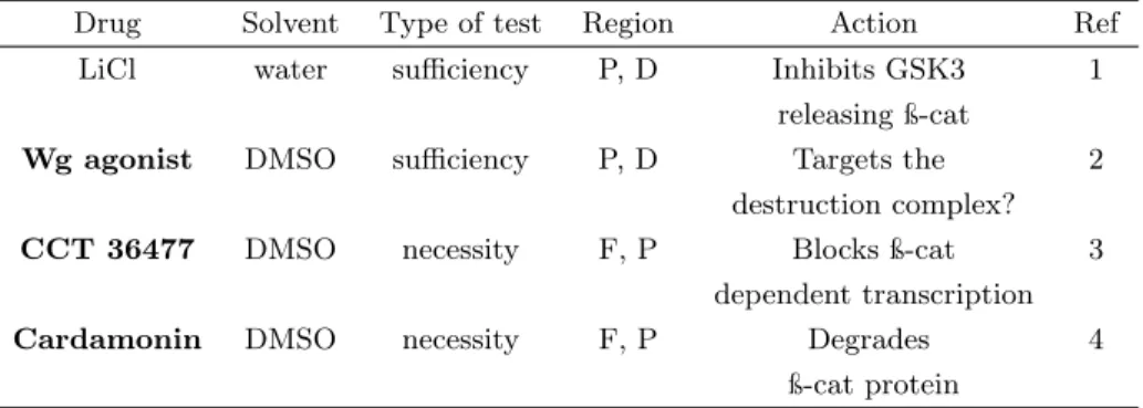
A central aspect about unraveling the molecular mechanisms underlying evolutionarily relevant phenotypic variation is to know the role of partic-ular genes in the formation of morphologies. In Chapter 3, we attempted to establish functional tools, still scarce in butterflies despite their unique advantages for the study of Evo-Devo. We focused on two stages, ring estab-lishment and pigment synthesis. For the first, microbeads soaked in agonists and antagonists drugs targeting components of the Wingless pathway were tested, respectively, for sufficiency and necessity in eyespot formation. De-spite hundreds of manipulations in two eyespot-bearing species, there was no difference between treatment and control for both tests, which indicated that either Wg is not necessary nor sufficient for ring establishment, or that the method failed. We speculated that, if the method was not effective, a possible reason was that beads were being melanized and encapsulated, typical insect immune responses to foreign bodies, isolating the content of bead from the wing tissue.
For the second stage of pigment synthesis, we optimized a tissue culture protocol and tested the necessity of melanin synthesis enzymes by a phar-macological approach. We found that enzymes phenoloxidase and dopa de-carboxylase are necessary for the progression of pigmentation inB. anynana. In a melanic mutant of this species, higher amounts of drug were required to arrest pigmentation of its overall darkened wings. Dopa decarboxylase may not be necessary for deposition of the yellow color, but may be necessary for scale development.
37
B. anynana. To explore how developmental and phenotypic variation relate in this final stage, in Chapter 4 we looked at the temporal development of pigment deposition. The appearance of colors, i.e., differentiation, always occurs in the same sequence across species. We looked whether the sequence and also the timing of differentiation was conserved in three satyrine species. Similarities of both temporal aspects for similar phenotypes across species was found, despite an accelerated onset of differentiation in one of them. Complementary, we asked whether timing of pigment deposition was robust to altered phenotypes of genetic variants within a species. In two mutants, one with overall enlarged eyespots and another with overall darkened wings, the timing as well as the duration of pigment deposition were not different from the wild-type condition. Timing for the black middle ring was not significantly different for both wing surfaces across species, and in these two genetic variants. The duration of black deposition, quantified in vitro for mutants, was also similar across phenotypes, suggesting that black eyespot rings, regardless of phenotypic variation in size and color intensity, have a conserved and robust time for deposition.
In the two mutants, cell fate of colored rings was not altered. In another, heterotopic, mutant where the typically black middle disc has the same color as the external, yellow ring, timing of deposition was significantly different from the wild-type. Cells at particular locations can have distinct fates and achieve those fates at different times. We further tested in this heterotopic mutant whether the timing of differentiation follows cell identity or cell loca-tion. We asked whether the yellow color at an unusual location differentiated at the time that is characteristic of that color, or its new location. Timing followed the differentiation of the color, suggesting that timing of pigment deposition is instructed by cell identity and not ring position.
species, we need to dissect the molecular mechanisms behind pigment de-position. In Chapter 5, we looked at changes in global expression levels of candidate genes and at the temporal dynamics of gene expression of this stage, and compared to what is found during the previous developmental stage of ring establishment. We expected to find higher dynamism and gene enrichment for gene classes related with patterning genes at the early, ring establishment stage. Similarly, the late stage should be over-represented by effector genes related with pigment synthesis. This prediction was not con-firmed because we found patterning and effector genes expressed throughout pupal life. While most patterning genes did not change their expression lev-els in seven time points of wing development transcriptomes, effector genes increased their expression at the time they are expected to be active. This result reveals the pleiotropic nature of pathways involved in wing, and wing pattern, development.
39
1.7
Acknowledgements
I would like to thank Suzanne Saenko for allowing to use the B. anynana
Chapter 2
Origin and diversification of
recruited circuitries for
organizer and color ring
establishment
2.1
Summary
dis-43
cussed in the context of inferring homology. Similar flexibility has been reported for the ring establishment stage during early pupal wings of very distantly related nymphalids. Here we looked at expression of three genes implicated in this stage in the satyrine Lasiommata megera, finding devel-opmental variation for the gene combination associated with black middle rings. Our study underscores the importance of widening the representation of phylogenetic, morphological, and genetic diversity in order to establish general principles about the mechanisms behind the evolution of novel traits.
Authors’ contributions
Part of this chapter has been published with co-authors S.V. Saenko, R.A. Keller, M.A. Jerónimo, P.M. Brakefield, H. Descimon, N. Wahlberg, and P. Beldade (Shirai et al. 2012). It was chosen as Editor’s pick and under the "Highly accessed" tag. It was also awarded with a Student Poster Prize at the 4th Meeting of the European Society for Evolutionary Developmental Biology (EuroEvoDevo) in July 2012.
The additional part, presented here in the Annex, has been funded by the Rosemary Grant Award (Society for the Study of Evolution) to LTS and was done in collaboration with the Butterfly House of the University of Lisbon. LTS coordinated and co-wrote the published manuscript, and wrote this chapter. SVS collected the bulk of larval expression data, MAJ collected individuals and larval expression data for Pieris, LTS collected individuals and expression data for pupal wings. PMB and HD provided Papilio and
"But has selection truly acted alone as the sole source of order in the
emergence of life and its subsequent evolution? I do not think so. From my
gut, from my dreams, from my work of three decades, from the work of a
growing number of other scientists, I do not think so".
Stuart Kauffman,1995p.98
2.2
Introduction
The origin and diversification of novel traits are central and longstanding issues in Evolutionary Biology (Muller and Wagner 1991). Evolutionary novelties are lineage-restricted traits often associated with new adaptive functions (Muller and Wagner 1991, Pigliucci 2008). Compelling examples include angiosperm flowers, beetle horns, bird feathers, and butterfly wing color patterns. Studies in Evolutionary Developmental Biology have shown that the origin of novel traits often involves the recruitment, or co-option, of conserved genetic circuitries. This idea is captured in the expression "teach-ing old genes new tricks" (True and Carroll 2002), used to explain the genetic mechanisms through which novel traits arise.
45
for the development of lineage-restricted traits, key questions remain unan-swered. For example, are entire pathways recruited as a whole or are indi-vidual genes co-opted and re-wired de novo (Monteiro and Podlaha 2009)? How do recruited or rebuilt pathways diversify along with trait diversifica-tion? Widening the representation of both phylogenetic and morphological diversity, together with focus on genetic networks rather than single genes, will be crucial to solving these issues (see Kopp 2009). In this study, we provide a taxonomically and genetically wide survey of a model evolution-ary novelty, butterfly eyespots, to investigate the origin and diversification of the genetic circuitry associated to its development.
to encompass all diversity, we assayed a number of species across three but-terfly families. This broad phylogenetic coverage of phenotypic diversity is presented along with data on the putative genetic circuitry associated to early eyespot specification.
Butterfly eyespots provide a good illustration of the recruitment of genetic circuitry implicated in developmental processes shared by all insects for the formation of novel traits. This includes commonalities between eyespot velopment - exclusive of butterflies - and processes such as embryonic de-velopment (Saenko et al. 2008, 2010), appendage formation (Carroll et al. 1994, Keys et al. 1999), and wound healing (Monteiro et al. 2006, Saenko et al. 2008) - conserved across insects. The colored rings that make up eyespots are sequentially formed in pupal wings (Brunetti et al. 2001, Wit-tkopp and Beldade 2009), around organizing centers which are themselves specified earlier in larval wing discs (reviewed inBeldade and Saenko 2010). Recently, examination of the expression of conserved genes Antennapedia
(Antp), Notch (N), and Distal-less (Dll)1 during the initial stages of
or-ganizer establishment revealed intriguing differences among lineages within nymphalids (Hombría 2011,Saenko et al. 2011). However, the lack of gene expression data outside this clade prevented the reconstruction of the evo-lutionary history of the recruitment of those genes for expression in larval eyespot fields. Here, we increased the taxonomic sampling by including representatives of an additional nymphalid clade and two non-nymphalid families. We also examined the expression of another transcription factor in the presumptive organizer, Spalt (Sal, Monteiro et al. 2006), in all species sampled. Phylogenetic analysis of this comprehensive dataset revealed great
1
47
flexibility in which genes, and combinations of genes, are expressed in asso-ciation with this novel trait in different lineages.
2.3
Material and Methods
2.3.1 Biological material
Thirteen species of three butterfly families were assayed in this study. The nymphalid data on Antp, N, and Dll was obtained fromSaenko et al. (2011, see reference for details of origin and maintenance of larvae). Additional species, stained for all genes, were obtained from the Lagartagis Butterfly House (Lisbon, Portugal) or field caught and kept as follows: Danaus plex-ippus (room temperature, and natural light (L) and dark (D) cycle, fed on milkweed), Pieris rapae (18/23°C at 6D:18 L, fed on cabbage),Parnassius apollo (27°C at 12D:12 L, fed on stonecrop), and Papilio machaon (27°C at 12D:12 L, fed on fennel). The staging of larval wing development of all families was done following the tracheal extension into the vein lacunae (cf.
Reed et al. 2007).
2.3.2 Immunohistochemistry
dilution), rabbit anti-Sal (de Celis et al. 1999, 1:500 dilution), and guinea pig anti-Sal GP66-2 (1:1000 dilution, used forP. rapae) were provided by other labs. Alexa Fluor 488 anti-mouse, Texas Red anti-rabbit, and Alexa Fluor 594 anti-guinea pig (Molecular Probes) were used as secondary antibodies (1:200 dilution). Images were collected on a BioRad MRC 1024 or a Zeiss Imager M1 laser scanning confocal microscope.
2.3.3 Ancestral character reconstruction and correlation of protein recruitment history
49
for the superfamily Papilionoidea. Branch length estimates were calculated as described in Heikkilä et al. (2012). ML reconstructions were performed in Mesquite 2.74 (Maddison and Maddison 2001) choosing the Mk1 model (Lewis 2001).
To assess whether there is significant correlation between evolutionary histo-ries of pair of genes, pairwise Likelihood Ratio Tests were performed compar-ing the likelihood of an independent versus a dependent model of evolution (Pagel 1994,Barker and Pagel 2005). The likelihood for each model was cal-culated with BayesDiscrete in the BayesTraits package (Pagel 2007), using the branch length estimates and character coding as above. The likelihood ratio was calculated as 2[log-likelihood (Dependent Model) - log-likelihood (Independent Model)], and is expected to follow aχ² distribution with four
degrees of freedom (Pagel 1994,Barker and Pagel 2005).
2.4
Results and Discussion
To investigate the evolutionary history of the co-option of conserved genes to the location of a developing novel trait, we analyzed expression patterns in larval wings of multiple species in different butterfly families. We targeted four genes involved in transcription regulation: transcription factors Antp, Dll, and Sal, and the transmembrane receptor N. The latter, when bound to its ligands (Delta/Serrate/LAG-2 family of proteins), releases an intracellu-lar domain that regulates gene expression when associated to DNA-binding CSL proteins (Kimble and Simpson 1997).
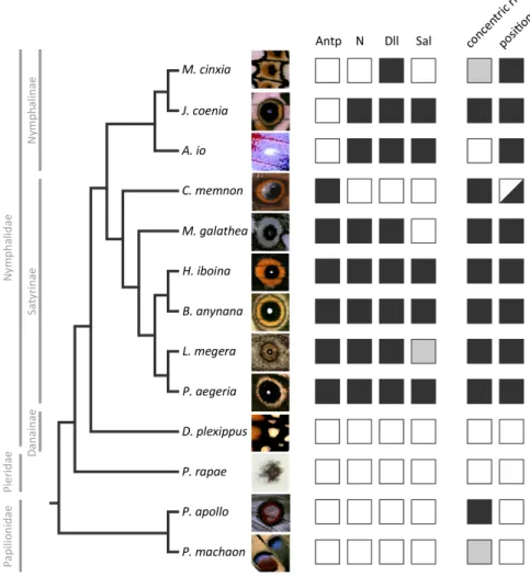
(Pieridae and Papilionidae; Fig. 2.2). Based on the complete dataset for all four proteins in the 13 representative species (Fig. 2.3), we investigated the evolutionary history of the recruitment of these genes. We mapped the local-ization of transcription regulators in presumptive eyespot centers onto the species tree, and performed ancestral character reconstructions using both parsimony and ML methods (Fig. 2.4). The species chosen in this study rep-resent diversity in (eye)spot morphology and position on the wing (cf. the conserved venation pattern), allowing for discussions about the inference of homology (Fig. 2.5).
2.4.1 Taxonomically wide sampling of genes expressed in the developing eyespot field
51
Antp expression during early organizer establishment clearly distinguished Satyrinae and Nymphalinae clades, being present only in the former (Saenko et al. 2011). In contrast, N and Dll showed no clear dichotomy between those clades, being expressed in association to most, but not all, developing organizers (Saenko et al. 2011). Our new data onSalshow that its expression is also variable within nymphalids, in a pattern which does not follow that of the other genes (see discussion about gene co-recruitment below) nor that of any particular aspect of eyespot morphology, such as the size, color, shape, or number of rings (see Fig. 2.3).
To infer the evolutionary history of gene recruitment to presumptive eyespot centers, we examined the expression of the four selected genes in a more dis-tantly related nymphalid (D. plexippus) and in three non-nymphalid species (P. rapae, P. apollo, and P. machaon). The monarch butterfly, D. plexip-pus, has series of white spots along the antero-posterior margin of its wings. These appear as multiple single-color spots on each wing compartment bor-dered by veins, instead of one single element with multiple concentric rings as is characteristic of nymphalid border ocelli (Figs. 2.2 and 2.3). These single-color spots are generally not considered homologous to border ocelli (Brakefield et al. 1996), even though wing patterns of the Danainae subfam-ily can be described in terms of the Nymphalid Groundplan (Nijhout 1991). On the other hand, many non-nymphalid species have diverse types of spot-like elements that diverge to different degrees from typical eyespots both in morphology (e.g. in the number and color of rings) and position; illustrated here by P. rapae’s single black spot, P. machaon’s quasi-concentric rings, and P. apollo’s concentric rings around a white center (Figs. 2.2 and 2.3,
53
Figure 2.3: Summary of expression data for the four developmental genes and adult (eye)spot traits. Complete dataset of the four transcription regulators targeted in this study for all 13 species coded as the presence (black) or absence (white) of expression (Lasiommata megera’s Sal expression could not be determined, grey box). Data for Nymphalidae Antp, N, and Dll expression were obtained from Saenko et al. (2011),
55
we show that none of four transcription regulators associated to eyespot organizers in nymphalids localizes to the regions of the presumptive eyespot-like elements in the outgroup species (Fig. 2.2). Also, with the exception of Dll forP. rapae, we could not detect any of those proteins at the intervein region, where N and Dll are found in some butterfly species (Reed and Serfas 2004,Monteiro et al. 2006).
The absence of all four transcription regulators analyzed from the posi-tion of presumptive eyespots in the outgroup species suggests that different mechanisms might be at play in the formation of their spots, as previously suggested for P. rapae (Monteiro et al. 2006). Possible scenarios include that 1. the same genes are associated with presumptive organizers but at a stage other than the last larval instar which we analyzed, when nymphalids specify their organizers (Beldade and Saenko 2010), or 2. other genes are specifying organizers in different butterfly clades, or 3. the spots in these lin-eages are formed by developmental mechanisms that do not involve central organizers. The latter possibility could be experimentally tested by the same type of tissue transplant or damage approaches that established nymphalid eyespot centers as organizers (Nijhout 1980a,French and Brakefield 1995), in which the transplantation of such cells to other competent regions of the wing lead to the production of an ectopic eyespot at the host site.
2.4.2 Ancestral reconstruction of gene recruitment
of the evolutionary history of gene recruitment to that location. Ancestral character reconstructions with both methods showed an unambiguous evo-lutionary history only for the expression of Antp (Fig. 2.4), found at the location of presumptive eyespot organizers of satyrines but not nymphalines (Saenko et al. 2011). Our sampling of outgroup species supports that the novelAntp expression is in fact exclusive to satyrines and originated in the common ancestor of the group (Fig. 2.4, see Annex).
Ancestral reconstructions of the recruitment of other three transcription reg-ulators resulted in an evolutionary history that is less clear. There are two equally parsimonious scenarios of losses (Fig. 2.4A top) and gains (Fig.
2.4A bottom) of eyespot-related expression for each of those genes, with many instances of homoplastic events. This ambiguity is mainly due to the character states of Caligo memnon (absence of N, Dll, and Sal) and Meli-taea cinxia (absence of N and Sal) in relation to all other members of their respective subfamilies (presence of N, Dll, and Sal). Given the phylogenetic positions of these species, it is not possible to recover a single scenario for the recruitment of the three transcription regulators to the presumptive eye-spot organizers. Worthy of special attention is the case of the satyrine C. memnon, in which onlyAntpis expressed in the area of presumptive eyespot centers (Fig. 2.3). The forewing eyespot of this species is composed of rings of different colors and placed at the typical location of Nymphalid Ground-plan’s border ocelli (Fig. 2.1). According to the parsimony reconstructions, eitherC. memnon represents a secondary loss ofN andDll expression (Fig.
2.4A top), or the absence of expression of these genes, together with that of
57