Susana Margarida Pires Gonçalves
Dissertation presented to obtain the Ph.D degree in Biochemistry
Instituto de Tecnologia Química e Biológica | Universidade Nova de Lisboa
Oeiras,
May, 2012
Structure-function relationships in a
Susana Margarida Pires Gonçalves
Dissertation presented to obtain the Ph.D degree in Biochemistry
Instituto de Tecnologia Química e Biológica | Universidade Nova de Lisboa
Oeiras, May, 2012
Structure-function relationships in a
i A thesis submitted in conformity with the requirements for the degree of Doctor of Instituto de Tecnologia Química Biológica, Universidade Nova de Lisboa.
v
ACKOWLEDGMENT
I wish to express my sincere gratitude to my supervisor Dr. Pedro
Matias for his careful guidance, his extended patience, his invariable
availability and most of all his encouragement throughout my
doctoral studies.
I extend my appreciation to my advisory committee; Dr. Helena Santos
and Dr. Peter Donner, and to Dr. Maria Arménia Carrondo, the three of whom
provided me with sound advice.
Special thanks to Dr. Antony Dean for its valuable recommendations on
the elucidation of the catalytic mechanism of
-oxidative dehydrogenation by
isocitrate dehydrogenase, as well as for his support and encouragement.
Dr. Nuno Borges and Dr. Helena Santos, in helpful discussions of the
biochemistry behind the “two
-
step” pathway of mannosylglycerate.
I wish to extend my appreciation to my labmates for their friendship
and for their precious hints on science and on life.
vii
THESIS ABSTRACT
Enzyme evolution is often constrained by aspects of catalysis. Mechanistically diverse enzymes evolved from a common ancestor still preserve those structural signatures essential to the core chemistry retained by all members of the superfamily. Indeed, these shared features allow superfamilies to be accurately classified, while derived features allow nested families and subfamilies to be identified in a hierarchical fashion. Accurate classification has helped elucidate mechanisms promoting functional diversification, for example catalytic promiscuity, and protein engineering by rational design.
Nowadays, a holistic view of enzymes` regulatory mechanisms and catalytic proficiency is provided by the identification of conserved features of molecular architecture in combination with aspects of reaction dynamics.
My work focused on the structural elucidation and analysis of three enzymes: a glycosyltransferase; a phosphatase and an oxidorreductase. “Snapshots” along the reaction coordinate of each enzyme were obtained by combining X-ray diffraction with “cryo-trapping” ligand-binding methods. These were used to characterize the molecular mechanisms involved in substrate recognition and binding. They were also used to distinguish between models proposed for the catalytic mechanisms of each enzyme, and provide insights into enzyme dynamics essential for catalysis and the stereo and regio-selective strategies at work.
mannosyl-3-viii phosphoglycerate. In the second step, mannosyl-3-phosphoglycerate is dephosphorylated to MG by mannosyl-3-phosphoglycerate phosphatase (MpgP; E.C. 3.1.3.70). The third enzyme studied is the metal dependent NADP+-linked isocitrate dehydrogenase (IDH; E.C.1.1.1.42) from Escherichia
coli. IDH catalyses the oxidative decarboxylation of isocitrate to -ketoglutarate. E. coli IDH is one of the pioneering case-studies of concerted protein dynamics with a productive Michaelis complex. However, a complete picture of these events has been hampered by limitations at the level of crystallographic packing.
Main conclusions from each study are:
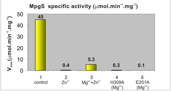
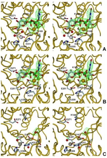
a) Structural elucidation shows T. thermophilus HB27 MpgS is a GT55 glycosyltransferase, a family that falls within the retaining GT-A enzymes (www.cazy.org). Two flexible loops involved in key interactions for the productive binding of substrates were identified by comparing the apo-form with the metal-substrate complex [Mg2+:GDP--D-mannose]. A second metal binding site was found about 6 Å away from the mannose moiety. Kinetic and mutagenesis studies provided evidence that this second metal site is indispensable for catalysis. Additionally, Asp167 of the conserved D-X-D motif was proposed as the catalytic nucleophile in light of the current mechanistic models for the retaining GTs. A survey of enzymes with the GT-A fold and with a bound sugar-donor, or a bound analogue, was used to identify the orientation of the scissile glycosidic bond of the sugar donor with respect to the D/E-X-D motif. The glycosidic bond is oriented towards the motif in the retaining GTs and away from it in the inverting GTs. This feature, which explains the stereochemistry of the reactions, provides a structural signature that will assist efforts in classifying the inverting and retaining GTs.
ix substrates while preserving core reaction chemistry. The domains of the “open” apo- and the “closed” holo- forms of MpgP are related to each other by a hinge rotation. The dynamics of the catalytic machinery were followed using cryo-trapped reaction species to provide “crystallographic snapshots” along the reaction cycle. Results suggest that phosphoryl-transfer by MpgP from T. thermophilus HB27 involves a concerted DNAN mechanism with Asp8 acting as a catalytic acid in the formation of a short-lived metaphosphate intermediate that is immediately subjected to nucleophilic attack by water. These structures identify the principle mechanistic features of phosphoryl monoester transfer catalysis in members of the HAD superfamily. More generally, they suggest a possible continuum of phosphoryl transfer mechanisms, ranging from those that are purely associative to those that are purely dissociative.
c) Comparison of the crystallographic structures of a pseudo -Michaelis-Menten complex of the wildtype EcoIDH, the K100M mutant trapped with its reaction products and several other EcoIDH structures identifies three distinct conformational states: open, quasi-closed and closed. Structural comparisons suggest substrate binding initiates domain closure, with hinge dynamics that span the central -sheet of each monomer in the biological homodimer. Conserved catalytic residues binding the nicotinamide ring of the NADP+ coenzyme and the metal-bound substrate move as rigid bodies together with the hinge rotation. The closed conformations of both the wildtype
xi
SUMÁRIO DA TESE
A evolução enzimática é na maioria dos casos determinada por aspectos associados à catálise. Enzimas que são funcionalmente distintas, mas que partilham uma origem ancestral comum, preservam assinaturas estruturais que asseguram a viabilidade da química base, comum a todos os membros de uma super-família. Estas assinaturas são fundamentais para a classificação de enzimas em diferentes super-famílias, assim como variantes destas assinaturas são indicações para a hierarquização de membros homólogos em famílias e sub-famílias. Tal sistema permite por sua vez a identificação dos mecanismos responsáveis pela diversidade funcional, entre os quais os factores estruturais que favorecem a promiscuidade catalítica e que são relevantes para a indústria biotecnológica e na engenharia de proteínas. Hoje em dia, os conceitos de estrutura e dinâmica estão correlacionados com o de catálise enzimática através da identificação de um conjunto de “movimentos-chave” com uma arquitectura molecular típica.
O meu trabalho centrou-se na elucidação da estrutura tridimensional e na análise estrutural de três enzimas: uma glicosiltransferase, uma fosfatase e uma oxidorreductase. Combinando técnicas de difracção de raios-X com métodos de crio-conservação de ligandos com diferentes tempos de incubação nos cristais das enzimas estudadas, obtiveram-se “instantâneos fotográficos” das diferentes etapas das coordenadas de reacção para cada enzima em estudo. A análise destas permitiu a identificação de mecanismos moleculares intervenientes no reconhecimento e ligação do substrato, a análise dos diferentes mecanismos de reacção com base nos modelos correntes, a aquisição de novos conceitos da relação entre a catálise e a dinâmica enzimáticas, e a identificação de determinantes que contribuem para a regio- e estereo-selectividades das enzimas estudadas.
xii ambientes extremos. O MG é um soluto compatível canónico, da classe dos derivados de açúcares, e que protege as células contra as tensões osmótica e térmica. A enzima manosil-3-fosfoglicerato sintetase (MpgS; E.C. 2.4.1.217), catalisa a transferência do grupo manosilo da GDP--D-manose para o aceitador nucleofílico 3-D-fosfoglicerato, produzindo o intermediário manosil-3-fosfoglicerato (MPG). No segundo passo da reacção, o MPG é desfosforilado a MG pela manosil-3-fosfoglicerato fosfatase (MpgP; E.C. 3.1.3.70). O segundo tópico abordou novos avanços no estudo da isocitrato desidrogenase dependente de NADP+ (IDH; E.C. 1.1.1.42) de Escherichia coli. A IDH catalisa a oxidação descarboxilativa do isocitrato em -cetoglutarato e é um dos casos de estudo pioneiros na demonstração da acção concertada entre dinâmica e catálise enzimáticas. No entanto, limitações devidas ao arranjo cristalográfico das moléculas tinham impedido até agora o seu completo esclarecimento.
As principais conclusões deste estudo são:
xiii açúcar ou um seu análogo. Nas GTs retentoras, a ligação glicosídica está orientada para aquele motivo, enquanto nas GTs inversoras esta encontra-se orientada no sentido oposto. Esta característica ajuda a explicar a natureza estereoquímica das reacções, e constitui uma assinatura estrutural na distinção entre GTs retentoras e inversoras.
b) a elucidação estrutural da MpgP de T. thermophilus HB27, permitiu a sua classificação como uma fosfatase metalo-dependente pertencente à super-família das “Haloalcanoic Acid Dehalogenase-like phosphatase” (HAD -like), incluída numa família das fosfatases do tipo “Cof”: a família das fosfatases de manosilo-3-fosfoglicerato. Esta estrutura é um excelente exemplo da combinação entre diferentes módulos funcionais numa nova família de enzimas com especificidade dirigida para novos substratos, mas preservando a base química da reacção. As formas estruturais “aberta” apo- e “fechada” holo- da MpgP estão relacionadas por rotação do módulo tipo “Cof” em torno de dois laços flexíveis da enzima, que ligam o módulo anterior ao módulo base “HAD-like”. A dinâmica do centro catalítico foi estudada através de “instantâneos fotográficos cristalográficos”, obtidos por crio-preservação de espécies representativas das várias etapas da coordenada da reacção. Os resultados sugerem que a hidrólise do grupo fosforilo do MPG ocorre através de um mecanismo concertado DNAN, onde o residuo Asp8 é o ácido catalítico para a formação do intermediário, sendo este subsequentemente atacado por uma molécula de água nucleofílica. Estas estruturas mostram aspectos mecanísticos chave na elucidação molecular da catálise de transferência de fosfomonoésteres que poderão ser extrapolados para outros membros da superfamília HAD. Genéricamente, estas características sugerem um
continuum de mecanismos alternativos, que variam entre os que são exclusivamente dissociativos e os que são associativos.
xv
TABLE OF CONTENTS
CHAPTER 1 _______________________________________________ 1 Introduction ______________________________________________ 1 1.1 Outline of the current dynamics and structural aspects behind the
catalytic power ________________________________________ 1 1.2 Molecular adaptations to extreme environments: compatible
compounds in osmo- and thermostabilization ________________ 3 1.3 Accumulation of -mannosylglycerate in Thermus thermophilus HB27
____________________________________________________ 6 1.4 Glycosyltransferases; fold, mechanism and biological implications 9 1.5 Mechanistic alternatives in phosphate monoester transfer catalysis 18 1.6 Isocitrate dehydrogenase; a paradigm for the concerted structural
dynamics with catalytic proficiency _________________________ 21 References ______________________________________________ 25
CHAPTER 2 _______________________________________________ 37 Structural analysis of Thermus thermophilus HB27
mannosyl-3--phosphoglycerate synthase. Evidence for a second catalytic metal ion and new insight into the retaining mechanism of
glycosyltransferases
xvi CHAPTER 3 _____________________________________________ 94 Structural analysis of Thermus thermophilus HB27
mannosyl-3--phosphoglycerate phosphatase. Evidence for a concerted DNAN
mechanism in phosphoryl-transfer catalysis of MpgP
3.1 Abstract _____________________________________________ 96 3.2 Introduction __________________________________________ 97 3.3 Material and methods ___________________________________ 100 3.4 Results _____________________________________________ 111 3.5 Discussion __________________________________________ 125 3.6 References ___________________________________________ 155 3.7 Acknowledgments _____________________________________ 162
CHAPTER 4 _______________________________________________ 163 Reaction-coupled dynamics and insights into mechanistic aspects of
the -oxidative decarboxylation reaction by isocitrate
dehydrogenase in Escherichia coli.Direct evidence for the
Try160-Lys230* diad in the assisted Brönsted acid-base catalysis
4.1 Abstract _____________________________________________ 164 4.2 Introduction __________________________________________ 165 4.3 Material and methods ___________________________________ 167 4.4 Results _____________________________________________ 172 4.5 Discussion __________________________________________ 190 4.6 References ___________________________________________ 201 4.7 Acknowledgments _____________________________________ 208
xvii LIST OF FIGURES
CHAPTER 1
Figure 1.1 The two pathways for the synthesis of mannosylglycerate 8 Figure 1.2 Overall reactions catalyzed by (a) glycosidases, (b)
glycosyltransferases, and (c) phosphorylases ________________ 10 Figure 1.3 Ribbon diagram of three glycosyltransferases (GTs)
representative of the different folds ________________________ 13 Figure 1.4 Established and proposed mechanisms for glycosidases,
transglycosidases and glycosyltransferases __________________ 16 Figure 1.5 Three possible limiting chemical mechanisms for phosphoryl
transfer ______________________________________________ 20 Figure 1.6 Isocitrate dehydrogenase (IDH) catalytic mechanism ____ 24
CHAPTER 2
Figure 2.1A SDS-PAGE 12% with pure recombinant MpgS from Thermus thermophilus HB27 _____________________________________ 43 Figure 2.1B Elution profile of MpgS loaded into an analytical 2.4 mL
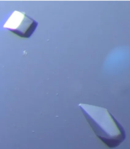
Superdex 200 3.2/30 PC column _________________________ 43 Figure 2.2 Prismatic crystals of T. thermophilus HB27 MpgS _______ 45 Figure 2.3 The structure of MpgS _____________________________ 54 Figure 2.4 Topology diagram of the MpgS monomer The structural role of Zn2+ _________________________________________________ 55 Figure 2.5 The structural role of Zn2+ __________________________ 57 Figure 2.6 The NDP-sugar binding pocket in MpgS and structurally related
enzymes _____________________________________________ 61 Figure 2.7 MpgS and its structural homologues __________________ 63 Figure 2.8 Two tunnels lead from the NDP-sugar binding pocket to the
xviii Figure 2.11 Column-chart of the MpgS maximum specific activities __ 70 Figure 2.12The Zn/M2+ binding site in the MpgS structures ________ 71 Figure 2.13 Detail of the alternative conformations adopted by loop 1 and loop 2 _______________________________________________ 74 Figure 2.14 Structural signatures in retaining and inverting
glycosyltransferases with GT-A fold ________________________ 77
CHAPTER 3
Figure 3.1A 15% SDS–PAGE of pure recombinant T. thermophilus HB27 MpgP _______________________________________________ 102 Figure 3.1B Elution profile of MpgP loaded onto an analytical 2.4 mL
Superdex 200 3.2/30 precision column _____________________ 102 Figure 3.2 Native MpgP crystals used for data collection __________ 106 Figure 3.3 DSF thermal denaturation curves for MpgP ____________ 112 Figure 3.4 MpgP elution profiles from the SEC analysis with a Superdex
75 3.2/30 precision column _______________________________ 114 Figure 3.5 Three-dimensional structure of MpgP from T.thermophilus
HB27 ________________________________________________ 117 Figure 3.6 Mean main chain thermal B-factors for chains A and B of
apo-MpgP and holo-apo-MpgP (control [Mg2+-MG-HPO
42-]) structures ____ 120 Figure 3.7 The electrostatic potential at the molecular surfaces for the
apo-MpgP and holo-MpgP structures _______________________ 122 Figure 3.8 Hinge rotations of the C2B cap domain from the open to the
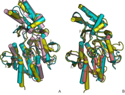
closed conformations of MpgP ____________________________ 124 Figure 3.9 Structural comparisons of T. thermophilus MpgP with its
orthologous members of the MPGP family ___________________ 127 Figure 3.10 Structure-based sequence alignment of MpgP with its
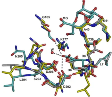
structural homologues __________________________________ 128 Figure 3.11 The MpgP catalytic pocket ________________________ 130 Figure 3.12 Stereoview of the [Gd3+-MPG] crystal 1 structure showing the
xix Figure 3.13 Unbiased sigmaA-weighted |Fo| - |Fc| electron density map
covering the ligands in the catalytic pockets of structures [Gd3+-MPG] crystal 2, [Mg2+-MPG] crystal 1 and crystal 2, [MPG-VO2+] and the control structure [Mg2+-MG-VO
43-] __________________________ 133 Figure 3.14 Alternative conformations of the catalytic HAD-core in the
apo-state ________________________________________________ 135 Figure 3.15 Enzyme activation and closure via hinge bending ______ 136 Figure 3.16 The catalytic events in the proposed phosphoryl-transfer
mechanism for MpgP ___________________________________ 139
CHAPTER 4
Figure 4.1 Tetragonal bipyramidal crystals of EcoIDH _____________ 169 Figure 4.2 The E. coli IDH structure and its ligands _______________ 174 Figure 4.3 Interdomain hinge dynamics in EcoIDH _______________ 177 Figure 4.4 The catalytic pocket of EcoIDH ______________________ 178 Figure 4.5 The ligands in the binding pocket of the EcoIDH crystal
structures ____________________________________________ 180 Figure 4.6 Bound ligands in the catalytic pocket of the “closed”and “quasi
-closed” structuresof EcoIDH _____________________________ 182 Figure 4.7 The NADP binding pocket __________________________ 184 Figure 4.8 Structural comparison of the ternary complexes at the NMN
and [Mg2+:ICT] binding sites ______________________________ 187 Figure 4.9 The substrate binding pocket _______________________ 190 Figure 4.10 The catalytic triad and the proton relay _______________ 196
CHAPTER 5
xx LIST OF TABLES
CHAPTER 2
Table 2.1 Data collection statistics for the three MpgS crystals ______ 80 Table 2.2 Phase refinement statistics for wtMpgS:GDP-Man:Mg2+ _____ 81 Table 2.3 Final refinement statistics for the three MpgS crystal structures ____________________________________________________ 82 Table 2.4 Hydrogen bond and metal ion coordination distances in the
MpgS NDP-sugar binding pocket __________________________ 83 Table 2.5 Representative structures of inverting and retaining classes of
glycosyltransferases with GT-A fold ________________________ 85
CHAPTER 3
Table 3.1 Solutions used in the preparation of the [Gd3+] crystals by soaking ______________________________________________ 147 Table 3.2 Summary of crystallization and crystal soaking procedures for
all datasets with their respective bound ligands _______________ 148 Table 3.3 Crystallographic data collection, processing and phase
refinement statistics for the [Gd3+] dataset ___________________ 149 Table 3.4 Data collection and processing statistics _______________ 150 Table 3.5 Final refinement statistics __________________________ 151
CHAPTER 4
xxi
LIST OF ABREVIATIONS
3D Three-dimensional
3-PG D-glycerate-3-phosphate
a.d.p. atomic displacement parameters
A.U. asymetric unit
A5P Adenosine 2',5'-biphosphate
AKG -ketoglutarate
AperIDH Aeropyrum pernix IDH
-PGM Lactococcus lactis -phosphoglucomutase BsubIDH Bacillus subtilis IDH
DN*ANss-like dissociative-associative solvent separated nucleophilic substitution reaction like
DLS Diamond Light Source
EcoIDH Escherichia coli IDH
ESRF European Synchrotron Radiation Facility
F.O.M figure of merit
FBPase Sus scrofa (Pig) fructose-1,6-bisphosphatase
GDP-Man GDP--D-mannose
GH glycosyl hydrolases
GpgS glucosyl-3-phosphoglycerate synthase
GT glycosyltransferase
HAD-like Haloalkanoid Acid Dehalogenase-like phosphatase
HPP Bacteroides thetaiotaomicron predicted hexose phosphate phosphatase
ICT 2R,3S-isocitrate
IDH isocitrate dehydrogenase
IDH (K100M) Escherichia coli IDH K100M mutant
IMDM isopropylmalate dehydrogenase
MAD Multiple-wavelength Anomalous Dispersion
MgS Mannosylglycerate synthase
MMca molecular mass
MPG 2-(-D-mannosyl)-3-phosphoglycerate
MpgP mannosyl-3-phosphoglycerate phosphatase MPGP family Mannosyl-3-phosphoglycerate phosphatase
family
MpgS mannosyl-3-phosphoglycerate synthase
xxii NADPH β-Nicotinamide Adenine Dinucleotide Phosphate
reduced
NCS non-crystallographic symmetry
NDPsugar nucleoside-diphospho-sugar
NMP ribosylnicotinamide-5'-phosphate
OXA oxalosuccinate
PDB Protein Data Bank
PO3- intermediate metaphosphate intermediate
r.m.s root-mean-square
R132H HsapIDH1 R132H mutant of humanIDHisoform 1 ScerIDH1 Saccharomyces cerevisiae IDH
mitochondrial NADP-dependent
SLS Swiss Light Source
SN1-like nucleophilic substitution unimolecular -like reaction
SN2-like nucleophilic substitution bimolecular-like reaction SNi-like nucleophilic substitution internal-like reaction
SscrIDH Sus scrofa (porcine) IDH
thio-NADP+ β-thio-Nicotinamide Adenine Dinucleotide Phosphate
thio-NMP ribosyl-thio-nicotinamide-5´-phosphate
Tm melting temperature
TS Transition state
w.r.t with respect to
wtIDH Escherichia coli IDH (wild type)
1
CHAPTER 1
INTRODUCTION
1.1. Outline on the current dynamics and structural aspects behind the catalytic power
Proteins play a myriad of roles such as: enzymatic catalysis, mechanical support, immune system protection, generation and transmission of nerve impulses. The understanding of protein structure-function relationships is one of the main tools for their rational classification into families and nested subfamilies.
Particularly, the role of protein structure in enzyme catalysis is well established, and conservation of structural features provides vital clues to their role in function (1). Proteins are inherently dynamical molecules that undergo structural fluctuations over a wide range of timescales, from femtoseconds to milliseconds or longer (2, 3). Conformational flexibility of enzymes has been associated with substrate (and cofactor) binding and product release, and is currently being topic of debate regarding to its relevance with the chemical steps and in the enzyme`s proficiency (1, 2, 4). In this view, flexible enzyme regions are found to be connected by conserved networks of coupled interactions that connect surface regions to active-site residues. In fact, different from the flexible structural regions, backbone flexibility profiles diverge slowly, being conserved both in protein family and superfamily (5-7).
2 Also, new models for computational analysis are being developed, in order to overcome the experimental limitations imposed by the time space where harmonic vibrations occur, and obtain insights into the molecular variables involved in the protein`s intramolecular signal transduction (1, 3, 6, 11).
However, these current experimental methods cannot simultaneously reveal the detailed atomic structures of the rare states and rationalize these findings with the intrinsic enzymes` motions that occur in a time scale as that of the catalytic turnover (12). With the advent of time-resolved crystallography (13-16), and with combined strategy of ambient-temperature X-ray crystallographic data collection, automated electron-density sampling and NMR analysis (12) steps are being made in this direction.
Other factors also play a role in enzyme catalysis, such as: the electrostatic effect of solvent reorganization; the active-site architecture which is set up to bind the substrate in a conformation approaching its transition state (TS), while at the same time provide a suitable environment for the stabilization of the TS species (1). In this context, the current view on the relationships between protein dynamics and function suggests that these are rooted in the free energy landscape and that fluctuations at equilibrium can influence biological functions (6, 7, 17).
3 domains, which include large displacements in surface loops, as well as, the coordinated movement of -strands and -helices by hinge motion. This will provide additional measurable data on the alternative enzymatic conformational states during the course of the reaction coordinate, and solidify the experimental background for its application into theoretical calculations on proteins` structural dynamics.
1.2 Molecular adaptations to extreme environments; compatible compounds in osmo- and thermostabilization
During these last 20 years, the interest in organisms that drive at extreme environmental conditions have caught the attention of many researchers, oriented towards uncovering the fundamental principles behind the strategies of adaptation and growth under such harsh conditions, as well as in finding novel resources tailored for biotechnological and medical applications.
Particularly, thermophiles have optimum growth temperatures between 65°C and 80°C, while hyperthermophilic organisms are those with optimum growth temperatures above 80°C (18). Many of these organisms are isolated from continental geothermal or artificial thermal environments, but some thermophiles have been isolated from marine hydrothermal environments, the best known of which are Rhodothermus marinus and Thermus thermophilus
4 for now only able to examine halotolerant and slightly halophilic organisms (21).
One major finding brought from physiological cell response studies was the accumulation the small organic compounds, designated compatible solutes that preserve cell viability by balancing the fluctuations in the osmotic pressure of the external milieu (22-24). Initially classified as neutral or zwitterionic compounds, negatively charged compounds were identified in several (hyper)thermophiles, a feature which has been linked to their superior ability to
act as protein stabilizers (25, 26). A wide variety of compounds is grouped into:
amino acids and amino acid derivatives, sugars, sugar derivatives (heterosides) and polyols, betaines and the ectoines (23). Some are widespread in microorganisms, namely trehalose, glycine betaine and -glutamates, while others are restricted to a few organisms. Polyols, for example, are widespread among fungi and algae but are very rare in bacteria and unknown in archaea. Ectoine and hydroxyectoine are examples of compatible solutes found only in bacteria (21). Storage of compatible solutes takes place by synthesis or by downregulation from the medium by means of special transport systems, activated by mechanical stimuli (27). Given that compatible solutes are implicated in defence against a variety of insults, e.g., too high or too low temperature, low water activity, high level of reactive species, high-dose of ionizing radiation, there is a demanding will for the elucidation of their protective role at the molecular level. This is particularly
true in respect to “thermolecular” mechanisms underlying the stabilization of
proteins by compatible solutes, a topic of great current interest as protein
misfolding and aggregation is associated with a number of debilitating
diseases, like Parkinson’s, Alzheimer’s, or Huntington’s (28, 29). Another pertinent question, is to know the specific effectiveness of each compound in the preservation/protection of the cell components. For example, while trehalose serves as a general stress protector, being implicated in a variety of
stress responses (osmotic, heat, acid and oxidative stress),
5
(hyper)thermophiles, is far better than trehalose for the preservation of protein
structure against thermal denaturation (25, 26, 30-33). Understanding the
nature of the molecular interactions, water/solute/protein, that promote the
native fold, is a challenging issue that fuels vivid debate in the scientific
community and demands powerfulcross-disciplinary approaches (33).
Parallel to the urge in unveiling the molecular basis of thermo- and osmostabilization by these compounds, the knowledge on the osmosensing-regulatory mechanisms, and on the enzymology behind solute accumulation, are also far to be fully elucidated. With the advent of sequencing genomic projects, the number of putative genes involved in the biosynthesis or uptake of these compounds in response to stress is increasing at faster pace. This provides means for a clearer distribution of a particular set of genes as either being clustered within restricted phylogenetic branches, or alternatively,
ubiquitously present among the three main Life Super-kingdoms (33-35). This
comprises a “fingerprint” on the prevalence of a kind of strategy to cope with
the environmental constrains and stress conditions. On the view of the applied research field, such distribution spectrum enlarges the scope of the applicability of these compounds, as well as, of potential biocatalysts with engineered superior catalytic efficiency to work under extreme reaction conditions. Thus, interest on the pathways for compatible solute biosynthesis is becoming current focus in the biotechnological industry.
A vast amount of data on the benefits and effectiveness of these compounds, as well as on their potential applications in the biotechnological
industry have been reviewed elsewhere (33, 35, 36). Furthermore, the
thermostability feature that accompanies mostly these enzymes opens new doors in the exploration of novel engineered enzymes, tailored for the specific needs of their industrial use.
6
1.3 Accumulation of -mannosylglycerate in Thermus thermophilus HB27 The solute -mannosylglycerate (MG), was initially identified in red
algae of the order Ceramiales (37). More recently, this solute has been found in thermophiles, such as the bacteria Rhodothermus marinus, Thermus thermophilus, and Rubrobacter xylanophilus, and in many hyperthermophilic archaea (38). The effect of heat stress on the “solute pool” of T. thermophilus
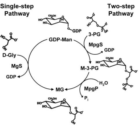
has not been studied, but knock-out mutants were used to demonstrate the role of MG during osmoadaptation of this extreme thermophile (39). Mannosylglycerate accumulates in R. marinus not only in response to osmotic stress, but also in response to supra-optimal growth temperature (40), by distinctively up-regulating one of the two pathways of MG biosynthesis (41) (Fig. 1.1).
The finding that MG is widespread in marine hyperthermophiles, and rarely found in mesophiles, led to the view that this osmolyte is involved not only in osmoprotection but also in thermoprotection of cellular components. In fact, MG possesses a superior ability to protect enzymes against thermal denaturation in vitro, and its potential usefulness in biotechnological and pharmaceutical applications has been often claimed (26, 30).
7
Inspection of public databases resulted in the identification of MPGS
homologues in the mesophilic bacteria Dehalococcoides ethenogenes, and
Verrucomicrobiae bacterium, and also in several fungi. The MPGS activity of
the respective gene products has been confirmed in Neurospora crassa and
Dehalococcoides ethenogenes (46, 47). Therefore, the synthesis of mannosylglycerate seems not restricted to (hyper)thermophiles, but its physiological role in algae is unknown, and the accumulation of this solute in other mesophiles awaits demonstration. It is expected that the investigation of the molecular evolution of mannosylglycerate synthesis will shed light on the origin and dissemination of this trait in the Tree of Life (33).
Biochemical characterization of the enzyme led to the classification of the MpgS in the retaining Glycosyltransferase family 55 (GT55; www.cazy.org). Thus far, this family comprises only proteins homologous to T. thermophilus
MpgS, whereas MgS is classified into the retaining GT78 family, and a “ front-face” SNi-like mechanism for glycosyl transfer with retention of anomeric configuration was proposed1 .
However, the catalytic mechanisms of MpgS and MpgP enzymes, were not been investigated at the beginning of this thesis, and were taken part of the global study on the molecular determinants that dictate the fundamental mechanisms of enzymatic catalysis.
1The crystallographic structure elucidation of MgS from R. marinus, was carried out by
8
9 1.4 Glycosyltransferases; fold, mechanism and biological implications.
10 Figure 1.2 Overall reactions catalyzed by (a) glycosidases, (b)
glycosyltransferases, and (c) phosphorylases (49).
Sequence-based classification spreads GTs in many families by amino acid sequence similarities, [(http://afmb.cnrs-mrs.fr/CAZY) (51)], thus reflecting the variety of molecules that can be used as acceptors. In addition to nucleotide-diphospho sugar-donor (NDP-sugar) dependent enzymes, they also utilize dolichol-phospho-sugars, sugar-1-phosphates and lipid diphospho-sugars as activated donors.
11 mechanistic features within the same family. Yet, the promiscuity for either an inverting or retaining 2 mechanism present within the same family is becoming
a current feature, and thus novel approaches for the rational classification of the GTs are mandatory .
In contrast to GHs that adopt a large variety of folds, including all all , or mixed / structures, GTs folds have been observed to consist primarily of // sandwiches similar or very close to the Rossmann type fold, a classical structural motif (six-stranded parallel -sheet with 321456 topology) found in many nucleotide binding proteins (53). Until recently only two structural superfamilies have been described for GTs, named GT-A and GT-B (Fig. 1.3), and which were first observed in the original Spore coat polysaccharide biosynthesis protein and β-glucosyltransferase structures, respectively (54, 55). A third family has recently emerged which comprises the bacterial sialyltransferase (CstII) belonging to family GT42 [based on the -strand connectivity (56)]. GT-A and GT-B folds are also shared by non-GT enzymes, such as nucleotidyltransferases and sugar epimerases, respectively. The GT-B fold consists of two separate Rossmann domains with a connecting linker region and a catalytic site located between the domains. There is an excellent structural conservation between protein members of the GT-B family, particularly in the C-terminal domain which corresponds to the nucleotide-binding domain. Variations are more pronounced in the N-terminal domains, in the loops and helices which point towards the active site, which have evolved to accommodate very different acceptors. The structural features of GT-A fold enzymes will be herein focused, whereas an extended structural survey describing GT-B fold enzyme features can be found elsewhere (57, 58).
12 DxD motif is shown in all crystal structures to interact primarily with the phosphate groups of nucleotide donor through the coordination of a divalent cation, typically Mn2+ (58). Comparison of the catalytic domains of enzymes of the GT-A family revealed the presence of two regions that are structurally well conserved in all members of the GT-A family, including inverting and retaining enzymes. This suggests that common structural elements are necessary for the glycosyl transfer reaction, irrespective of the stereochemistry of the reaction. The first region mostly corresponds to the Rossmann-type nucleotide-binding domain, encompassing the first 100-120 residues, and that is terminated by the DxD motif. The key amino acids that interact with NDP are mainly found at the C-terminal of strands β1 and β4. In some crystal structures, residues in the C-terminus of the catalytic domain were shown to make additional contacts with the NDP moiety.
While the mechanistic strategies used by GlycosylHydrolases (GHs), to catalyse glycosidic bond hydrolysis are fairly well understood on both a structural and chemical level (61, 62), the mechanistic understanding of the GTs responsible for glycoside bond formation has lagged far behind. Despite a lack of evolutionary relatedness, by simple chemical analogy, GTs are thought to use mechanistic strategies that directly parallel those used by GHs and transglycosidases (63, 64).
2 By analogy with glycosidases, two main stereochemical outcomes exist for
13 Figure 1.3 Ribbon diagram of three glycosyltransferases (GTs) representative of the different folds. Bound nucleotide sugar are represented with stick model, and manganese, when present, by a ball. (A) GT-A fold, mouse α-1,4-N-acetylhexosaminyltransferase (EXTL2) complexed with UDP-Gal-NAc (PDB code 1OMZ) (59), (B) GT-B fold, Escherichia coli
MurG complexed with UDP-GlcNAc (PDB code 1NLM) (60), and (C)
Campylobacter jejuni sialyltransferase CstII complexed with cytidine monophospho 3-fluoro N-acetyl neuraminic acid (CMP-3FNeuAc) (PDB code 1RO7) (56). Figure obtained from reference (57).
A
C
14 Nucleophilic substitution at the anomeric carbon of the transferred glycosyl residue proceeds with either inversion or retention of configuration of the donor substrate and each stereochemical course necessitates a distinct catalytic mechanism (Fig. 1.4). It is currently not clear how stereochemical control in glycosyltransferase-catalyzed group transfer is achieved. In fact, one of the fundamental challenges in the field of glycobiology remains the dissection of the catalytic mechanism of nucleotide-sugar dependent GTs, especially those that act with retention of anomeric configuration (65).
Inverting GTs are proposed to utilize a direct-displacement SN2-like reaction mechanism in which departure of the leaving group is facilitated by a Lewis acid, and nucleophilic attack is assisted by a catalytic base of the enzyme. Drawing analogy to GHs for which reaction coordinates leading to inversion or retention have been very well-characterized (63) a double displacement-like reaction involving a covalent glycosyl-enzyme intermediate appeared to be the preferred choice of mechanism for the retaining GTs. However, GTs crystal structures revealed a surprising lack of conserved architecture in the region of the active site where the catalytic nucleophile would have to be positioned (58). In addition, despite exhaustive biochemical studies with techniques that have been successfully used in the characterization of retaining glycoside hydrolases (66, 67) evidence supporting a covalent intermediate for retaining GTs has remained elusive. Major barrier to the study of these enzymes is the lack of non-hydrolysable substrates or fluoro-sugar type inhibitors with affinities similar to that of the substrates, has rendered ineffective the ability in altering the relative rates of glycosylation versus deglycosylation steps, thus in trapping intermediate species on retaining GTs (67), and in allowing the structural access to the ternary complex or something that resembles it.
15 (69). Partially as a consequence of this lack of suitable compounds, there are no 3-D structures of intact ternary complexes with which to describe the catalytic centre and reveal geometry of the transfer process (65). An alternative mechanism, often referred to as internal return SNi-like (IUPAC defenition DNANss), was therefore considered. Chemical precedent for this type of a mechanism comes from detailed kinetic and conformational studies of the solvolysis of glucose derivatives in mixtures of ethanol and trifluoroethanol (70). The SNi-like mechanism has since been proposed for other structurally defined retaining transferases based on the lack of appropriately positioned nucleophiles in their active sites (59, 71) and had previously been proposed for the structurally similar glycogen phosphorylase (72). This is proposed to involve a short-lived ion pair intermediate, the formation of which arguably requires electrostatic front-side stabilization from the departing (substituted) phosphate and perhaps a certain amount of nucleophilic “push” from an enzyme group positioned on the backside of the glycosyl ring. In addition to stabilizing the intermediate, hydrogen bonding between the incoming nucleophile and the leaving group would play an important base catalytic role during activation of the acceptor hydroxyl group for nucleophilic attack.
18 1.5 Mechanistic alternatives in phosphate monoester transfer catalysis.
The hydrolysis of phosphate esters is an important reaction in many biological systems. DNA and RNA are phosphate diesters, while many intermediates in the metabolism exist as phosphate monoesters. Phosphorylation of proteins is an important control mechanism. While triesters are not naturally occurring biological molecules, enzymes have evolved to hydrolyse these man-made toxic compounds. However, monoesters and diesters have well-known roles in genetic materials, in coenzymes and in energy reservoirs, and as intermediates in biological transformations, while monoesters formed by protein phosphorylation have key roles in the regulation of host processes (73).
Phosphoryl transfer reactions are substitution reactions at phosphorus, and three limiting mechanisms exist (Fig. 1.5). One is a dissociative, SN1-type mechanism (DN + AN in the IUPAC nomenclature). In such a mechanism, a stable metaphosphate ion (PO3-) is attacked by a nucleophile in a subsequent rate-determining step. Other phosphoryl transfer mechanisms are an associative, two-step addition-elimination mechanism (AN + DN) with a phosphorane intermediate, and a concerted mechanism (ANDN) with no intermediate. In the concerted ANDN mechanism, bond formation to the nucleophile and bond fission to the leaving group both occur in the transition state. The prevailing chemical pathway is determined by the nature of the nucleophile, electrophile and leaving group and by the solvent (74-76).
19 nucleophile (73, 78). It would then be expected that the total axial phosphorous-oxygen bond order is lower in the TS than in the reactants and products (79, 80). Metaphosphate has been observed in the X-ray structure of fructose-1,6-bisphosphatase grown in an equilibrium mixture of substrate and product (81), and a moiety that could be described as a stabilized metaphosphate has been observed in the structure of -phosphoglucomutase from Lactococcus lactis obtained at cryogenic temperature (82). This dogma is, however, repeatedly challenged by enzyme studies in which phosphate monoester cleavage is often interpreted in terms of more or less associative bimolecular mechanisms (73, 83, 84). It is also possible that the energetics of the associative and dissociative mechanisms are similar in solution and that enzymes catalyzing phosphate monoester (P-O bond) cleavage actually can alter the mechanism of their uncatalyzed reaction counterparts (85). What then controls the chemical pathway of the enzymes that catalyze phosphoryl-transfer reaction?
21 1.6 Isocitrate dehydrogenase: a paradigm for a concerted structural dynamics with catalytic proficiency
Isocitrate dehydrogenase [IDH; 2R, 3S-isocitrate:NADP+ oxidoreductase (decarboxylating); EC 1.1.1.42] belongs to a large family of pyridine nucleotide-linked -hydroxyacid decarboxylating dehydrogenases. IDH converts isocitrate into -ketoglutarate, and is the first enzyme in the CO2 -evolving steps of the tricarboxylic acid cycle with concomitant reduction of NAD(P)+ to NAD(P)H. The overall reaction catalyzed by IDH involves two steps: the activation of isocitrate into its oxaloacetate intermediate and -decarboxylation followed by hydrogenation yielding -ketoglutarate (Fig. 1.6).
Eukaryotic cells express two distinct IDH classes: the NAD+-dependent enzyme (EC 1.1.1.41) found only in mitochondria and displaying allosteric properties, and a non-allosteric NADP+-dependent enzyme (EC 1.1.1.42) that is found in both mitochondria and cytoplasm. In humans, the impaired ability of NADP+-dependent IDHs (IDH1 and IDH2 isoforms) to convert isocitrate into -ketoglutarate is known to be related with metabolic disorders having severe phenotypic outcomes, such as the cancer-associated IDH1 malignant progression of gliomas (87, 88). In Escherichia coli, as well as other bacterial cells, only a single NADP+-dependent IDH is found (89) and it is a key regulator in the bypass of the TCA cycle to the glyoxylate pathway when acetate is used as carbon source (90, 91). The IDH regulatory activity mechanisms are thus of major interest as they act as checkpoints for modulating cell homeostasis.
22 the lactate dehydrogenase-like classes of nucleotide-binding protein, thus a potential alternative solution for the same evolutionary challenge (93, 94). This has led to the inclusion of the prior into the dimeric isocitrate/isopropylmalate dehydrogenase family (SCOP accession 53660).
IDH activation and inactivation in E. coli is controlled by a unique bifunctional regulatory enzyme, the IDH kinase/phosphatase AceK (EC 2.7.11.5), via “on-off switch” mechanism as the phosphorylated form of EcoIDH at Ser113 residue has no activity (96-98).
The AceK-IDH complex has long been considered a prototypic model system for protein phosphorylation in prokaryotes (96, 99) and recently, the structure of the complex AceK-IDH of E. coli has been determined shedding light into the molecular basis for AceK multifunctionality. The AMP-mediated conformational changes act as a switch between the kinase and phosphatase activities, in addition with the high order of interaction and recognition between AceK and IDH(99). On the other hand, eukaryotic NADP+-linked IDHs rely, in a self-regulatory based electrostatic repulsive mechanism that mimics the prokaryotic regulation by phosphorylation (89, 100) while NAD+-linked IDHs are allosterically regulated (101).
23 remained still to be answered, such as the identities of the assisting Brönsted acid/base catalyst.
Chapter 4 describes novel structures of EcoIDH that mirror the dehydrogenation and -decarboxylation steps of the reaction cycle. Together with the subtract-induced protein dynamics, details on the chemistry behind the -decarboxylation reaction are explained in light of our structural data and which can be generalised to all homodimeric NADP+-linked IDHs.
25
REFERENCES
1. Ramanathan, A., and Agarwal, P. K. (2011) Evolutionarily conserved linkage between enzyme fold, flexibility, and catalysis, PLoS Biol 9, e1001193.
2. Agarwal, P. K. (2006) Enzymes: An integrated view of structure, dynamics and function, Microb Cell Fact5, 2.
3. Ma, C. W., Xiu, Z. L., and Zeng, A. P. (2011) A new concept to reveal protein dynamics based on energy dissipation, PLoS One6, e26453. 4. Schwartz, S. D., and Schramm, V. L. (2009) Enzymatic transition states
and dynamic motion in barrier crossing, Nat Chem Biol5, 551-558. 5. Law, A. B., Fuentes, E. J., and Lee, A. L. (2009) Conservation of
side-chain dynamics within a protein family, J Am Chem Soc 131, 6322-6323.
6. Papaleo, E., Tiberti, M., Invernizzi, G., Pasi, M., and Ranzani, V. (2011) Molecular determinants of enzyme cold adaptation: comparative structural and computational studies of cold- and warm-adapted enzymes, Curr Protein Pept Sci12, 657-683.
7. Villali, J., and Kern, D. (2010) Choreographing an enzyme's dance,
Curr Opin Chem Biol14, 636-643.
8. Henzler-Wildman, K., and Kern, D. (2007) Dynamic personalities of proteins, Nature450, 964-972.
9. Mittermaier, A., and Kay, L. E. (2006) New tools provide new insights in NMR studies of protein dynamics, Science312, 224-228.
10. Teodoro, M. L., Phillips, G. N., Jr., and Kavraki, L. E. (2003) Understanding protein flexibility through dimensionality reduction, J Comput Biol10, 617-634.
26 12. Fraser, J. S., Clarkson, M. W., Degnan, S. C., Erion, R., Kern, D., and Alber, T. (2009) Hidden alternative structures of proline isomerase essential for catalysis, Nature462, 669-673.
13. Bolduc, J. M., Dyer, D. H., Scott, W. G., Singer, P., Sweet, R. M., Koshland, D. E., Jr., and Stoddard, B. L. (1995) Mutagenesis and Laue structures of enzyme intermediates: isocitrate dehydrogenase, Science 268, 1312-1318.
14. Cho, H. S., Dashdorj, N., Schotte, F., Graber, T., Henning, R., and Anfinrud, P. (2010) Protein structural dynamics in solution unveiled via 100-ps time-resolved x-ray scattering, Proc Natl Acad Sci U S A 107, 7281-7286.
15. Genick, U. K., Borgstahl, G. E., Ng, K., Ren, Z., Pradervand, C., Burke, P. M., Srajer, V., Teng, T. Y., Schildkamp, W., McRee, D. E., Moffat, K., and Getzoff, E. D. (1997) Structure of a protein photocycle intermediate by millisecond time-resolved crystallography, Science275, 1471-1475. 16. Moffat, K. (1989) Time-resolved macromolecular crystallography, Annu
Rev Biophys Biophys Chem18, 309-332.
17. Nashine, V. C., Hammes-Schiffer, S., and Benkovic, S. J. (2010) Coupled motions in enzyme catalysis, Curr Opin Chem Biol 14, 644-651.
18. Blöchl, E., Burggraf, S., Fiala, G., Lauerer, G., Huber, G., and Huber, R. (1995) Isolation, taxonomy and phylogeny of hyperthermophilic microorganisms, World J Microbiol Biotechnol11, 9-16.
19. Alfredsson, G. A., Kristjánsson, J. K., Hjörleifsdottir, S., and Stetter, K. O. (1988) Rhodothermus marinus, gen. nov., sp. nov., a thermophilic, halophilic bacterium from submarine hot springs in Iceland, J Gen Microbiol 134, 299-306.
20. da Costa, M. S., Nobre, M. F., and Rainey, F. A. (2001) The genus
27 21. Santos, H., and da Costa, M. S. (2002) Compatible solutes of organisms that live in hot saline environments, Environ Microbiol 4, 501-509.
22. Brown, A. D. (1976) Microbial water stress, Bacteriol Rev40, 803-846. 23. da Costa, M. S., Santos, H., and Galinski, E. A. (1998) An overview of
the role and diversity of compatible solutes in Bacteria and Archaea,
Adv Biochem Eng/Biotechnol61, 117-153.
24. Roberts, M. F. (2005) Organic compatible solutes of halotolerant and halophilic microorganisms, Saline Systems1, 5.
25. Faria, T. Q., Knapp, S., Ladenstein, R., Macanita, A. L., and Santos, H. (2003) Protein stabilisation by compatible solutes: effect of mannosylglycerate on unfolding thermodynamics and activity of ribonuclease A, ChemBioChem4, 734-741.
26. Faria, T. Q., Mingote, A., Siopa, F., Ventura, R., Maycock, C., and Santos, H. (2008) Design of new enzyme stabilizers inspired by glycosides of hyperthermophilic microorganisms, Carbohydr Res 343, 3025-3033.
27. Sochocka, M., and Boratynski, J. (2011) [Osmoregulation--an important parameter of bacterial growth], Postepy Hig Med Dosw (Online) 65, 714-724.
28. Lee, V. M., and Trojanowski, J. Q. (2006) Mechanisms of Parkinson's disease linked to pathological alpha-synuclein: new targets for drug discovery, Neuron52, 33-38.
29. Skovronsky, D. M., Lee, V. M., and Trojanowski, J. Q. (2006) Neurodegenerative diseases: new concepts of pathogenesis and their therapeutic implications, Annu Rev Pathol1, 151-170.
30. Borges, N., Ramos, A., Raven, N. D., Sharp, R. J., and Santos, H. (2002) Comparative study of the thermostabilizing properties of mannosylglycerate and other compatible solutes on model enzymes,
28 31. Faria, T. Q., Lima, J. C., Bastos, M., Macanita, A. L., and Santos, H. (2004) Protein stabilization by osmolytes from hyperthermophiles: effect of mannosylglycerate on the thermal unfolding of recombinant nuclease a from Staphylococcus aureus studied by picosecond time-resolved fluorescence and calorimetry, J Biol Chem 279, 48680-48691.
32. Ramos, A., Raven, N., Sharp, R. J., Bartolucci, S., Rossi, M., Cannio, R., Lebbink, J., Van Der Oost, J., De Vos, W. M., and Santos, H. (1997) Stabilization of Enzymes against Thermal Stress and Freeze-Drying by Mannosylglycerate, Appl Environ Microbiol63, 4020-4025.
33. Santos, H., Lamosa, P., Borges, N., Gonçalves, L. G., Pais, T. M., and M. V. Rodrigues, M. V. (2011) Organic Compatible Solutes of Prokaryotes that Thrive in Hot Environments: The Importance of Ionic Compounds for Thermostabilization, in Extremophiles Handbook
(Horikoshi, K., Antranikian, G., Bull, A. T., Robb, F. T., and Stetter, K. O., Eds.), pp 497-520, Springer, Tokyo.
34. Empadinhas, N., and da Costa, M. S. (2008) To be or not to be a compatible solute: bioversatility of mannosylglycerate and glucosylglycerate, Syst Appl Microbiol31, 159-168.
35. Empadinhas, N., and da Costa, M. S. (2011) Diversity, biological roles and biosynthetic pathways for sugar-glycerate containing compatible solutes in bacteria and archaea, Environ Microbiol13, 2056-2077. 36. Luley-Goedl, C., and Nidetzky, B. (2011) Glycosides as compatible
solutes: biosynthesis and applications, Nat Prod Rep28, 875-896. 37. Bouveng, H., Lindberg, B., and Wickberg, B. (1995) Low-molecular
carbohydrates in algae, Acta Chem Scand9.
29 39. Alarico, S., Empadinhas, N., Mingote, A., Simoes, C., Santos, M. S., and da Costa, M. S. (2007) Mannosylglycerate is essential for osmotic adjustment in Thermus thermophilus strains HB27 and RQ-1,
Extremophiles11, 833-840.
40. Silva, Z., Borges, N., Martins, L. O., Wait, R., da Costa, M. S., and Santos, H. (1999) Combined effect of the growth temperature and salinity of the medium on the accumulation of compatible solutes by Rhodothermus marinus and Rhodothermus obamensis, Extremophiles 3, 163-172.
41. Borges, N., Marugg, J. D., Empadinhas, N., da Costa, M. S., and Santos, H. (2004) Specialized roles of the two pathways for the synthesis of mannosylglycerate in osmoadaptation and thermoadaptation of Rhodothermus marinus, J Biol Chem 279, 9892-9898.
42. Empadinhas, N., Albuquerque, L., Henne, A., Santos, H., and da Costa, M. S. (2003) The bacterium Thermus thermophilus, like hyperthermophilic archaea, uses a two-step pathway for the synthesis of mannosylglycerate, Appl Environ Microbiol69, 3272-3279.
43. Empadinhas, N., Marugg, J. D., Borges, N., Santos, H., and da Costa, M. S. (2001) Pathway for the synthesis of mannosylglycerate in the hyperthermophilic archaeon Pyrococcus horikoshii. Biochemical and genetic characterization of key enzymes, J Biol Chem 276, 43580-43588.
44. Martins, L. O., Empadinhas, N., Marugg, J. D., Miguel, C., Ferreira, C., da Costa, M. S., and Santos, H. (1999) Biosynthesis of mannosylglycerate in the thermophilic bacterium Rhodothermus marinus. Biochemical and genetic characterization of a mannosylglycerate synthase, J Biol Chem 274, 35407-35414.
30 thermoadaptation in the order thermococcales, Appl Environ Microbiol 71, 8091-8098.
46. Empadinhas, N. (2005) Pathways for the synthesis of mannosylglycerate in prokaryotes: genes, enzymes and evolutionary implications, PhD thesis University of Coimbra, Coimbra, Portugal. 47. Empadinhas, N., Albuquerque, L., Costa, J., Zinder, S. H., Santos, M.
A., Santos, H., and da Costa, M. S. (2004) A gene from the mesophilic bacterium Dehalococcoides ethenogenes encodes a novel mannosylglycerate synthase, J Bacteriol186, 4075-4084.
48. Flint, J., Taylor, E., Yang, M., Bolam, D. N., Tailford, L. E., Martinez-Fleites, C., Dodson, E. J., Davis, B. G., Gilbert, H. J., and Davies, G. J. (2005) Structural dissection and high-throughput screening of mannosylglycerate synthase, Nat Struct Mol Biol 12, 608-614.
49. Lairsonb, L. L., and Withers, S. G. (2004) Mechanistic analogies amongst carbohydrate modifying enzymes, Chem Commun (Camb, U.K.), 2243-2248.
50. Pesnot, T., Jorgensen, R., Palcic, M. M., and Wagner, G. K. (2010) Structural and mechanistic basis for a new mode of glycosyltransferase inhibition, Nat Chem Biol6, 321-323.
51. Coutinho, P. M., Deleury, E., Davies, G. J., and Henrissat, B. (2003) An evolving hierarchical family classification for glycosyltransferases, J Mol Biol328, 307-317.
52. Davies, G., and Henrissat, B. (1995) Structures and mechanisms of glycosyl hydrolases, Structure3, 853-859.
53. Lesk, A. M. (1995) NAD-binding domains of dehydrogenases, Curr Opin Struct Biol5, 775-783.
54. Charnock, S. J., and Davies, G. J. (1999) Structure of the nucleotide-diphospho-sugar transferase, SpsA from Bacillus subtilis, in native and nucleotide-complexed forms, Biochemistry38, 6380-6385.
beta-31 glucosyltransferase in the presence and absence of the substrate uridine diphosphoglucose, EMBO J13, 3413-3422.
56. Chiu, C. P., Watts, A. G., Lairson, L. L., Gilbert, M., Lim, D., Wakarchuk, W. W., Withers, S. G., and Strynadka, N. C. (2004) Structural analysis of the sialyltransferase CstII from Campylobacter jejuni in complex with a substrate analog, Nat Struct Mol Biol 11, 163-170.
57. Breton, C., Snajdrova, L., Jeanneau, C., Koca, J., and Imberty, A. (2006) Structures and mechanisms of glycosyltransferases,
Glycobiology16, 29R-37R.
58. Lairson, L. L., Henrissat, B., Davies, G. J., and Withers, S. G. (2008) Glycosyltransferases: structures, functions, and mechanisms, Annu Rev Biochem77, 521-555.
59. Heightman, T. D., and Vasella, A. T. (1999) Recent Insights into Inhibition, Structure, and Mechanism of Configuration-Retaining Glycosidases, Angew. Chem, Int Ed38, 750-770.
60. Zechel, D. L., Reid, S. P., Stoll, D., Nashiru, O., Warren, R. A., and Withers, S. G. (2003) Mechanism, mutagenesis, and chemical rescue of a beta-mannosidase from cellulomonas fimi, Biochemistry 42, 7195-7204.
61. Davies, G. J., Sinnott, M. L., and Withers, S. G. (1998) In
Comprehensive Biological Catalysis (Sinnott, M. L., Ed.), pp 119-208, Academic Press, San Diego.
62. Sinnott, M. L. (1990) Catalytic mechanism of enzymic glycosyl transfer,
Chem. Rev. (Washington, DC, U. S.) 90, 1171-1202.
32 64. Hu, Y., Chen, L., Ha, S., Gross, B., Falcone, B., Walker, D., Mokhtarzadeh, M., and Walker, S. (2003) Crystal structure of the MurG:UDP-GlcNAc complex reveals common structural principles of a superfamily of glycosyltransferases, Proc Natl Acad Sci U S A 100, 845-849.
65. Errey, J. C., Lee, S. S., Gibson, R. P., Martinez Fleites, C., Barry, C. S., Jung, P. M., O'Sullivan, A. C., Davis, B. G., and Davies, G. J. (2010) Mechanistic insight into enzymatic glycosyl transfer with retention of configuration through analysis of glycomimetic inhibitors, Angew Chem, Int Ed Engl49, 1234-1237.
66. Lairson, L. L., Chiu, C. P., Ly, H. D., He, S., Wakarchuk, W. W., Strynadka, N. C., and Withers, S. G. (2004) Intermediate trapping on a mutant retaining alpha-galactosyltransferase identifies an unexpected aspartate residue, J Biol Chem 279, 28339-28344.
67. Ly, H. D., Lougheed, B., Wakarchuk, W. W., and Withers, S. G. (2002) Mechanistic studies of a retaining alpha-galactosyltransferase from Neisseria meningitidis, Biochemistry41, 5075-5085.
68. Compain, P., and Martin, O. R. (2001) Carbohydrate mimetics-based glycosyltransferase inhibitors, Bioorg Med Chem9, 3077-3092.
69. Waldscheck, B., Streiff, M., Notz, W., Kinzy, W., and Schmidt, R. R. (2001) alpha(1-3)-Galactosyltransferase Inhibition Based on a New Type of Disubstrate Analogue, Angew Chem, Int Ed Engl 40, 4007-4011.
70. Sinnott, M. L., and Jencks, W. P. (1980) J. Am.Chem. Soc. 102, 20-26. 71. Gibson, R. P., Turkenburg, J. P., Charnock, S. J., Lloyd, R., and
Davies, G. J. (2002) Insights into trehalose synthesis provided by the structure of the retaining glucosyltransferase OtsA, Chem Biol 9, 1337-1346.
33 73. Cleland, W. W., and Hengge, A. C. (2006) Enzymatic mechanisms of
phosphate and sulfate transfer, Chem Rev106, 3252-3278.
74. Anderson, M. A., Shim, H., Raushel, F. M., and Cleland, W. W. (2001) Hydrolysis of phosphotriesters: determination of transition states in parallel reactions by heavy-atom isotope effects, J Am Chem Soc 123, 9246-9253.
75. Catrina, I. E., and Hengge, A. C. (2003) Comparisons of phosphorothioate with phosphate transfer reactions for a monoester, diester, and triester: isotope effect studies, J Am Chem Soc125, 7546-7552.
76. Grzyska, P. K., Czyryca, P. G., Purcell, J., and Hengge, A. C. (2003) Transition state differences in hydrolysis reactions of alkyl versus aryl phosphate monoester monoanions, J Am Chem Soc 125, 13106-13111.
77. Lad, C., Williams, N. H., and Wolfenden, R. (2003) The rate of hydrolysis of phosphomonoester dianions and the exceptional catalytic proficiencies of protein and inositol phosphatases, Proc Natl Acad Sci U S A100, 5607-5610.
78. Admiraal, S. J., and Herschlag, D. (1995) Mapping the transition state for ATP hydrolysis: implications for enzymatic catalysis, Chem Biol 2, 729-739.
79. Cleland, W. W. (1990) Secondary 18O isotope effects as a tool for studying reactions of phosphate mono-, di-, and triesters, FASEB J 4, 2899-2905.
80. Cleland, W. W., and Hengge, A. C. (1995) Mechanisms of phosphoryl and acyl transfer, FASEB J9, 1585-1594.
81. Choe, J. Y., Iancu, C. V., Fromm, H. J., and Honzatko, R. B. (2003) Metaphosphate in the active site of fructose-1,6-bisphosphatase, J Biol Chem 278, 16015-16020.
alpha-34 galactose 1-phosphate in the active site of beta-phosphoglucomutase form a transition state analogue of phosphoryl transfer, J Am Chem Soc 131, 16334-16335.
83. Lahiri, S. D., Zhang, G., Dunaway-Mariano, D., and Allen, K. N. (2003) The pentacovalent phosphorus intermediate of a phosphoryl transfer reaction, Science299, 2067-2071.
84. Williams, N. H. (2004) Models for biological phosphoryl transfer,
Biochim Biophys Acta1697, 279-287.
85. Aqvist, J., Kolmodin, K., Florian, J., and Warshel, A. (1999) Mechanistic alternatives in phosphate monoester hydrolysis: what conclusions can be drawn from available experimental data?, Chem Biol6, R71-80. 86. Allen, K. N., and Dunaway-Mariano, D. (2004) Phosphoryl group
transfer: evolution of a catalytic scaffold, Trends Biochem Sci 29, 495-503.
87. Dang, L., White, D. W., Gross, S., Bennett, B. D., Bittinger, M. A., Driggers, E. M., Fantin, V. R., Jang, H. G., Jin, S., Keenan, M. C., Marks, K. M., Prins, R. M., Ward, P. S., Yen, K. E., Liau, L. M., Rabinowitz, J. D., Cantley, L. C., Thompson, C. B., Vander Heiden, M. G., and Su, S. M. (2009) Cancer-associated IDH1 mutations produce 2-hydroxyglutarate, Nature462, 739-744.
88. Reitman, Z. J., and Yan, H. (2010) Isocitrate dehydrogenase 1 and 2 mutations in cancer: alterations at a crossroads of cellular metabolism,
J Natl Cancer Inst102, 932-941.
89. Yates, S. P., Edwards, T. E., Bryan, C. M., Stein, A. J., Van Voorhis, W. C., Myler, P. J., Stewart, L. J., Zheng, J., and Jia, Z. (2011) Structural basis of the substrate specificity of bifunctional isocitrate dehydrogenase kinase/phosphatase, Biochemistry50, 8103-8106. 90. Cozzone, A. J., and El-Mansi, M. (2005) Control of Isocitrate
Dehydrogenase Catalytic Activity by Protein Phosphorylation in

![Figure 3.6 Mean main chain thermal B-factors for chains A and B of apo- apo-MpgP and holo-apo-MpgP (control [Mg 2+ -MG-HPO 4 2- ]) structures](https://thumb-eu.123doks.com/thumbv2/123dok_br/15769984.641180/146.748.134.620.406.711/figure-mean-chain-thermal-factors-chains-control-structures.webp)