Arq Neuropsiquiatr 2008;66(2-A):251-253
251
MYASTHENIA GRAVIS ANd MULTIPLE SCLEROSIS
An uncommon presentation
Paulo J. Lorenzoni, Rosana H. Scola, Cláudia S. Kamoi Kay, Lineu C. Werneck
MIASTENIA GRAVIS E ESCLEROSE MúLTIPLA: UMA APRESENTAçãO INCOMUM
Neurology Division, Internal Medicine Department, Hospital de Clínicas da Universidade Federal do Paraná (UFPR), Curitiba PR, Brazil. Received 6 September 2007, received in inal form 2 January 2008. Accepted 16 February 2008.
Dra. Rosana Herminia Scola – Serviço de Doenças Neuromusculares / Hospital de Clínicas da UFPR - Rua General Carneiro 181 / 3º andar - 80060-900 Curitiba PR - Brasil. E-mail: [email protected]
Myasthenia gravis (MG) is an autoimmune disease that compromises neuromuscular transmission and is mediat-ed by autoantibodies against acetylcholine receptors on the postsynaptic membrane1. In its usual form it leads to
symptoms of decreased muscle strength and fatigue1.
Mul-tiple sclerosis (MS) is an immune-mediated demyelinating disease of the central nervous system that shows a wide range of clinical features and a variable natural history2.
There is some evidence that patients with MG or MS have a higher risk of developing autoantibodies and other neu-roimmune disorders than normal controls3,4.
An autoimmune pathogenesis is implicated in both MG and MS, but the coexistence of the two disorders has rarely been documented, and for this reason we re-port this case4.
CASE
A 28-year-old woman presented with diplopia, dysphagia and mild limitation of ocular movements, with progressive palpebral ptosis and weakness, which changed in intensity from day to day and during the day according to the patient’s physical activity.
Physical examination did not reveal any abnormalities. On neurological examination she was found to have asymmetri-cal palpebral ptosis (left>right); symmetrical muscle weakness (grade 4 on the MRC scale) in the proximal upper and lower limbs; and deep tendon relexes. Gait and all sensory examina-tions were normal.
The investigation yielded the following results: (1) positive anti-acetylcholine receptor antibody test (14.91 nmol/L; normal
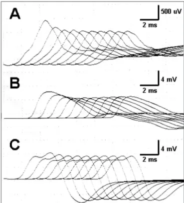
< 0.20 nmol/L); (2) normal needle electromyography and nerve conduction studies; (3) repetitive stimulation of the facial, spinal accessory and ulnar nerves at 3 Hz with a decrement of more than 10% in compound muscle action potential amplitude (Fig 1); and (4) improvement of symptoms after treatment with pyridostigmine (180 mg daily). Chest computed tomography scan was normal.
A diagnosis of MG was made, and the patient showed an im-provement in symptoms after she started to receive prednisone and pyridostigmine. Prednisone was discontinued after one year
and was followed by oral administration of azathioprine (100 mg/day), with an improvement in palpebral ptosis and muscle strength (grade 5 on the MRC scale).
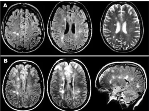
When she was 32 years old and still on azathioprine treat-ment, the patient developed a sudden weakness in her left low-er limb and was treated with prednisone (60 mg/day). Aftlow-er this new neurological manifestation suggestive of MS, the patient was submitted to brain magnetic resonance imaging (MRI), which revealed multiple areas of high signal on FLAIR and T2-weight-ed images in the periventricular and subcortical white matter of the brain hemispheres (Fig 2A).
Arq Neuropsiquiatr 2008;66(2-A)
252
Myasthenia gravis and multiple sclerosis Lorenzoni et al.
Three years later, she presented with a sudden episode of deafness, paresthesia and weakness in her left lower limb. Neu-rological examination showed nystagmus; left facial paresis; bi-lateral hearing loss; muscle strength grade 4 (MRC scale) in the left lower limb; bilateral increased deep tendon relexes in the lower limbs; left Babinski sign; gait ataxia; and impaired pain, temperature, pinprick and light touch sensory examinations in the left lower limb. Vibration sensibility and joint position sense also revealed impairment in the left lower limb.
MRI showed new areas of high signal on FLAIR and low sig-nal on T1-weighted images in the periventricular and subcorti-cal white matter of the brain hemispheres as well as in the cer-ebellum (Fig 2B).
Hematological tests, biochemistry screening and thyroid function were normal, and serological tests for HIV and HTLV were negative. Anti-nuclear antibody test (1:640 with diffuse pattern; normal <1:40) and anti-SSB/La (13.8 U/mL; normal <10 U/mL) were positive, but the other serum antibody tests (anti-RNP, Sm and SSA/Ro) were negative. Cerebrospinal luid anal-ysis was normal with a normal electrophoretic protein pattern. The patient was diagnosed as having had another MS relapse (her disability EDSS score was 5.5) combined with MG. Intrave-nous methylprednisolone pulse therapy was started, with sub-stantial improvement in muscle strength in the left lower limb. Currently the patient is receiving azathioprine, and her disabili-ty EDSS score is 4.5 after ive years of follow-up.
All studies were carried out following informed consent.
dISCUSSION
MG is a rare disease with a prevalence ranging between 0.5 and 15.0 cases per 100000 inhabitants and an incidence of 0.4 to 1.1 cases per 100000 inhabitants1,5. In Brazil there
are probably 15500 persons affected by MG6. In a number
of studies of MS, a variation in prevalence and clinical pat-tern with geographical location was observed, probably related to ethnic and environmental factors2,7. With 15.0
cases of MS per 100000 inhabitants, Brazil is considered to have a low prevalence, particularly compared with other countries at similar latitudes7. The probability of the same
patient developing both of these disorders is extremely low, although it should be remembered that these pa-tients are more likely to develop other autoimmune dis-eases than patients without immune-mediated disorders8.
However, the combination of these two diseases oc-curs at rates higher than those expected by random asso-ciation and appears to be more common than estimated. An epidemiological survey in Finland found that two per-sons in a population of 1.5 million had combined MG and MS. As the expected combined prevalence was 3.6 cases per 100 million people, the prevalence identiied in the study was 37 times greater than that expected3. A similar
study in Canada found a signiicantly higher prevalence than that predicted by estimates of the prevalence of this combination of disorders4.
This nonrandom association of MG and MS in patients could support the hypothesis of an immunological mech-anism of pathogenesis common to both disorders4,9. The
Finnish study referred to above speculated that a similar immunogenetic background predisposes to susceptibility to these two disorders but that unknown genetic factors and different triggering factors result in two different clin-ical diseases3,9. The same might be said of the increased
occurrence of other autoimmune disorders such as sys-temic lupus erythematosus in patients with MG8.
Arq Neuropsiquiatr 2008;66(2-A)
253
Myasthenia gravis and multiple sclerosis Lorenzoni et al.
The clinical onset of MS can be observed before or after the development of MG, and the time to onset of this association can vary from 1 to 28 years3,9. The patients
were young and predominantly female and appear to it the typical demographic characteristics for both MS and the younger peak of the bimodal age distribution in MG, as in our case4. According to case reports, the
clini-cal course of both MG and MS is mild in most patients with this combination of neuroimmunological disorders, but the onset of MG could cause an exacerbation of MS, whereas MG can be relatively unaffected by luctuations in the clinical course of MS3,4. Neurologists must bear in
mind that patients with MG who present with atypical clinical characteristics or evolution can have other asso-ciated autoimmune disorders, such as MS, and, as in our case, these patients should be submitted to others tests, such as brain imaging, for differential diagnosis.
Antinuclear antibodies (ANAs) occur more frequently in patients with MS or MG than in the general popula-tion, and their presence often causes uncertainty in the diagnosis of these two diseases10-12. ANAs usually occur in
20 to 30% of MS patients and can be observed in almost 40% of MG patients11-13. The high frequency of ANAs in
MS and MG probably relects ongoing systemic immune dysregulation11-13.
The most recent descriptions of this association re-fer to the onset of MG in MS patients during immuno-modulatory drug treatment4,14,15. The development of MG
during interferon-β or glatiramer acetate treatment has two possible explanations: irst, it may be a coincidental autoimmune disorder, as sporadically described in the lit-erature; or, second, it may be triggered by the treatment with these drugs as a result of deviation of the immune response towards enhanced Th2 cell reactions14,15.
The patients reported in the international literature in whom MG occurred before MS were not on immunosup-pressive therapy at the onset of MS4. Azathioprine is
of-ten considered for the treatment of patients with MG and can be used for the management of MS. The development of central nervous system demyelinating diseases, such as acute myelitis or disseminated encephalomyelitis, after the start of MG treatment with azathioprine has been re-ported previously, supporting the possibility that immune modulating treatments may play a role in the immunologi-cal mechanism of pathogenesis of this combination10.
How-ever, the development of MS in a patient with MG during treatment with azathioprine has not been reported to date.
REFERENCES
1. Cunha FMB, Scola RH, Werneck LC. Myasthenia gravis: clinical eval-uation of 153 patients. Arq Neuropsiquiatr 1999;57:457-464. 2. Arruda WO, Scola RH, Teive HAG, Werneck LC. Multiple sclerosis:
re-port on 200 cases from Curitiba, Southern Brazil and comparison with others Brazilian series. Arq Neuropsiquiatr 2001;59:165-170. 3. Somer H, Muller K, Kinnunen E. Myasthenia gravis associated with
multiple sclerosis, epidemiological survey and immunological ind -ings. J Neurol Sci 1989;89:37-48.
4. Isbister CM, Mackenzie PJ, Anderson D, Wade NK, Oger J. Co-occur-rence of multiple sclerosis and myasthenia gravis in British Columbia. Mult Scler 2003;9:550-553.
5. Sanchez JL, Uribe CS, Franco A, Jimenez ME, Arcos-Burgos M, Pala-cio LG. Prevalence of myasthenia gravis in Antioquia, Colômbia. Rev Neurol 2002;34:1010-1012.
6. Assis JL. Miastenia gravis. São Paulo: Savier, 1990: 10.
7. Callegaro D, Goldbaum M, Morais L, et al. The prevalence of multi-ple sclerosis in the city of São Paulo, Brazil, 1997. Acta Neurol Scand 2001;104:208-213.
8. Carvalho MF, Abrahão TCM, Assaf M. Systemic lupus erythematosus and myasthenia gravis: case report. Arq Neuropsiquiatr 1998;56:137-140. 9. Keesey JC. Does myasthenia gravis affect the brain? J Neurol Sci
1999;170:77-89.
10. Gotkine M, Fellig Y, Abramsky O. Occurrence of CNS demyelinating disease in patients with myasthenia gravis. Neurology 2006;67:881-883. 11. Collard RC, Koehler RPM, Mattson DH. Frequency and signiicance of
antinuclear antibodies in multiple sclerosis. Neurology 1997;49:857-861. 12. Sthoeger Z, Neiman A, Elbirt D, et al. High prevalence of systemic lu-pus erythematosus in 78 myasthenia gravis patients: a clinical and se-rologic study. Am J Med Sci 2006;331:4-9.
13. Barned S, Goodman AD, Mattson DH. Frequency of nuclear anti-bodies in multiple sclerosis. Neurology 1995;45:384-385.
14. Dionisiotis J, Zoukos Y, Thomaides T. Development of myasthenia gra-vis in two patients with multiple sclerosis following interferon b treat-ment. J Neurol Neurosurg Psychiatry 2004;75:1076-1079.