270
Radiol Bras. 2017 Jul/Ago;50(4):266–276 Letters to the EditorGiant cyamella: a rare sesamoid bone
Dear Editor,
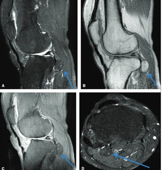
Here, we report the case of a 46-year-old male who present-ed with a three-year history of pain in the right knee. He statpresent-ed that he had experienced no trauma or torsion, had not under-gone any surgery, and did not engage in sports. In the physical examination, he tested positive for a meniscal tear. Magnetic resonance imaging (MRI) revealed a giant cyamella; complex rupture of the body and anterior horn of the lateral meniscus; and chondral lesions in the lateral femorotibial compartment (Figure 1).
Sesamoid bones are accessory ossicles located in the ten-dons and muscles; their function is to facilitate the physiologi-cal movement of the tendon, although they can, in some cases, cause disease(1). Most sesamoid bones are located in the lower limbs(2). Embryologically, sesamoid bones are generally more common in the fetus; with skeletal growth and bone maturation, many sesamoid bones fuse(2,3). In humans, the largest sesamoid bone is the patella(2).
The popliteal tendon typically originates at the lateral fem-oral condyle, its muscle inserting into the posterior surface of the tibia above the soleal line(4). The sesamoid bone that can exist in the tendon of the popliteal muscle is known as the
cya-mella, popliteal fabella, fabella distalis, or sesamoideum genu inferius laterale(5). It is often confused with the fabella, which is within the lateral head of the gastrocnemius muscle(5).
Although the cyamella is common in other primates, it is quite rare in humans, and, when it occurs, it can articulate with the lateral condyle of the tibia and be quite near the head of the ibula(3,4). However, it does not have a well-deined function(6). It resides as an accessory ossicle in the popliteal tendon itself or at the intersection between the tendon and the muscle(6,7); its size can vary considerably(3), and it should be clearly distin-guished from free bodies, calciications, osteophytes, and the fabella, as well as from osteochondromatosis(3) and avulsion of the popliteal tendon(7).
A diagnosis of cyamella can be established through various imaging modalities, such as X-ray, computed tomography, and MRI(3). On T1-, T2-, and T2*-weighted MRI scans, a cyamella appears as an ossicle with low signal intensity along its bor-ders(6). A computed tomography scan can reveal fat within the ossicle(6). Due to the rarity of cyamella, its characterization and the exclusion of other potential diagnoses are of particular clini-cal relevance(3).
In patients with lateral knee pain, physicians should bear in mind the possibility of cyamella as the cause of the pain(1). Cyamella typically does not have pathological implications,
Figure 1. Non-contrast-enhanced MRI scans. T2-weighted spectral presatu-ration inversion recovery (SPIR) se-quence (A) and proton-density (PD) sequence (B), both acquired in the sagittal plane, showing a voluminous ossiied mass, measuring 2.2 × 1.7 ×
1.5 cm (arrows), in the popliteal ten-don. Sagittal PD-SPIR sequence (C) and axial T2-weighted SPIR sequence (D) showing a voluminous ossiied mass in the popliteal tendon (arrows).
A
B
271
Radiol Bras. 2017 Jul/Ago;50(4):266–276Letters to the Editor
Hemangioma of the urinary bladder: an atypical location
Dear Editor,
A two-year-old male patient was referred to the pediatric emergency room with persistent gross hematuria. Abdominal ultrasound (data not shown) revealed an echogenic formation within the urinary bladder, the formation remaining ixed during changes in decubitus. Contrast-enhanced computed tomogra-phy of the abdomen showed a partially delimited, solid-to-cystic expansile urinary bladder lesion with a vegetative component, presenting lobulated contours, a small focal calciication, and although cyamella-associated pain has been described(1). Be-cause of the rarity of the diagnosis, there is no consensus re-garding the treatment of cyamella, which should therefore be treated on a case-by-case basis, the symptoms and imaging ind-ings being taken into account(3).
REFERENCES
1. Rehmatullah N, McNair R, Sanchez-Ballester J. A cyamella causing pop
-liteal tendonitis. Ann R Coll Surg Engl. 2014;96:91E–93E.
2. Akansel G, Inan N, Sarisoy HT, et al. Popliteus muscle sesamoid bone (cyamella): appearance on radiographs, CT and MRI. Surg Radiol Anat. 2006;28:642–5.
3. Khanna V, Maldjian C. The cyamella, a lost sesamoid: normal variant or posterolateral corner anomaly? Radiol Case Rep. 2015;9:e00031. 4. Reddy S, Vollala VR, Rao R. Cyamella in man – its morphology and review
of literature. Int J Morphol. 2007;25:381–3.
Márcio Luís Duarte1, André de Queiroz Pereira Silva2, Simone Botelho
Alvarenga3, José Luiz Masson de Almeida Prado4, Luiz Carlos Donoso
Scoppetta4
1. WebImagem, São Paulo, SP, Brazil. 2. CADI Diagnóstico, Imperatriz, MA, Bra-zil. 3. Axial Medicina Diagnóstica, Belo Horizonte, MG, BraBra-zil. 4. Hospital São Camilo, São Paulo, SP, Brazil. Mailing address: Dr. Márcio Luís Duarte. Avenida Marquês de São Vicente, 446, Barra Funda. São Paulo, SP, Brazil, 01139-020. E-mail: [email protected].
5. Keats TE, Anderson MW. Atlas of normal roentgen variants that may sim
-ulate disease. 9th ed. Philadelphia: Mosby Elsevier; 2007.
6. Munk PL, Althathlol A, Rashid F, et al. MR features of a giant cyamella in a patient with osteoarthritis: presentation, diagnosis and discussion. Skeletal Radiol. 2009;38:69, 91–2.
7. Jadhav SP, More SR, Riascos RF, et al. Comprehensive review of the anat -omy, function, and imaging of the popliteus and associated pathologic
conditions. Radiographics. 2014;34:496–513.
http://dx.doi.org/10.1590/0100-3984.2015.0240
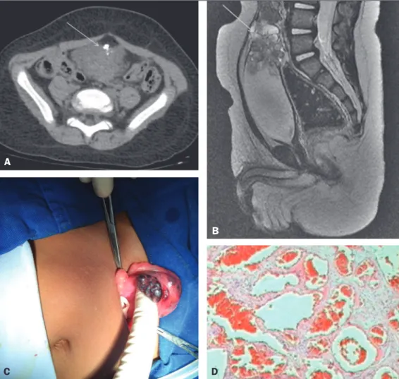
enhancement of the solid component; the epicenter of the lesion was at the bladder dome (Figure 1A). A subsequent contrast-enhanced magnetic resonance imaging scan of the abdomen revealed a formation with intermediate signal intensity on T1-weighted images, heterogeneous signal intensity with a predom-inance of hyperintensity on T2-weighted images, and marked enhancement of the lesion (Figure 1B). Partial cystectomy was performed (Figure 1C), and the histopathological analysis dem-onstrated a lesion characterized by proliferation of vein-like ves-sels of different calibers, with intense congestion and without atypia, consistent with cavernous hemangioma (Figure 1D).
Figure 1. A: Axial computed tomog-raphy scan of the abdomen, showing a partially delimited, solid-to-cystic expansile urinary bladder lesion with a vegetative component, presenting lobulated contours, a small focal cal-ciication, and enhancement of the solid component; the epicenter of the lesion was at the bladder dome. B: Sagittal reconstruction of contrast-enhanced magnetic resonance im-aging of the abdomen, revealing an expansile formation, with intermedi-ate signal intensity on T1-weighted images, heterogeneous signal inten-sity with a predominance of hyperin-tensity on T2-weighted images, the le-sion showing marked enhancement. C: Partial cystectomy demonstrating a tumor. D: Histopathological section showing a lesion consistent with cav-ernous hemangioma.