271
Radiol Bras. 2017 Jul/Ago;50(4):266–276Letters to the Editor
Hemangioma of the urinary bladder: an atypical location
Dear Editor,
A two-year-old male patient was referred to the pediatric emergency room with persistent gross hematuria. Abdominal ultrasound (data not shown) revealed an echogenic formation within the urinary bladder, the formation remaining ixed during changes in decubitus. Contrast-enhanced computed tomogra -phy of the abdomen showed a partially delimited, solid-to-cystic expansile urinary bladder lesion with a vegetative component, presenting lobulated contours, a small focal calciication, and although cyamella-associated pain has been described(1). Be -cause of the rarity of the diagnosis, there is no consensus re-garding the treatment of cyamella, which should therefore be treated on a case-by-case basis, the symptoms and imaging ind -ings being taken into account(3).
REFERENCES
1. Rehmatullah N, McNair R, Sanchez-Ballester J. A cyamella causing pop
-liteal tendonitis. Ann R Coll Surg Engl. 2014;96:91E–93E.
2. Akansel G, Inan N, Sarisoy HT, et al. Popliteus muscle sesamoid bone (cyamella): appearance on radiographs, CT and MRI. Surg Radiol Anat. 2006;28:642–5.
3. Khanna V, Maldjian C. The cyamella, a lost sesamoid: normal variant or posterolateral corner anomaly? Radiol Case Rep. 2015;9:e00031. 4. Reddy S, Vollala VR, Rao R. Cyamella in man – its morphology and review
of literature. Int J Morphol. 2007;25:381–3.
Márcio Luís Duarte1, André de Queiroz Pereira Silva2, Simone Botelho Alvarenga3, José Luiz Masson de Almeida Prado4, Luiz Carlos Donoso Scoppetta4
1. WebImagem, São Paulo, SP, Brazil. 2. CADI Diagnóstico, Imperatriz, MA, Bra-zil. 3. Axial Medicina Diagnóstica, Belo Horizonte, MG, BraBra-zil. 4. Hospital São Camilo, São Paulo, SP, Brazil. Mailing address: Dr. Márcio Luís Duarte. Avenida Marquês de São Vicente, 446, Barra Funda. São Paulo, SP, Brazil, 01139-020. E-mail: marcioluisduarte@gmail.com.
5. Keats TE, Anderson MW. Atlas of normal roentgen variants that may sim
-ulate disease. 9th ed. Philadelphia: Mosby Elsevier; 2007.
6. Munk PL, Althathlol A, Rashid F, et al. MR features of a giant cyamella in a patient with osteoarthritis: presentation, diagnosis and discussion. Skeletal Radiol. 2009;38:69, 91–2.
7. Jadhav SP, More SR, Riascos RF, et al. Comprehensive review of the anat -omy, function, and imaging of the popliteus and associated pathologic
conditions. Radiographics. 2014;34:496–513.
http://dx.doi.org/10.1590/0100-3984.2015.0240
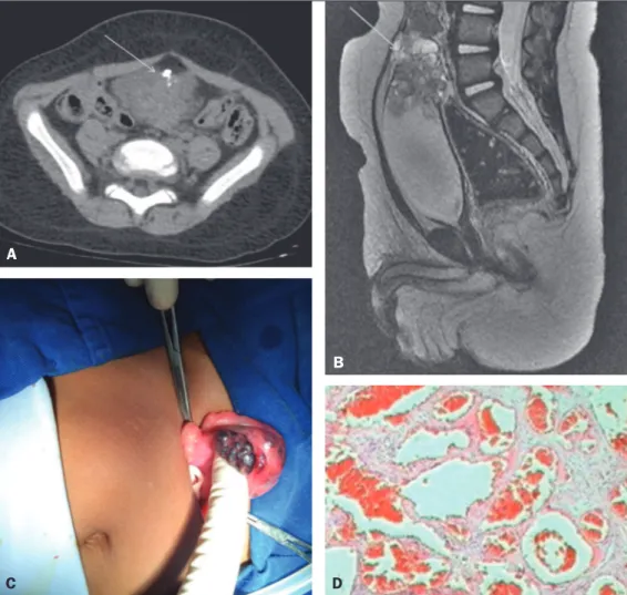
enhancement of the solid component; the epicenter of the lesion was at the bladder dome (Figure 1A). A subsequent contrast-enhanced magnetic resonance imaging scan of the abdomen revealed a formation with intermediate signal intensity on T1-weighted images, heterogeneous signal intensity with a predom-inance of hyperintensity on T2-weighted images, and marked enhancement of the lesion (Figure 1B). Partial cystectomy was performed (Figure 1C), and the histopathological analysis dem -onstrated a lesion characterized by proliferation of vein-like ves -sels of different calibers, with intense congestion and without atypia, consistent with cavernous hemangioma (Figure 1D).
Figure 1. A: Axial computed tomog-raphy scan of the abdomen, showing a partially delimited, solid-to-cystic expansile urinary bladder lesion with a vegetative component, presenting lobulated contours, a small focal
cal-ciication, and enhancement of the
solid component; the epicenter of the lesion was at the bladder dome.
B: Sagittal reconstruction of contrast-enhanced magnetic resonance im-aging of the abdomen, revealing an expansile formation, with intermedi-ate signal intensity on T1-weighted images, heterogeneous signal inten-sity with a predominance of hyperin-tensity on T2-weighted images, the
le-sion showing marked enhancement. C: Partial cystectomy demonstrating a tumor. D: Histopathological section showing a lesion consistent with cav-ernous hemangioma.
A
B
272
Radiol Bras. 2017 Jul/Ago;50(4):266–276 Letters to the EditorHemangiomas are benign tumor formations of capillaries and blood vessels, common in various organs; they are extremely rare in the urinary bladder, accounting for only 0.6% of all uri -nary bladder tumors(1,2). There have been fewer than 100 re -ported cases of histologically proven hemangioma of the urinary bladder(1).
Most urinary bladder hemangiomas are solitary and smaller than 3 cm in diameter, affecting the dome, posterior wall, or tri -gone of the bladder. Although hemangiomas can occur in indi -viduals of any age, they are seen most often in indi-viduals under 30 years of age and are slightly more common among males(2). A hemangioma usually presents as an incidental inding during the investigation of hematuria. The most common symptom is gross hematuria, which can be accompanied by irritative urinary symptoms and abdominal pain. Urinary bladder hemangiomas occasionally coexist with cutaneous hemangioma or are asso -ciated with one of two conditions(3–5): Sturge-Weber syndrome and Klippel-Trenaunay-Weber syndrome.
In young patients, endoscopic indings of a bluish, ses -sile mass and gross hematuria are highly suggestive of heman-gioma(1). The main differential diagnoses for pigmented lesions seen on endoscopy include endometriosis, melanoma, and sar-coma(6). Imaging tests, such as ultrasound, computed tomogra -phy, and magnetic resonance imaging, are useful in deining the location and extent of a hemangioma(2).
For individuals with hemangioma, the treatment is contro-versial. Although there are many options available, partial cys -tectomy is currently the most widely used treatment for heman-gioma of the urinary bladder(3,6,7). Although hemangioma has a benign course, follow-up is mandatory in order to detect recur-rence or residual disease(3,7,8). The purpose of this case report
was to underscore the importance of early diagnosis of heman-gioma of the urinary bladder and of differentiating it from ma-lignant neoplasms, which would affect the therapeutic strategy and patient survival.
REFERENCES
1. Cheng L, Nascimento AG, Neumann RM, et al. Hemangioma of the uri
-nary bladder. Cancer. 1999;86:498–504.
2. Stimac G, Dimanovski J, Katusic J, et al. A large cavernous hemangioma of the urinary bladder: imaging of possible spontaneous regression. Eur J Radiol Extra. 2007;61:61–3.
3. Jibhkate S, Sanklecha V, Valand A. Urinary bladder hemangioma – a rare urinary bladder tumor in a child. APSP J Case Rep. 2015;6:6.
4. Kim YY, Kim MJ, Lee MJ, et al. Multiple hemangiomas of the urinary bladder in a child with gross hematuria. Ultrasonography. 2015;34:231–4. 5. Ikeda T, Shimamoto K, Tanji N, et al. Cavernous hemangioma of the uri
-nary bladder in an 8-year-old child. Int J Urol. 2004;11:429–31. 6. Wong-You-Cheong JJ, Woodward PJ, Manning MA, et al. From the Ar
-chives of the AFIP: Neoplasms of the urinary bladder: radiologic-patho
-logic correlation. Radiographics. 2006;26:553–80.
7. Lahyani M, Slaoui A, Jakhlal N, et al. Cavernous hemangioma of the blad
-der: an additional case managed by partial cystectomy and augmentation cystoplasty. Pan Afr Med J. 2015;22:131.
8. Castillo OA, Foneron A, Sepúlveda F, et al. Bladder hemangioma: case report. Arch Esp Urol. 2012;65:623–5.
Camila Soares Moreira de Sousa1, Ivo Lima Viana1, Carla Lorena Vasques Mendes de Miranda1, Breno Braga Bastos2, Ilan Lopes Leite Mendes1
1. Medimagem, Teresina, PI, Brazil. 2. UDI 24 horas, Teresina, PI, Brazil. Mailing address: Dra. Camila Soares Moreira de Sousa. Medimagem – Ra-diologia. Rua Paissandu, 1862, Centro. Teresina, PI, Brazil, 64001-120. E-mail: camilasoares__@hotmail.com.
http://dx.doi.org/10.1590/0100-3984.2015.0231
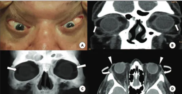
Figure 1. A: Fatty mass in the lateral corner of the orbits, best characterized by the retropul-sion of the globes. CT of the orbits in the coronal plane (B), with volumetric reconstructions in the coronal plane (C) and axial plane (D), showing the masses in the lateral corners (straight arrows), contiguous with the intraconal fat (arrow-heads) and pushing aside the lacrimal glands (curved arrows).
Subconjunctival fat prolapse: a disease little known to radiologists
Dear Editor,
A 69-year-old male patient sought outpatient treatment with a 10-year history of fatty masses in the lateral corners of his eyes, best characterized as retropulsion of the globes. He underwent computed tomography (CT) of the orbits, which re -vealed intraconal fat proliferation in the lateral corners of the
eyes, from the orbits to the epibulbar region (Figure 1). Given the clinical presentation and imaging indings, a diagnosis of subconjunctival fat prolapse was made.