Dosimetry in thyroid follicles due to low-energy
electrons of iodine using the Monte Carlo method*
Dosimetria em folículos tireoidianos devido aos elétrons de baixa energia do iodo usando o método Monte CarloLaélia Campos1, Frank da Silva2
OBJECTIVE: To evaluate the absorbed dose in thyroid follicles due to low-energy electrons such as Auger and internal conversion electrons, besides beta particles, for iodine radioisotopes (131
I, 132 I, 133
I, 134 I and 135
I) utilizing the Monte Carlo method. MATERIALS AND METHODS: The dose calculation was performed at follicular level, simulating Auger, internal conversion electrons and beta particles, with the MCNP4C code. The follicles (colloid and follicular cells) were modeled as spheres with colloid diameter ranging from 30 to 500 µµµµµm, and with the same density of water (1.0 g.cm–³). RESULTS: Considering low-energy particles, the contribution of 131
I for total absorbed dose to the colloid is about 25%, while the contribution due to short-lived isotopes is 75%. For follicular cells, this contribution is still higher achieving 87% due to short-lived iodine and 13% due to 131
I. CONCLUSION: The results of the present study demonstrate the importance of considering low-energy particles in the contribution for the total absorbed dose at follicular level (colloid and follicular cells) due to iodine radioisotopes (131
I, 132 I, 133
I, 134 I and 135
I).
Keywords: Internal dosimetry; Short-lived iodines; Thyroid cancer; Monte Carlo method; MCNP.
OBJETIVO: Avaliar a dose absorvida em folículos tireoidianos devido aos elétrons de baixa energia, como os elétrons Auger e os de conversão interna, além das partículas beta, para os radioisótopos de iodo (131
I, 132 I, 133
I, 134 I e 135
I) usando o método Monte Carlo. MATERIAIS E MÉTODOS: O cálculo da dose foi feito ao nível folicular, simulando elétrons Auger, conversão interna e partículas beta, com o código MCNP4C. Os folícu-los (colóide e células foliculares) foram modelados como esferas, com diâmetros do colóide variando de 30 a 500 µµµµµm. A densidade considerada para os folículos foi a da água (1,0 g.cm–³). RESULTADOS: Considerando partículas de baixa energia, o percentual de contribuição do 131
I na dose total absorvida pelo colóide é de aproximadamente 25%, enquanto os isótopos de meia-vida física curta apresentaram contribuição de 75%. Para as células foliculares, esse percentual é ainda maior, chegando a 87% para os iodos de meia-vida curta e 13% para o 131
I. CONCLUSÃO: Com base nos resultados obtidos, pode-se mostrar a importância de se considerar partículas de baixa energia na contribuição para a dose total absorvida ao nível folicular (colóide e células foliculares) devido aos radioisótopos de iodo (131
I, 132 I, 133
I, 134 I e 135
I).
Unitermos: Dosimetria interna; Iodos de meia-vida curta; Câncer de tireóide; Método Monte Carlo; MCNP.
Abstract
Resumo
* Study developed in the Department of Statistics and Infor-mation Technology at Universidade Federal Rural de Pernambuco (UFRPE), Recife, PE, Brazil.
1. PhD, Associate Professor II, Department of Statistics and Information Technology, Universidade Federal Rural de Pernam-buco (UFRPE), Recife, PE, Brazil.
2. Master, Professor, Department of Statistics and Informa-tion Technology, Universidade Federal Rural de Pernambuco (UFRPE), Recife, PE, Brazil.
Mailing address: Dra. Laélia Campos. Departamento de Esta-tística e Informática, UFRPE. Rua Dom Manoel de Medeiros, s/ nº, Dois Irmãos. Recife, PE, Brazil, 52171-900. E-mail: [email protected] / [email protected]
Received March 31, 2008. Accepted after revision July 31, 2008.
that concentrates the iodines circulating in the blood, and incorporates thyroid hor-mones whose production is associated to the iodine intake by the organism(2). Iodine is an essential element in the composition of hormones secreted by the thyroid, that play a determinant role in the metabolism of all cells in the organism(3) and, equally, in the growth process and development of the majority of organs(4), particularly the brain(5).
In case of accidents involving nuclear plants, large amounts of radioactive iodine isotopes are released in the environment and, due to its volatility and mobility, the exposure to these radioisotopes requires special attention regarding
radioprotec-Campos L, Silva F. Dosimetry in thyroid follicles due to low-energy electrons of iodine using the Monte Carlo method. Radiol Bras. 2008;41(6):403–407.
man(1). Each of the lateral thyroid lobes, connected by the isthmus, has a height of 4 to 6 cm, 2 cm transversely and 2 cm in thickness(2). The functional unit of the thy-roid is the follicle, formed by follicular cells and colloid. The follicle is sphere-shaped and its average diameter, in an adult man, is 200 µm, presenting however, an important variation in size, proportional to its functional activity. At rest, the follicles are large, with 200 to 500 µm in diameter, and in a hyperactivity state they are smaller, with 30 to 50 µm in diameter(2). Follicular cells are joined by a basal membrane con-stituting a single layer that limits the col-loid volume and have an average diameter of 10 µm. Colloid is a proteic substance
INTRODUCTION
tion(6,7). In fact, the thyroid capacity of con-centrating iodine makes such organ one of the most vulnerable in accidents of this nature(8). Amongst the main types of thy-roid cancer, papillary carcinoma represents from 60% to 80% of cases, and follicular carcinoma, from 10% to 20%(9). These two types of carcinomas originate from the fol-licular cells. From the radioprotection standpoint, the most important radioactive iodine isotopes are 131I and short half- life
isotopes as follows: 132I (T
1/2 = 2.38 h), 133I
(T1/2 = 20.8 h), 134I (T
1/2 = 52.5 min) and 135I
(T1/2 = 6.7 h). Due to these iodine
radioiso-topes, dosimetry is of paramount impor-tance(10,11).
Thus, the absorbed dose, defined as the average energy deposited by radiation per tissue mass unit or irradiated organ(12), is the most important physical magnitude in the assessment of risks associated with ion-izing radiation contamination or exposure. However, retrospective dosimetry involv-ing accidental internal contamination is not an easy task, generally due to the lack of information about exposure conditions. In the Chernobyl accident, adopted as a com-parative example in the present study, the majority of direct dose measurements were made only approximately one month after the accident. Moreover, these measure-ments were only made for 131I, while the
short half-life iodine radioisotopes were not taken into consideration in the estima-tion of dose absorbed by the thyroid(8).
Some studies have reported results of dosimetry at cellular level of 131I beta
par-ticles utilizing deterministic methods(13–15), while others have utilized Monte Carlo simulations for 131I(16) and short half-life iodines(11). However none of these studies has taken low energy particles, such as Auger electrons or internal conversion electrons, into consideration.
The adoption of Monte Carlo codes for calculating absorbed dose in organs and/or tissues of the human body(17,18) as well as in the cellular field(11) has been diffused worldwide, serving professionals involved in internal or external dosimetry, utilizing for such purpose, geometrical models (or phantoms) of organs and/or tissues to be studied(19).
In this context, the present study is aimed at assessing the absorbed dose in
thyroid follicles due to low energy elec-trons, such as the Auger and internal con-version electrons, besides the beta particles for iodine radioisotopes (131I, 132I, 133I, 134I
and 135I) by means of the Monte Carlo
method.
MATERIALS AND METHODS
Modeling for the thyroidal follicle
The modeling of the thyroid follicles (consisting of colloid and follicular cells) was made with two concentrical spheres, with the internal sphere representing the colloid, and the region between the inter-nal and exterinter-nal spheres (whose distance between internal and external radii was 10
µm) representing the follicular cells. Diam-eters utilized for the internal sphere (col-loid) were the following: 30, 50, 80, 100, 200, 300, 400 and 500 µm. These values are justified by the diameter of the human thyroid follicles, that vary between 30 and 50 µm, when in hyperactivity state, while in rest state this variation is between 200 and 500 µm(2).
Simulation of thyroid follicle with the MCNP code
The code MCNP (Monte Carlo N-Par-ticle) version 4C(20), which utilizes the Monte Carlo method for particles transpor-tation, was adopted to simulate thyroid follicles contaminated by iodine radioiso-topes. In the code input, the following pa-rameters were described: follicle geometry, radioactive source distribution, energy de-cay spectrums of the Auger and internal conversion electrons, as well as the beta particles for each simulated iodine isotope. The water density (1.0 g.cm–³) was
consid-ered as a parameter for colloid and follicu-lar cells. For each diameter, the iodines (131I, 132I, 133I, 134I and 135I) were simulated
in a uniform manner within the colloid. The tables with the iodine energy decay spec-trums and energy abundancy rates were obtained at the Brookhaven National Laboratory’s (BNL) National Nuclear Data Center (http://www.nndc.bnl.gov; July/ 2008).
In the MCNP output, one has the energy deposited both in the colloid and in the follicular cells due to the iodine isotopes. Photons were not utilized in the simulation,
as they do not contribute to the absorbed dose at cellular level.
RESULTS
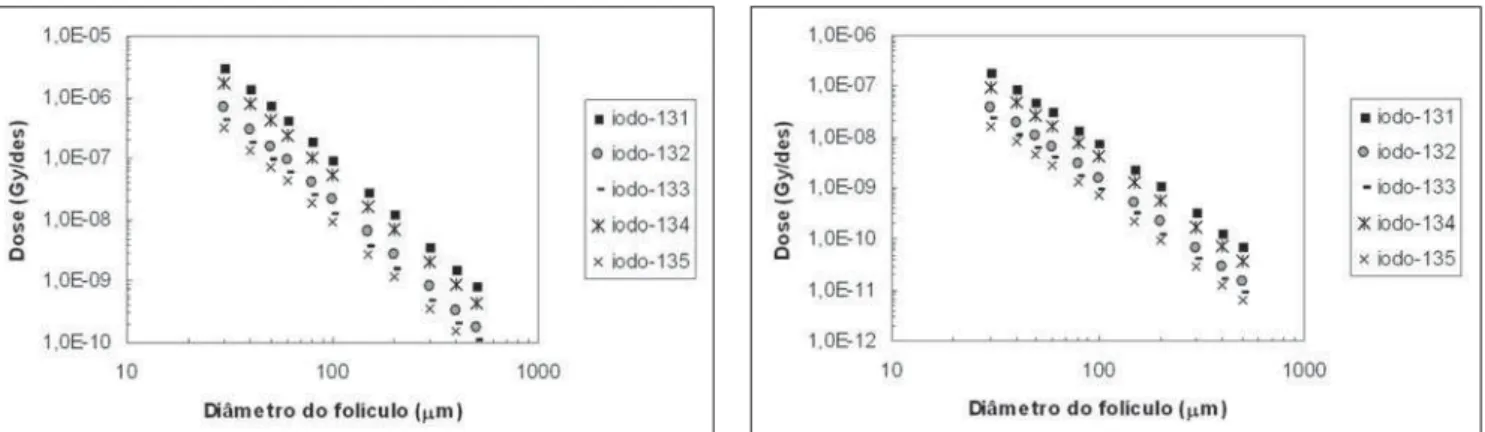
The results of the simulations for the thyroid follicle model (colloid and follicu-lar cells) are presented on Figures 1 thru 6. The graphs are plotted in logarithmic scale and correspond to dose in Gray per disin-tegration (Gy/dis) as a function of the simu-lated colloid diameter (µm). The points plotted in the graphs were individually obtained by means of a single simulation with specific physical and geometrical pa-rameters.
Figures 1 and 2 present the results for the absorbed dose, respectively, in the col-loid and follicular cells due to Auger elec-trons. Based on the graphs, one can observe that greatest contribution of absorbed dose due to Auger electrons comes from 131I and
next from 134I, for all simulated diameters.
The results of dose due to internal con-version electrons are shown on Figures 3 and 4. One observes that the greatest dose contribution due to internal conversion electrons is initially due to 131I, and as the
follicle diameter increases, the 131I
contri-bution percentage tends to reach that of 134I,
until it becomes practically equal. Figures 5 and 6 show absorbed doses due to beta particles. The contribution of the dose in the colloid is almost the same for all iodine isotopes (Figure 5) due to the beta particles, with a slightly higher con-tribution from 131I, when compared with
other isotopes. Observing Figure 6, one can affirm that all iodine isotopes have the same contribution to the deposited dose on follicular cells due to beta particles for all the simulated diameters.
The estimated relative errors generated by the MCNP code ranged from 0.06% to 1.7%, thus being within the confidence range, as established by this code.
Table 1 shows the total dose absorbed by the colloid (Gy/dis), considering the Auger electrons, internal conversion elec-trons and beta particles for all iodine iso-topes and all simulated diameters, besides presenting the percentage of dose due to
131I and short half-life iodines. Table 1
colloid is, on average, 25%, while for the short physical half-life iodines this total percentage is 75% on average.
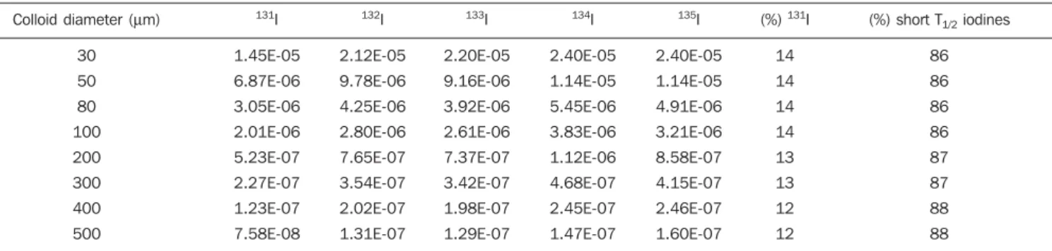
Table 2 shows the values of total dose absorbed by follicular cells (Gy/dis), con-sidering Auger electrons, internal conver-sion electrons and beta particles, besides presenting the dose percentages due to 131I
(13%, on average) and percentages due to
short physical half-life isotopes (87%, on average).
DISCUSSION
Figures 1 and 2 demonstrate the doses absorbed due to Auger electrons, with em-phasis on the greater contribution for 131I.
The relevance of the Auger electrons in
dosimetry has been neglected for several years, to a large extent because the energy absorbed by tissues due to these electrons is normally insignificant as compared with the total energy released in the radionuclide decay(21).
Auger electrons emissions produce a cascade of low energy electrons. Most of this energy is deposited within a few
na-Figure 1. Absorbed dose in colloid due to Auger electrons. Figure 2. Absorbed dose in follicular cells due to Auger electrons.
Figure 3. Absorbed dose in colloid due to internal conversion electrons. Figure 4. Absorbed dose in follicular cells due to internal conversion
elec-trons.
nometers (nm), with a very high local dose. The most energetic Auger electron results from the transition to the K layer (25 to 27 keV), but most electrons are produced by transitions between exterior orbits, and therefore, present energies < 500 eV, with corresponding ranges < 25 nm. Auger elec-tron emitting radionuclides are widely uti-lized in nuclear medicine and biomedical research. The Auger electrons effects have been evaluated by means of microdosime-try techniques(22).
Electrons carry only a small fraction of the energy released by decay, and make only a minor contribution for the total dose in the organ; however, Auger electrons may play a crucially significant role the deter-mination of the cell damage magnitude, as the biological risks associated with Auger emission depend to a great extent on the local accuracy decay within the cell. Extra-cellular Auger electrons would be relatively harmless because of their limited range, but they may produce irreparable damage to any radiosensitive(21) structure.
The dose contributions to colloid and follicular cells due to internal conversion electrons are shown on Figures 3 and 4. Because of their lower range in the energy
deposition, low energy electrons, such as the internal conversion ones, are widely utilized in the treatment of superficial tu-mors, and are also appropriate for intraop-erative radiotherapy. The dense shower of short-range Auger electrons released by radionuclides, which decay by means of electron capture or internal conversion, results in biological damage, which is very dependent of the site of of decay within the cell(23).
Figures 5 and 6 show the doses ab-sorbed by colloid and by follicular cells due to the beta particles. In radioimmunother-apy, preference is given to the treatment of deep tumors by beta particle emitting radio-nuclides. However, for the erradication of small groups of cancerigenous cells, Auger electron or alpha particle emitting radionu-clides are considered as advantageous in the erradication of small cluster of cancer cells because of their ability to deposit ra-diation energy locally(14).
The percentage value of 75% of short physical half-life isotopes presented on Table 1, confirms the high contribution of dose absorbed by the colloid due to these isotopes, besides demonstrating that this percentage is even higher than the one
re-ported in the literature(11), where no calcu-lation with Auger and internal capture elec-trons is found. For the total dose absorbed by follicular cells (see Table 2), the percent-age of the dose due to short half-life iodines is even higher, representing on average 87% of the total dose, while 131I contributes
with 13% on average. This result highlights the significant role of low energy particles in dose calculations at cellular levels, con-sidering that in the present study the simu-lated Auger electrons present an energy range from 0.08 to 32 keV.
For low-energy beta particles emitting radionuclides, the conventional dosimetry frequently establishes an inappropriately estimated dose, i.e., the dose in individual cells within an organ may be much higher or much lower than the mean dose calcu-lated for the organ as a whole, as conven-tional dosimetry establishes average radia-tion doses for specific organs or tissues, allowing a gross under- or overestimation of radiation exposure for individual cells. Because of the low energy of Auger electrons and, correspondingly, their low range (from 1 nm to 1 µm), the biological effects of Auger emissions are highly de-pendent on their cellular and subcellular
Table 1 Total dose absorbed by colloid due to iodine isotopes.
Colloid diameter (µm)
30 50 80 100 200 300 400 500 131I 6.37E-05 2.10E-05 7.77E-06 4.80E-06 1.07E-06 4.38E-07 2.32E-07 1.40E-07 132I 3.70E-05 1.44E-05 5.76E-06 3.60E-06 8.05E-07 3.45E-07 1.91E-07 1.20E-07 133I 4.23E-05 1.53E-05 5.59E-06 3.45E-06 8.13E-07 3.51E-07 1.94E-07 1.22E-07 134I 3.39E-05 1.26E-05 5.15E-06 3.28E-06 7.70E-07 3.30E-07 1.83E-07 1.16E-07 135I 4.20E-05 1.67E-05 6.82E-06 4.25E-06 9.22E-07 3.87E-07 2.11E-07 1.31E-07
(%) 131I
29 26 25 25 24 24 23 22
(%) short T1/2 iodines
71 74 75 75 76 76 77 78
Table 2. Total dose absorbed by follicular cells due to iodine isotopes.
Colloid diameter (µm)
30 50 80 100 200 300 400 500 131I 1.45E-05 6.87E-06 3.05E-06 2.01E-06 5.23E-07 2.27E-07 1.23E-07 7.58E-08 132I 2.12E-05 9.78E-06 4.25E-06 2.80E-06 7.65E-07 3.54E-07 2.02E-07 1.31E-07 133I 2.20E-05 9.16E-06 3.92E-06 2.61E-06 7.37E-07 3.42E-07 1.98E-07 1.29E-07 134I 2.40E-05 1.14E-05 5.45E-06 3.83E-06 1.12E-06 4.68E-07 2.45E-07 1.47E-07 135I 2.40E-05 1.14E-05 4.91E-06 3.21E-06 8.58E-07 4.15E-07 2.46E-07 1.60E-07
(%) 131I
14 14 14 14 13 13 12 12
(%) short T1/2 iodines
distribution. Conventional dosimetry uti-lized for a specific organ does not take into account the cell-by-cell heterogeneity, where the Auger emission will concentrate in the cell nucleus(24).
CONCLUSION
Based on the results obtained by Monte Carlo simulation, the importance of consid-ering the contribution of low-energy par-ticles such as Auger and internal conver-sion electrons to the total absorbed dose at follicular level (colloid and follicular cells) due to short half-life iodine isotopes (132I, 133I, 134I and 135I) and due to 131I can be
demonstrated. For the same number of dis-integrations, the contribution percentage was at the order of 75% for the total ab-sorbed dose by the colloid due to the short half-life iodines, and 25% for 131I. For the
follicular cells, these values reached 87% for the short half-life iodine isotopes and 13% for 131I.
Acknowledgements
The authors wish to thank the Post Graduation Program in Biometry and Ap-plied Statistics of the Universidade Federal Rural de Pernambuco (UFRPE), for its lo-gistical support, and the Coordenação de Aperfeiçoamento de Pessoal de Nível Su-perior (Capes), within the Capes/Cofecub agreement, for the financial support.
REFERENCES
1. Cristy M. Reference man anatomical model. In: Raabe OG, editor. Internal radiation dosimetry.
Madison: Medical Physics Pub Corp; 1994. p. 217–38.
2. Leclère J, Orgiazzi J, Rousset B, et al. La thyroïde: des concepts à la pratique clinique. 2nd ed. Paris: Elsevier; 2001.
3. Sterling K. Thyroid hormone action at the cellu-lar level. In: Ingbar SH, Braverman LE, editors. Werner’s The thyroid: a fundamental and clini-cal text. Philadelphia: JB Lippincott; 1986. p. 219–33.
4. Fisher DA. Thyroid hormone effects on growth and development. In: Delange F, Fisher DA, Malvaux P, editors. Pediatric thyroidology. Basel: Karger; 1985. p. 75–9.
5. Dussault JH. Action of thyroid hormones on brain development. In: Delange F, Fisher DA, Glinoer D, editors. Research in congenital hypothyroid-ism. New York: Plenum Press; 1989. p. 95–102. 6. Galle P. Toxiques nucléaires. 2nd ed. Paris:
Mas-son; 1998.
7. International Commission on Radiological Pro-tection (ICRP). Human respiratory tract model for radiological protection. ICRP Publication 66. Oxford: Pergamon Press; 1994.
8. United Nations Scientific Committee on the Ef-fects of Atomic Radiation (UNSCEAR). Sources, effects and risks of ionizing radiation. UNSCEAR Report 2000 to the General Assembly, with An-nexes. New York: United Nations; 2000.
9. Schlumberger MJ. Papillary and follicular thyroid carcinoma. N Engl J Med. 1998;338:297–306.
10. Balonov M, Kaidanovsky G, Zvonova I, et al. Contributions of short-lived radioiodines to thy-roid doses received by evacuees from the Chernobyl area estimated using early in vivo ac-tivity measurements. Radiat Prot Dosimetry. 2003;105:593–9.
11. Campos L, Amaral A, Colas-Linhart N, et al. Evaluation of absorbed dose in thyroid follicles due to short-lived iodines irradiation using the Monte Carlo method. Journal of Radioanalytical and Nuclear Chemistry. 2006;269:635–8. 12. International Commission on Radiation Units &
Measurements (ICRU). Quantities and units in radiation protection dosimetry. ICRU Report 51. Bethesda: ICRU; 1993.
13. Bardiès M, Chatal JF. Absorbed doses for
inter-nal radiotherapy from 22 beta-emitting radionu-clides: beta dosimetry of small spheres. Phys Med Biol. 1994;39:961–81.
14. Goddu SM, Rao DV, Howell RW. Multicellular dosimetry for micrometastases: dependence of self-dose versus cross-dose to cell nuclei on type and energy of radiation and subcellular distribu-tion of radionuclides. J Nucl Med. 1994;35:521– 30.
15. Li WB, Friedland W, Pomplun E, et al. Track structures and dose distributions from decays of
131I and 125I in and around water spheres
simulat-ing micrometastases of differentiated thyroid can-cer. Radiat Res. 2001;156:419–29.
16. Champion C, Zanotti-Fregonara P, Hindié E. CELLDOSE: a Monte Carlo code to assess elec-tron dose distribution – S values for 131I in spheres
of various sizes. J Nucl Med. 2008;49:151–7. 17. Campos L, Stabin M. Intravascular brachytherapy
to prevent restenosis: dosimetric considerations. Cell Mol Biol (Noysi-le-grand). 2002;48:429–39.
18. Campos L, Stabin M. Internal dosimetry for ra-diation therapy in coronary arteries. Radiat Prot Dosimetry. 2002;101:423–6.
19. Fisher DR. From “micro” to “macro” internal dosimetry. In: Raabe OG, editor. Internal radia-tion dosimetry. Madison: Medical Physics Pub Corp; 1994. p. 61–80.
20. Briesmeister JF. MCNP™ – a general Monte Carlo N-particle transport code, version 4C. Manual LA-13709-M. Los Alamos: Los Alamos National Laboratory; 2000.
21. Ftá…niková S, Böhm R. Monte Carlo calculations of energy deposition on cellular, multicellular and organ level for auger emitters. Radiat Prot Dosim-etry. 2000;92:279–88.
22. Persson L. Radiation protection issues of Auger electron emitters. Radiat Prot Dosimetry. 1996; 64:189–91.
23. Humm JL, Howell RW, Rao DV. Dosimetry of Auger-electron-emitting radionuclides: report No. 3 of AAPM Nuclear Medicine Task Group No. 6. Med Phys. 1994;21:1901–15.