Fluid of Hyper-IgE and CVID Patients
Andreas Cederlund1, Marie Olliver2, Rokeya Sultana Rekha1,3, Monica Lindh1, Lennart Lindbom4, Staffan Normark2, Birgitta Henriques-Normark2, Jan Andersson5, Birgitta Agerberth1, Peter Bergman5,6*
1Department of Medical Biochemistry and Biophysics, Karolinska Institutet, Stockholm, Sweden,2Department of Microbiology, Tumor and Cellbiology, Karolinska Institutet, Stockholm, Sweden,3International Centre for Diarrheal Diseases Research, Bangladesh, Dhaka, Bangladesh,4Department of Physiology and Pharmacology, Karolinska Institutet, Stockholm, Sweden,5Department of Medicine, Center for Infectious Medicine, Karolinska Institutet, Stockholm, Sweden,6Department of Laboratory Medicine, Division of Clinical Microbiology, Karolinska University Hospital, Huddinge, Sweden
Abstract
Background:Patients with primary immunodeficiency (PID) often suffer from frequent respiratory tract infections. Despite standard treatment with IgG-substitution and antibiotics many patients do not improve significantly. Therefore, we hypothesized that additional immune deficits may be present among these patients.
Objective:To investigate if PID patients exhibit impaired production of antimicrobial peptides (AMPs) in nasal fluid and a possible link between AMP-expression and Th17-cells.
Methods:Nasal fluid, nasopharyngeal swabs and peripheral blood mononuclear cells (PBMCs) were collected from patients and healthy controls. AMP levels were measured in nasal fluid by Western blotting. Nasal swabs were cultured for bacteria. PBMCs were stimulated with antigen and the supernatants were assessed for IL-17A release by ELISA.
Results:In healthy controls and most patients, AMP levels in nasal fluid were increased in response to pathogenic bacteria. However, this increase was absent in patients with common variable immunodeficiency (CVID) and Hyper-IgE syndrome (HIES), despite the presence of pathogenic bacteria. Furthermore, stimulation of PBMCs revealed that both HIES and CVID patients exhibited an impaired production of IL-17A.
Conclusion:CVID and HIES patients appear to have a dysregulated AMP response to pathogenic bacteria in the upper respiratory tract, which could be linked to an aberrant Th17 cell response.
Citation:Cederlund A, Olliver M, Rekha RS, Lindh M, Lindbom L, et al. (2011) Impaired Release of Antimicrobial Peptides into Nasal Fluid of Hyper-IgE and CVID Patients. PLoS ONE 6(12): e29316. doi:10.1371/journal.pone.0029316
Editor:Jo¨rg Hermann Fritz, McGill University, Canada
ReceivedAugust 23, 2011;AcceptedNovember 24, 2011;PublishedDecember 27, 2011
Copyright:ß2011 Cederlund et al. This is an open-access article distributed under the terms of the Creative Commons Attribution License, which permits unrestricted use, distribution, and reproduction in any medium, provided the original author and source are credited.
Funding:PB holds a PostDoc position financed by Karolinska Institutet and the Stockholm County Council. PB also receives grants from DA Hagelen, A˚ke Wiberg and Magnus Bergwall foundations as well as Karolinska Institutet and Swedish Association for Medical Microbiology. BA is funded by the Swedish Research Council (58X-11217-14-3), the Torsten and Ragnar So¨derberg Foundation, the Swedish Cancer Society and Karolinska Institutet. The Swedish Foundation for Strategic Research supports BA, JA and LL with a joint grant where PB is included as a junior scientist and coordinator. BH-N and SN are supported by Torsten and Ragnar So¨derberg foundation, the Swedish Research Council, the Royal Swedish Academy of Sciences, the Swedish foundation for Strategic Research and Karolinska Institutet. The funders had no role in study design, data collection and analysis, decision to publish, or preparation of the manuscript.
Competing Interests:The authors have declared that no competing interests exist. * E-mail: [email protected]
Introduction
Diseases that are characterized by lack of antibodies (hypogam-maglobulinemias, HGGs) include syndromes such as selective IgA-deficiency, IgG-deficiency and common variable immunodeficien-cy (CVID). Common symptoms include frequent bacterial res-piratory tract infections (RTIs), fatigue as well as autoimmune manifestations [1]. Bacteria are the most common infectious agent, although viral, fungal and protozoal infections may also occur [2– 5]. Standard treatment involves antibiotic therapy and patients with low levels of IgG and/or IgA are administered IgG prepara-tions, which in most cases reduce symptoms and improve overall health [6]. Nevertheless, many patients still experience frequent infections. Hence, it is possible that immune deficits other than lack of immunoglobulins may be involved in some of these disorders. Such immune deficits could include two key
compo-nents of the innate and adaptive immune system, namely antimicrobial peptides (AMPs) and Th17 cells.
AMPs are expressed by immune and epithelial cells and constitute an essential part of innate immunity [7]. They are released early during infection and have potent antimicrobial activities against a wide range of microbes. AMPs also induce chemotaxis of neutrophils and macrophages, which are rapidly recruited to the site of infection [8]. AMPs have been implicated in several human diseases, including psoriasis [9,10], Crohn’s disease [11], cystic fibrosis [12] and tuberculosis [13].
Th17 cells constitute a recently discovered subset of T helper cells characterized by expression of the transcription factor RORct and
mediate autoimmunity in both rodents and humans [16]. Further-more, studies using animal models have demonstrated a role for Th17 cells in mediating protection against several different bacteria and fungi, suggesting dual and context-dependent roles for Th17 cells [17,18]. Recently, a number of human disorders characterized by infectious susceptibility and low numbers of Th17 cells have been described, including Hyper-IgE syndrome (HIES) [19] and chronic mucocutaneous candidiasis [20]. Special focus has been given to HIES, a PID characterized by frequent skin and RTIs together with distinct skeletal abnormalities and very high levels of serum-IgE. The HIES diagnosis is obtained by genetic analysis of the STAT3 gene [21].
Here we sought to investigate whether induction of AMPs and Th17 cell responses are impaired in PID patients with frequent RTIs. We collected nasopharyngeal (NPH) swabs, nasal fluid and peripheral blood mononuclear cells (PBMC) from patients with selective IgA-, or IgG-deficiencies, CVID, or with frequent RTIs without a defined diagnosis (Not Defined, ND group). In addition, two patients with HIES carrying mutations in the STAT3 gene were enrolled. NPH swabs were screened for bacterial growth while nasal fluid samples were investigated for AMP expression, IL-8 levels and induction of neutrophil chemotaxis. PBMCs were challenged with antigens and IL-17A was measured in the supernatant. Finally, a clinical scoring system was used to assess the clinical status of the patient at the time of inclusion.
Methods
Ethics statement
The study was approved by the Regional Ethical Committee in Stockholm, FE 289, Nobels va¨g 9, Karolinska Institutet, 171 77 Stockholm, Sweden (DNR 2007/751-31/1-4) and written in-formed consent was collected from all patients prior to inclusion and sample-collection.
Patients and healthy controls
All patients were recruited at the Immune Deficiency Unit, Karolinska University Hospital, Huddinge. The diagnostic criteria were the following: XLA (D80.0): a mutation in the btk-gene leading to a total lack of endogenous synthesis of immunoglobulins (n = 3). CVID (D83.0): serum-IgG,3 g/l, serum-IgA,0.07 g/l and a lack of IgG response to pneumo-coccal polysaccharide vaccination (Pneumovax). No other diagnosis should explain the lack of IgG and IgA (n = 18).IgG-subclass deficiency (D80.3): serum-IgG1,2.8 g/l; s-IgG2,1.15 g/l; s-IgG3,0.24 g/l (n = 55 for the whole group of subclass deficiencies) or total serum IgG,6.7 g/l (n = 9).Selective IgA-deficiency (D80.2): serum-IgA,0.07 g/l and normal IgM- and IgG-levels (n = 16). All IgA-deficient patients included in the study exhibited at least four bacterial sinopulmonary infections per year. To obtain sufficient numbers for statistical calculations XLA, IgG-deficiency and selective IgA-deficiency were analyzed together in the results-section.HIES (D82.4): mutation analysis of the STAT3 gene was performed by the Grimbacher lab (UCL School of Life and Medical Sciences, London), (n = 2). Patient HIES1 had a heterozygous mutation (Y657C) and serum IgE of 7600 units. Patient HIES2 had a heterozygous mutation (R382L) and serum IgE of 23000 units. Patients with increased numbers of respiratory tract infections (Not Defined, ND-group, D89.9): at least 4 RTIs per year during the last 2 years that required antibiotic treatment and/or recurrent episodes of virus-like symptoms with muscle pain, malaise and fever during at least 3 months per year during the last 2 years, with significant negative impact on quality of life (n = 30). 26 healthy controls were recruited as well as 5 control individuals with
ongoing infections and symptoms from the respiratory tract, such as runny nose, cough and malaise.
Clinical scoring system
On the day of study inclusion, the patients were investigated by a physician who recorded the following symptoms: symptoms from ears, sinuses, nose, bronchitis, pneumonia, cough, runny nose, sore throat, fever, fatigue or malaise. Each symptom was given 1 point and the scoring range was between 0 points (no symptoms) and 11 points (maximum score).
Sampling of nasal fluid, NPH swabs
A nasopharyngeal swab was taken from one nostril and sent to the Clinical Microbiology Laboratory at Karolinska University Hospital, Huddinge for bacterial culture. The bacterial content was evaluated as either ‘‘no growth of bacteria’’, ‘‘normal flora’’ (typical findings include alpha haemolytic streptococci, corynebacteria spp, commensal Neisseria spp. and other non-pathogenic strains) or ‘‘pathogenic growth’’ (defined here as H. influenzae, S. aureus, S. pneumoniae,M. catharralisandenterobacteriacaespp). Subsequently, nasal fluid was collected through a thin plastic tube carefully inserted into the back of the nose using the other nostril as entry port (10–12 cm from the nostril meatus). 5–10 ml of saline was administered prior to sampling in order to make the epithelial lining moist and to dissolve mucus depositions. A gentle vacuum was applied and 3–5 ml nasal fluid was collected and stored at220uC.
Extraction of peptides and proteins from nasal fluid Nasal fluid (3–5 ml) was extracted in an equal volume (1:1) of 60% acetonitrile (AcN) in 1% trifluoroacetic acid (TFA) over night at 4uC. The extract was centrifuged at 3500 g and the supernatant was lyophilized. The lyophilized extract was resuspended in 0.1% TFA and enriched for polypeptides using solid phase extraction as described in [22]. The lyophilized polypeptide extract was re-constituted in 0.1% TFA to a concentration of 5mg/ml as determined spectrophotometrically using a Nanodrop-system (Thermo Scientific, Wilmington, U.S.).
Lithium dodecyl sulfate polyacrylamide gel electrophoresis (LDS-PAGE) and Western blotting
The concentrated and lyophilized extract (25mg) was dissolved in lithium dodecyl sulphate (LDS) sample buffer, 50 mM Dithiothreitol (DTT) (Sigma-Aldrich, St. Louis, Missouri, USA) and incubated at 70uC for 10 min. The samples were then separated using LDS-PAGE and blotted onto PVDF membranes as described in [22]. Antibodies used were a LL-37 monoclonal [23] and a HNP1–3 goat polyclonal (sc-22916, Santa Cruz, Santa Cruz, Calif., USA). Proteins and peptides were visualized on chemiluminescence film with ECL plus Western blot detection system (GE Healthcare, Buckinghamshire, United Kingdom).
Concentrations of LL-37 and HNP1–3 in nasal fluid were determined by densitometry using the software ImageJ (http:// rsbweb.nih.gov/ij/). The intensity of each band was normalized to an external standard on each membrane and the total amount of LL-37 and HNP1–3 was determined by multiplying the densitometric result (ng peptide/mg extract) with the total amount of polypeptide-extract (mg). Thus, the values represent the total amount of LL-37 and HNP1–3 from each nasal fluid sample.
Enzyme-linked immunosorbent assay (ELISA) of IL-8 and HBD-2
OptEIA IL-8 (BD Biosciences, New Jersey, USA) ELISA respectively. ELISAs were performed according to the manufac-turers’ instructions and samples were run in triplicate in two independent experiments.
Isolation of polymorphonuclear leukocytes (PMNs) Human PMNs were isolated from freshly prepared buffy coats from healthy donors (Karolinska Hospital Blood Bank, Stockholm, Sweden) in accordance with [22]. PMNs were suspended to 106 cells/mL in HBSS (Invitrogen, Carlsbad, CA, USA). The viability of PMNs was determined with Trypan blue (Invitrogen, Carlsbad, CA, USA) staining.
Neutrophil chemotaxis
PMN chemotaxis was measured in a modified Boyden chamber assay. Briefly, 105human PMNs in HBSS (Invitrogen, Carlsbad, CA, USA) were added to the upper chamber of a 3mm pore polycarbonate Transwell culture insert plate (Corning, NY, USA) and incubated at 37uC for 2 hours in the absence or presence of 10mg concentrated nasal fluid extract in the lower chamber. IL-8 (10 nM) (BD Pharmingen, San Diego, CA, USA) was used as a positive control for migration across the filter. The filters had previously been blocked with 0.1% BSA in PBS for 30 min. To efficiently remove all transmigrated cells from the lower chamber, a final concentration of 10 mM EDTA was added to each well and cells were resuspended. Thereafter, the cell number was determined in a FACSCalibur (Beckton Dickinson, San Jose, CA, USA) by gating on the forward and side scatter character-istics.
Stimulation of PBMCs and IL-17A ELISA
PBMCs were isolated from whole blood using Lymphoprep, according to the manufacturer’s instructions. Cryopreserved PBMCs were quickly thawed at 37uC, washed twice and resuspended in IMDM (Sigma-Aldrich), supplemented with 10% heat-inactivated FCS and 2 mM L-glutamine (Invitrogen). PBMCs (56105 cells per well) were stimulated with 20mg/ml Candida albicans antigen (Greer Labs, Lenoir, NC, USA) or 100 ng/ml Staphylococcal enterotoxin B (SEB) (Sigma-Aldrich) in 96-well flat bottom plates. Experiments were performed in triplicates and stimulated cells were incubated at 37uC in a
humidified atmosphere with 5% CO2. Supernatants were collected after 5 days of stimulation and stored at220uC until assayed for IL-17A using a Ready-SET-Go! ELISA kit (eBioscience, San Diego, CA, USA), performed according to the manufacturer’s instructions.
Statistical methods
Data was processed using the softwares Microsoft Excel and Graph Pad Prism. Normal distribution tests were performed and most data sets were found to be non-normally distributed. Thus, non-parametric tests (Mann Whitney U-test) were used where applicable. Two-sided tests were used for all analyses and p-values,0.05 were considered significant.
Results
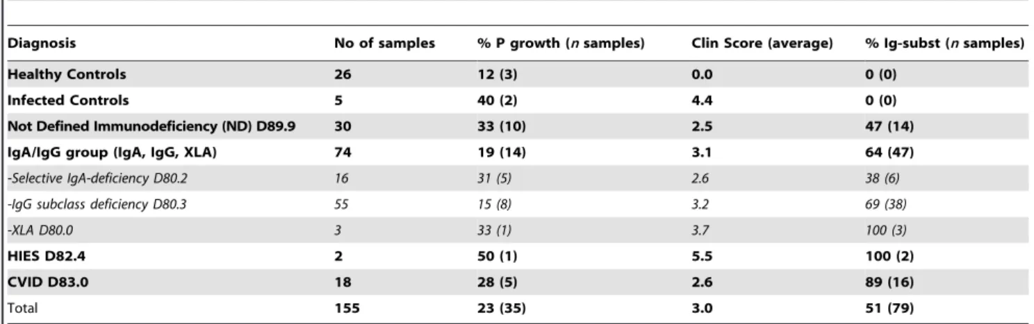
Clinical characteristics of patients and healthy controls A total of 155 samples were collected from patients with various PID diagnoses and healthy controls (Table 1). All patients submitted a clinical scoring card (see Methods for details) before sample collection. No significant differences were found in clinical score between the different groups (p = 0.23, Kruskal-Wallis test). Healthy individuals with an ongoing infection exhibited a clinical score of 4.4, however, a positive bacterial culture was only detected in 40% of these patients, indicating that the sampling method used has a low specificity [24] or that also viral infections could be involved [2]. A total of 23% (range 0–50%) of all individuals in the study exhibited growth of a primary pathogen in nasal swabs, including the healthy controls with a clinical score of zero. There was no correlation between the clinical score and the presence of potentially pathogenic bacteria in the NPH culture. Out of all patients, 51% were treated with IgG-substitution, with the highest treatment fraction in XLA, HIES- and CVID patients (Table 1), which is in line with current guidelines [25]. In fact, XLA- and CVID-patients are subjected to lifelong IgG replace-ment therapy based on the fact that they have an impaired endogenous production of immunoglobulins [26]. Also patients with IgG subclass deficiencies have been shown to benefit from IgG replacement therapy [27]. Moreover, in our centre we evaluate the efficacy of IgG substitution to selected patients with frequent bacterial infections but with normal IgG-levels [28].
Table 1.Clinical characteristics of patients and controls.
Diagnosis No of samples % P growth (nsamples) Clin Score (average) % Ig-subst (nsamples)
Healthy Controls 26 12 (3) 0.0 0 (0)
Infected Controls 5 40 (2) 4.4 0 (0)
Not Defined Immunodeficiency (ND) D89.9 30 33 (10) 2.5 47 (14)
IgA/IgG group (IgA, IgG, XLA) 74 19 (14) 3.1 64 (47)
-Selective IgA-deficiency D80.2 16 31 (5) 2.6 38 (6)
-IgG subclass deficiency D80.3 55 15 (8) 3.2 69 (38)
-XLA D80.0 3 33 (1) 3.7 100 (3)
HIES D82.4 2 50 (1) 5.5 100 (2)
CVID D83.0 18 28 (5) 2.6 89 (16)
Total 155 23 (35) 3.0 51 (79)
% P growth = Percent of samples with growth of bacterial pathogens in the nasopharynx. The values in brackets designate the number of samples with growth of a pathogen.
Clin Score = clinical score from 0 (no symptoms)2to 11 (for details see the Methods section). % Ig-subst = Percent of patients that receive immunoglobulin treatment.
The bacterial composition in the nasopharynx dictates levels of AMPs in nasal fluid
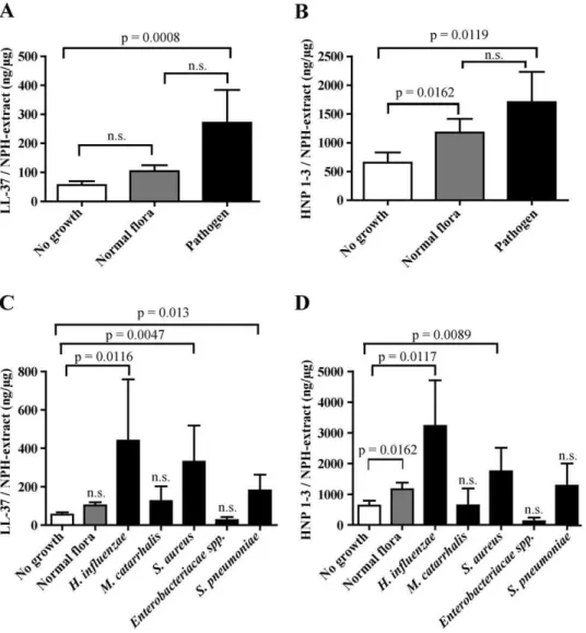
AMP levels in nasal fluid were determined by Western blot analysis and subsequent densitometric measurements. Interesting-ly, patients and healthy controls colonized with a primary pathogen, here defined asMoraxella catarrhalis, Staphylococcus aureus, Haemophilus influenzae, Streptococcus pneumoniae and Enterobacteriaceae spp., exhibited significantly increased levels of AMPs (LL-37, Fig. 1A; HNP1–3, Fig. 1B) compared to individuals with a negative bacterial culture. Furthermore, LL-37 levels in nasal fluid from individuals with normal flora were not significantly elevated compared to individuals where no bacteria were detected (Fig. 1A). However, HNP1–3 levels were significantly increased if normal flora was detected compared to samples from individuals with no growth (Fig. 1B). Bacteria associated with the highest AMP concentrations wereH. influenzae,S. aureusandS. pneumoniae(Fig. 1C and D, respectively). AMP levels associated with growth of
Enterobacteriacae spp. and M. catarrhalis were not significantly elevated (Fig. 1C and D).
Expression of AMPs in nasal fluid is the result of a dynamic response to bacteria
AMP levels were measured in nasal fluid samples from 26 healthy individuals and from 5 previously healthy individuals which were defined by the clinical score as having on-going infections, (See Materials & Methods for details and Table 1). For two healthy infected individuals (Control A, female, age 36 and Control B, male, age 38), samples were obtained on two consecutive days (Fig. 2A and B). Nasal swabs were cultured for the presence of bacteria and levels of LL-37 and HNP1–3 were determined. Control A had a negative bacterial culture day 1 (no growth, NG) and growth of normal flora (NF) day 2. This change in bacterial composition had only a moderate impact on LL-37 and HNP1–3, which is in line with previous results (Fig. 1A and B). Control B had no growth of bacteria
Figure 1. Bacterial composition and levels of AMPs in nasal fluid.AMP levels in nasal fluid samples were determined by Western blotting and subsequent densitometric measurements and the results were correlated to bacterial growth in the nasopharynx. The levels of LL-37 (A and C) and HNP1–3 (B and D) are expressed as nanogram peptide/total nasal fluid extract (mg). The group that exhibited growth of primary pathogens was
further subdivided according to bacterial species (C and D). The number of samples included (no growth, normal flora, and pathogen respectively) are n = 44, 43, and 30 (A), n = 39, 34, and 28 (B). The number of samples included (no growth, normal flora,H. influenzae,M. catarrhalis,S. aureus,
Enterobacteriacae,S. pneumoniaerespectively) are n = 44, 43, 8, 3, 11, 3, 5 (C), n = 39, 34, 7, 3, 11, 2, 5 (D). Statistical analyses were performed with Mann Whitney U-test and the standard error of the mean (SEM) was used to estimate sample variation.
day 1 (NG) while the primary pathogen S. pneumoniae(S. pn) was isolated day 2. In accordance with results presented in Figure 1A and B, this individual exhibited an increased production of both LL-37 and HNP1–3 in response to colonization with a pathogen. Notably, control A with a clinical score of 5 exhibited high levels of both LL-37 and HNP1-, despite the absence of pathogenic bacteria in the nasal swab. Thus, a possible explanation for this finding is that a viral infection caused control A’s symptoms through a local inflammation and subsequent AMP-release. In contrast, control B had an initial score of 3, which decreased to 2 on the second day of sampling, despite being culture-positive forS. pneumoniae. Therefore the rapid AMP release in control B is most likely explained by a transient pneumococcal colonization rather than a symptomatic infection. Again, these data imply that the regulation of AMPs occurs independently of clinical symptoms. When the total group of healthy and infected controls was evaluated, a significant induction of both LL-37 (n = 18, p = 0.0396, Fig. 2C) and HNP1–3 (n = 12, p = 0.0217, Fig. 2D) in response to primary pathogens was detected, supporting the results presented in Figure 2A and B. A nasal swab with growth of ‘‘normal flora’’ was not associated with a significant induction of LL-37 or HNP1–3 (Figure 2C and D) in these healthy controls.
Release of AMPs in response to pathogenic bacteria is significantly impaired in CVID and HIES patients
Next, the AMP response in the immunodeficient patient groups with regard to bacterial pathogens in the nasopharynx was studied
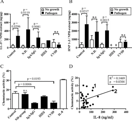
(Fig. 3A and B). A significant increase in LL-37 levels was observed in controls colonized by pathogens (same data as in fig. 2C). This increase in AMP-levels was also true for patients with no determined immune disorder (N.D. group) and for the IgA/IgG group (Fig. 3A). XLA-patients (n = 3) had an AMP-response to pathogenic bacteria in the same range as healthy controls (data not shown) and were presented together with the IgA/IgG-group in figure 3A and B. HNP1–3 levels in the control group and in IgA/ IgG patients were also significantly increased when pathogens were present, and while a similar trend was noted for the N.D. group, it did not reach statistical significance (Fig. 3B). Notably, there was no increase in neither LL-37 nor HNP1–3 in the CVID or HIES patients, despite bacterial growth (Fig. 3A and B, table 2). In fact, both HIES patients completely lacked HNP1–3 in the nasal fluid (Fig. 3B).
Chemotactic activity of nasal fluid towards healthy control neutrophils
We then wanted to investigate the reason for the dysregulated release of AMPs into nasal fluid of HIES and CVID patients. Nasal fluid derived from patients and controls was investigated for neutrophil chemotaxis using neutrophils isolated from healthy control individuals (Fig. 3C). These results revealed that nasal fluid collected from the N.D group exhibited a chemotactic activity that was indistinguishable from control individuals (Fig. 3C). In contrast, patients with IgA/IgG-deficiency and CVID showed Figure 2. AMP response in nasal fluid of healthy controls.Two previously healthy controls (A, female, age 36; B, male, age 38) were sampled for nasal fluid on two consecutive days and AMP levels were determined. The total levels of LL-37 (A) and HNP1–3 (B) are expressed as nanogram peptide/total nasal fluid extract (mg). The clinical score prior to sampling was also measured (A and B). The whole group of controls (both healthy and
significantly reduced chemotactic activity compared to controls (Fig. 3C). Unexpectedly, the two HIES-patients exhibited the same chemotactic activity as control individuals (table 2).
To further dissect the chemotactic activity of nasal fluid, the concentration of the main chemoattractant for neutrophils, IL-8, was determined by ELISA. There was a weak - although statistically significant - correlation between the chemotactic activity and IL-8 levels in nasal fluid, indicating a contribution of this cytokine to the observed chemotaxis (p = 0.0304 and R2= 0.1469) (Fig. 3D). Notably, HIES patients 1 and 2 appeared in the upper right quadrant of the figure, indicating high chemotactic activity as well as high IL-8 levels (Fig. 3D, marked with ‘‘1’’ and ‘‘2’’, respectively).
PBMCs from CVID and HIES patients release low levels of IL-17A
To investigate if AMP expression in nasal fluid could be correlated with the IL-17A response from circulating immune cells, we measured release of IL-17A in stimulated PBMCs from patients and healthy controls (Fig. 4). PBMCs were isolated from whole blood and stimulated for 5 days with Candida antigen (Fig. 4A) or Staphylococcal Enterotoxin B (SEB) (Fig. 4B). IL-17A production in PBMCs isolated from healthy controls, IgA/IgG patients as well as from patients of the N.D. group, exhibited a similar and robust increase in IL-17A after stimulation. As expected, samples from HIES patients contained very low levels Figure 3. AMP response and chemotaxis in relation to diagnosis.AMP levels were studied according to diagnosis and presence of bacterial pathogens. ‘‘No growth’’ designates negative bacterial cultures and ‘‘Pathogen’’ designates growth of a primary bacterial pathogen (A and B). The number of samples included (control, ND, IgA/IgG, HIES, and CVID respectively with and without growth) are n = 7, 3, 11, 9, 27, 14, 1,1, 6, 4 (A), n = 7, 2, 11, 10, 28, 13, 1,1, 6, 4 (B). The chemotactic capacity was determined for samples from each diagnostic group and is expressed as percent migrated neutrophils (C). The numbers of samples in C are: n = 5, 10, 44, 2, 10, 7. Error bars depict the SEM of a sample. Levels of IL-8 in nasal fluid (n = 32) were plotted against the chemotactic index and a weak positive correlation was found (p = 0.03, R2= 0.14) (D). HIES-patients 1 and 2 are marked ‘‘1’’ and ‘‘2’’. Statistical analyses in figure A, B and C were performed with Mann Whitney U-test and for figure D a correlation test was used.
doi:10.1371/journal.pone.0029316.g003
Table 2.Summary of results obtained in the study.
AMP induction
LL-37 HNP1–3 Chemotaxis PBMC (IL-17A)
Controls + + + +
ND group + + + +
IgA/IgG + + 2 +
HIES 2 2 + 2
CVID 2 2 2 +/2
A summary of the responses for the different patient groups. A ‘‘+’’-sign denotes a significant response with regards to AMP-induction, chemotaxis or IL17-A release from PBMC-cultures, respectively. A ‘‘2’’-sign represent a non-significant response. ‘‘+/2’’ means that CVID-patients respond with a significant increase in IL17-A after stimulation with SEB, but not candida. Moreover, CVID-patients responded significantly less than healthy controls (see also fig. 4A and B).
of IL-17A. PBMCs from CVID patients did also respond to antigen stimulation with release of IL-17A (n.s. forCandidaantigen and p = 0.0009 for SEB) (Fig. 4A and B). Interestingly, this release was significantly lower than the release observed for cells obtained from healthy controls (p = 0.0185 for Candidaand p = 0.0025 for SEB, table 2). Thus, these results imply that CVID-patients – similarly to HIES patients – have an impaired release of IL-17A from SEB- and Candida stimulated PBMCs. If this problem stems from aberrant recognition of antigen or intrinsic abnormalities in IL-17A production, remains to be elucidated.
The major findings of this report are summarized in table 2. A (+)-sign in columns ‘‘LL-37’’ and/or ‘‘HNP1–3’’ represent an increased release of LL-37 and/or HNP1–3 in response to pathogenic bacteria comparable to that of the control group, whereas those denoted by a (2)-sign exhibit an impaired release. Patient groups marked with (+) in the ‘‘Chemotaxis’’ column showed a chemotactic activity against healthy PMNs comparable to the control group and a (2)-sign denotes impaired or lack of chemotactic activity. A (+)-sign in the ‘‘PBMC’’ column refers to a
capacity of the PBMCs to release IL-17A in response to Candida or SEB antigens, a (2)-sign is indicative of an impaired IL-17A release, whereas +/2 is indicative of statistically significant response to SEB antigen, but not to Candida antigen (CVID-patients).
Discussion
In this study we investigated if altered levels of AMPs in nasal fluid may explain why PID patients frequently acquire RTIs. A key finding was that most patients and healthy controls responded to pathogenic bacteria with an increased AMP expression in nasal fluid. However, this response was impaired in CVID and HIES patients, which may be due to a low response from IL-17A producing cells, including CD4+ Th17-cells, NK-cells or mono-cytes [29]. However, IL-17A has been described as the ‘‘signature cytokine’’ for Th17-cells and is – together with IL-22 – implicated in mucosal immunity against bacterial and fungal infections [15]. The role of IL-17A and IL-22 producing immune cells in mucosal Figure 4. Stimulation of PBMCs with Candida antigen and SEB.PBMCs isolated from healthy controls and patients were stimulated with
Candidaantigen (A) and SEB (B), respectively. After 5 days, IL-17A levels were measured in the supernatants with ELISA. U; unstimulated, C; stimulated withCandidaantigen, and S; stimulated with SEB antigen. The number of samples are n = 15, 15, 9, 9, 17, 17, 2, 2, 8, 8 (A), n = 15, 9, 9, 6, 17, 16, 2, 1, 8, 8 (B). Bars represent the mean value. Mann Whitney U-test was used in A and B.
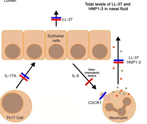
immunity is based on the assumption that these cells migrate to the mucosa during infection where they orchestrate local immune responses, such as neutrophil chemotaxis (via IL-8) or induction of AMP synthesis (direct effects of IL-17A and IL-22 on epithelial cells) [15]. Our data indicate that HIES and CVID patients exhibit a similar impaired release of AMPs into nasal fluid, but with separate immunological pathways involved (summarized in table 2 and figure 5).
The two HIES patients with STAT3 gene mutations responded with very low levels of IL-17A in antigen-stimulated PBMCs, which is in line with a previous study [19]. Nasal fluid from these two patients contained low levels of LL-37 and completely lacked HNP1–3. LL-37 is expressed by both immune and epithelial cells, whereas HNP1–3 is mainly present in neutrophils. Therefore, low or absent HNP1–3 in nasal fluid indicate an impaired recruitment of neutrophils to the nasal compartment. Indeed, HIES patients often suffer from ‘‘cold abscesses’’; bacterial skin infection without neutrophil infiltration and classical signs of inflammation [21]. In chemotaxis experiments, nasal fluid from the two HIES patients induced normal chemotaxis in neutrophils derived from healthy individuals. Indeed, IL-8 levels in nasal fluid from these two HIES-patients were similar to controls (Figure 3D, numbers ‘‘1’’ and ‘‘2’’), despite an almost complete lack of IL-17A production in PBMCs. This was unexpected but suggests that the low AMP levels in nasal fluid of HIES patients is not caused by a lack of chemotactic signals, but rather that neutrophils derived from HIES patients have impaired chemotaxis. In fact, it has been known since 1983 that neutrophils from HIES patients (Job’s syndrome) do not respond properly to chemotactic signals such as
complement (C5a) [30]. Moreover, deletion of the STAT3 gene in mice resulted in impaired neutrophil chemotaxis [31] and neutrophils from HIES patients display low levels of CXCR1, a receptor for IL-8 [32]. Thus, it is possible that the frequent mucosal infections among HIES patients is related to an impaired STAT3 mediated neutrophil migration, rather than impaired Th17 responses (Table 2 and Fig. 5).
In contrast, CVID patients were reported to have normally migrating neutrophils [30]. Nevertheless, we detected aberrant AMP responses to pathogenic bacteria in these patients. This could be explained by the significantly decreased chemotactic activity of nasal fluid from CVID patients (Fig. 3C). In addition, PBMCs derived from CVID patients responded with a weaker IL-17A response to SEB andCandidaantigen, compared to control PBMCs (Fig. 4A and B). Thus, these data are in line with the current hypothesis that lack of IL-17A leads to a lower expression of mucosal AMPs, possibly caused by an aberrant chemotactic signal (Table 2 and Fig. 5). A recent paper from Barbosa et al, describes in detail that both CVID-patients and agammaglobu-linemic patients (XLA-patients) exhibit Th17-cell disturbances, suggesting that B-cells are important for development of functional Th17-cells [33]. These data are in line with our results on CVID-patients but does not explain the mechanisms involved. Unfortu-nately, the low number of XLA-patients in our study precludes any conclusions regarding the role of Th17-cells in this disease. One possible explanation to the low IL-17A release from CVID-patients’ PBMCs is that it reflects a general T-cell defect among CVID-patients, rather than a specific Th17-cell problem. Previously, CVID patients were shown to have lower numbers
Figure 5. Impaired release of AMPs into nasal fluid of HIES- and CVID patients – A proposed model.HIES patients (blue lines) exhibit an impaired production of IL-17A and their nasal fluid contain very low levels of LL-37 and HNP1–3 despite normal chemotactic activity and IL-8 levels. This may be explained by defects in expression of the IL-8 receptor CXCR1, resulting in non-responding neutrophils. CVID patients (red lines) also exhibited a dysregulated release of AMPs, which may be explained by the low chemotactic activity of nasal fluid as well as an impaired Th17 response. However, CVID patients have, in contrast to HIES patients, normally responding neutrophils, but this may not be sufficient, since the chemotactic signal is too weak.
of CD4+T cells, from which Th17 cells differentiate [34,35]. This is in contrast to HIES-patients where a defined mutation in STAT3 is the underlying cause of the lack of Th17-cells, without general T-cell abnormalities [19]. SEB and candida induce Th17-cell differentiation by distinct pathways; SEB is a general T-Th17-cell activator by virtue of its superantigenic properties [36] whereas candida activates antigen presenting cells (APCs) via dectin-1 ligation [37]. Notably, SEB stimulation – but not candida stimulation – resulted in a small but statistically significant increase in IL-17A release from PBMCs of CVID-patients (figure 4A and B). In fact, it has been shown that CVID-patients have low numbers of dendritic cell subsets in peripheral blood [38]. Thus, it is possible that CVID-patients may have deficiencies both in the T- and dendritic cell compartments, although this speculation needs further work to become established. Interestingly, the CVID group contained one outlier with high IL-17A production. This patient was originally diagnosed with selective IgA-deficiency and has gradually declined in IgG-levels and was given the CVID diagnosis 2 years ago, which could explain the relatively high IL-17A levels (Fig. 4A and B, arrows).
We believe that the results presented here give further insight into the complex regulation of AMP production at mucosal
surfaces of the respiratory tract in general and among PID patients in particular. The data presented here could also serve as a starting point for future studies, since the detailed mechanism for impaired IL-17A release among CVID patients merits further investigations. Finally, our data show that certain primary immune disorders, such as CVID and HIES are associated with mucosal dysregu-lation of AMP release, which provide exciting new avenues for clinical trials with AMP-inducing agents, such as vitamin D.
Acknowledgments
The authors gratefully acknowledge nurses Kristina Johansson, Maria Linde´n and Susanne Hansen for their skilful work with the patients. In addition, we thank all participating patients and healthy volunteers for their contributions.
Author Contributions
Conceived and designed the experiments: AC ML RSR MO PB. Performed the experiments: AC ML RSR PB. Analyzed the data: LL SN BH-N JA BA. Contributed reagents/materials/analysis tools: LL BH-N BA. Wrote the paper: AC ML PB.
References
1. Wood PM (2010) Primary antibody deficiency syndromes. Curr Opin Hematol 17: 356–361.
2. Kainulainen L, Vuorinen T, Rantakokko-Jalava K, Osterback R, Ruuskanen O (2010) Recurrent and persistent respiratory tract viral infections in patients with primary hypogammaglobulinemia. J Allergy Clin Immunol 126: 120–126. 3. Mamishi S, Eghbali AN, Rezaei N, Abolhassani H, Parvaneh N, et al. (2010) A
single center 14 years study of infectious complications leading to hospitalization of patients with primary antibody deficiencies. Braz J Infect Dis 14: 351–355. 4. Lucas M, Lee M, Lortan J, Lopez-Granados E, Misbah S, et al. (2010) Infection
outcomes in patients with common variable immunodeficiency disorders: relationship to immunoglobulin therapy over 22 years. J Allergy Clin Immunol 125: 1354–1360 e1354.
5. Oksenhendler E, Gerard L, Fieschi C, Malphettes M, Mouillot G, et al. (2008) Infections in 252 patients with common variable immunodeficiency. Clin Infect Dis 46: 1547–1554.
6. Orange JS, Grossman WJ, Navickis RJ, Wilkes MM (2010) Impact of trough IgG on pneumonia incidence in primary immunodeficiency: A meta-analysis of clinical studies. Clin Immunol 137: 21–30.
7. Zasloff M (2002) Antimicrobial peptides of multicellular organisms. Nature 415: 389–395.
8. Agerberth B, Gudmundsson GH (2006) Host antimicrobial defence peptides in human disease. Curr Top Microbiol Immunol 306: 67–90.
9. Frohm M, Agerberth B, Ahangari G, Stahle-Backdahl M, Liden S, et al. (1997) The expression of the gene coding for the antibacterial peptide LL-37 is induced in human keratinocytes during inflammatory disorders. J Biol Chem 272: 15258–15263.
10. Ong PY, Ohtake T, Brandt C, Strickland I, Boguniewicz M, et al. (2002) Endogenous antimicrobial peptides and skin infections in atopic dermatitis. N Engl J Med 347: 1151–1160.
11. Koslowski MJ, Beisner J, Stange EF, Wehkamp J (2010) Innate antimicrobial host defense in small intestinal Crohn’s disease. Int J Med Microbiol 300: 34–40. 12. Goldman MJ, Anderson GM, Stolzenberg ED, Kari UP, Zasloff M, et al. (1997) Human beta-defensin-1 is a salt-sensitive antibiotic in lung that is inactivated in cystic fibrosis. Cell 88: 553–560.
13. Liu PT, Stenger S, Li H, Wenzel L, Tan BH, et al. (2006) Toll-like receptor triggering of a vitamin D-mediated human antimicrobial response. Science 311: 1770–1773.
14. Miossec P, Korn T, Kuchroo VK (2009) Interleukin-17 and type 17 helper T cells. N Engl J Med 361: 888–898.
15. Kolls JK, McCray PB, Jr., Chan YR (2008) Cytokine-mediated regulation of antimicrobial proteins. Nat Rev Immunol 8: 829–835.
16. Hemdan NY, Birkenmeier G, Wichmann G, Abu El-Saad AM, Krieger T, et al. (2010) Interleukin-17-producing T helper cells in autoimmunity. Autoimmun Rev 9: 785–792.
17. Lin L, Ibrahim AS, Xu X, Farber JM, Avanesian V, et al. (2009) Th1–Th17 cells mediate protective adaptive immunity against Staphylococcus aureus and Candida albicans infection in mice. PLoS Pathog 5: e1000703.
18. Saijo S, Ikeda S, Yamabe K, Kakuta S, Ishigame H, et al. (2010) Dectin-2 recognition of alpha-mannans and induction of Th17 cell differentiation is essential for host defense against Candida albicans. Immunity 32: 681–691.
19. Milner JD, Brenchley JM, Laurence A, Freeman AF, Hill BJ, et al. (2008) Impaired T(H)17 cell differentiation in subjects with autosomal dominant hyper-IgE syndrome. Nature 452: 773–776.
20. Ng WF, von Delwig A, Carmichael AJ, Arkwright PD, Abinun M, et al. (2010) Impaired T(H)17 responses in patients with chronic mucocutaneous candidiasis with and without autoimmune polyendocrinopathy-candidiasis-ectodermal dystrophy. J Allergy Clin Immunol 126: 1006–1015, 1015 e1001–1004. 21. Minegishi Y (2009) Hyper-IgE syndrome. Curr Opin Immunol 21: 487–492. 22. Cederlund A, Agerberth B, Bergman P (2010) Specificity in killing pathogens is
mediated by distinct repertoires of human neutrophil peptides. J Innate Immun 2: 508–521.
23. Yoshio H, Tollin M, Gudmundsson GH, Lagercrantz H, Jornvall H, et al. (2003) Antimicrobial polypeptides of human vernix caseosa and amniotic fluid: implications for newborn innate defense. Pediatr Res 53: 211–216.
24. Saukkoriipi A, Leskela K, Herva E, Leinonen M (2004) Streptococcus pneumoniae in nasopharyngeal secretions of healthy children: comparison of real-time PCR and culture from STGG-transport medium. Mol Cell Probes 18: 147–153.
25. Maarschalk-Ellerbroek LJ, Hoepelman IM, Ellerbroek PM (2011) Immuno-globulin treatment in primary antibody deficiency. Int J Antimicrob Agents 37: 396–404.
26. Orange JS, Hossny EM, Weiler CR, Ballow M, Berger M, et al. (2006) Use of intravenous immunoglobulin in human disease: a review of evidence by members of the Primary Immunodeficiency Committee of the American Academy of Allergy, Asthma and Immunology. J Allergy Clin Immunol 117: S525–553.
27. Olinder-Nielsen AM, Granert C, Forsberg P, Friman V, Vietorisz A, et al. (2007) Immunoglobulin prophylaxis in 350 adults with IgG subclass deficiency and recurrent respiratory tract infections: a long-term follow-up. Scand J Infect Dis 39: 44–50.
28. Berger M (2008) Principles of and advances in immunoglobulin replacement therapy for primary immunodeficiency. Immunol Allergy Clin North Am 28: 413–437, x.
29. McAleer JP, Kolls JK (2011) Mechanisms controlling Th17 cytokine expression and host defense. J Leukoc Biol 90: 263–270.
30. Van Epps DE, El-Naggar A, Ochs HD (1983) Abnormalities of lymphocyte locomotion in immunodeficiency disease. Clin Exp Immunol 53: 679–688. 31. Nguyen-Jackson H, Panopoulos AD, Zhang H, Li HS, Watowich SS (2010)
STAT3 controls the neutrophil migratory response to CXCR2 ligands by direct activation of G-CSF-induced CXCR2 expression and via modulation of CXCR2 signal transduction. Blood 115: 3354–3363.
32. Mintz R, Garty BZ, Meshel T, Marcus N, Katanov C, et al. (2010) Reduced expression of chemoattractant receptors by polymorphonuclear leukocytes in Hyper IgE Syndrome patients. Immunol Lett 130: 97–106.
33. Barbosa RR, Silva SP, Silva SL, Melo AC, Pedro E, et al. (2011) Primary B-cell deficiencies reveal a link between human IL-17-producing CD4 T-cell homeostasis and B-cell differentiation. PLoS One 6: e22848.
35. van de Ven AA, van de Corput L, van Tilburg CM, Tesselaar K, van Gent R, et al. (2010) Lymphocyte characteristics in children with common variable immunodeficiency. Clin Immunol 135: 63–71.
36. Dinges MM, Orwin PM, Schlievert PM (2000) Exotoxins of Staphylococcus aureus. Clin Microbiol Rev 13: 16–34, table of contents.
37. Cheng SC, van de Veerdonk FL, Lenardon M, Stoffels M, Plantinga T, et al. (2011) The dectin-1/inflammasome pathway is responsible for the induction of
protective T-helper 17 responses that discriminate between yeasts and hyphae of Candida albicans. J Leukoc Biol 90: 357–366.