Extracellular DNase (Spd-3) by Rgg in
Streptococcus
pyogenes
Srivishnupriya Anbalagan, Michael S. Chaussee*
Division of Basic Biomedical Sciences, The Sanford School of Medicine of the University of South Dakota, Vermillion, South Dakota, United States of America
Abstract
TheStreptococcus pyogenestranscriptional regulator Rgg controls the expression of virulence-associated genes encoded both within the core genome and within horizontally transmissible DNA such as temperate bacteriophage. Previously, we showed that Rgg binds to the non-coding DNA upstream of the bacteriophage gene encoding an extracellular DNase Spd-3. In the current study, we further characterized Rgg-mediated regulation ofspd-3expression. Twospd-3transcripts were identified by northern blotting. The 59ends were 27 and 594 nucleotides upstream of the start codon as determined with primer extension analysis and 59RACE (rapid amplification of c-DNA ends), respectively. Results obtained with gel shift assays showed that purified Rgg bound specifically to non-coding DNA containing the promoters of both transcripts. Transcriptional fusion analyses confirmed the presence of Rgg-repressible promoters within these DNA regions. In addition, repression was associated with direct DNA binding by Rgg as determined with chromatin immunoprecipitation (ChIP) coupled with quantitative PCR (qPCR). The results show that the chromosomally encoded transcriptional regulator, Rgg, directly represses both bacteriophage promoters controlling the expression of Spd-3. The results provide new information regarding the regulation of prophage encoded virulence factors ofS. pyogenesand highlight the complex evolutionary history ofS. pyogenesand temperate bacteriophage.
Citation:Anbalagan S, Chaussee MS (2013) Transcriptional Regulation of a Bacteriophage Encoded Extracellular DNase (Spd-3) by Rgg inStreptococcus pyogenes. PLoS ONE 8(4): e61312. doi:10.1371/journal.pone.0061312
Editor:Indranil Biswas, University of Kansas Medical Center, United States of America
ReceivedJanuary 2, 2013;AcceptedMarch 7, 2013;PublishedApril 17, 2013
Copyright:ß2013 Anbalagan, Chaussee. This is an open-access article distributed under the terms of the Creative Commons Attribution License, which permits unrestricted use, distribution, and reproduction in any medium, provided the original author and source are credited.
Funding:Funding was provided by the Sanford School of Medicine of the University of South Dakota and by National Institutes of Health grant 2 P20 RR016479 from the INBRE Program of the National Center for Research Resources. The funders had no role in study design, data collection and analysis, decision to publish, or preparation of the manuscript.
Competing Interests:Michael S. Chaussee serves as a PLOS ONE Editorial Board member. This does not alter the authors’ adherence to all the PLOS ONE policies on sharing data and materials.
* E-mail: [email protected]
Introduction
Streptococcus pyogenes causes several human diseases ranging in severity from self-limiting pharyngitis to life-threatening necrotiz-ing fasciitis and streptococcal toxic shock syndrome [1]. The virulence of the pathogen varies temporally over the course of decades due to changes in both the pathogen and human immunity [2,3]. The determination of the genome sequences of several isolates revealed a theoretically limitless pan-genome, or mobilome, comprised of mobile genetic elements (MGE) including bacteriophages, integrative conjugative elements, transposons, and insertion sequences [3–5]. MGEs significantly alter the composi-tion of the chromosome. For example, between two and eight complete or partial bacteriophage genomes are present in the genomes ofS. pyogenesand bacteriophage DNA can account for up to 12% of the chromosome [6,7]. Often, the bacteriophage encode virulence factors including superantigens [8] and extracellular nucleases [9,10], which profoundly influence interactions between the pathogen and its human host. Thus, chromosomal heteroge-neity, including variation in the number and types of prophage within a chromosome, is responsible for much of the genetic diversity observed among clinical isolates and contributes to the clinical and temporal variation in the outcome of human colonization withS. pyogenes [7,11–13].
S. pyogenescan produce up to four extracellular DNases [14,15]. One (MF-1/DNaseB) is chromosomally encoded and is adjacent to rgg (also known as ropB [16]), which encodes a global transcriptional regulator [17]. The remainder are encoded by prophage. Therefore, the number of extracellular DNases potentially produced by an isolate varies depending on the prophage content of the chromosome.
models of invasive infection [20] and in a cynomolgus macaque model of pharyngitis [21].
The serotype M49 strain NZ131 possesses three prophages [22], including one consisting of only 16 kb that presumably has decayed. The remaining two prophages, NZ131.2 and NZ131.3, are 37,895 and 47,501 bp, respectively. NZ131.2 encodes a super-antigen known as streptococcal pyrogenic exotoxin H (SpeH; [23]) and NZ131.3 encodes an extracellular nuclease known as Spd-3. Thus, strain NZ131 has two extracellular nucleases, the chromo-somally encoded SdaB (MF-1) and the prophage encoded Spd-3. Inactivation of the gene encoding the transcriptional regulator Rgg increased expression of both SdaB (Spy49_1692c; Mf-1) and Spd-3 (Spy49_1455) in the post-exponential phase of growth [17]. Subsequently, we found that Rgg binds to non-coding prophage DNA upstream ofspd-3[24]. The purpose of the current study was to characterize further the role of Rgg in the regulation of the prophage encoded DNase Spd-3.
Results
Identification of spd-3Transcripts
As an initial step to characterize the regulation of spd-3
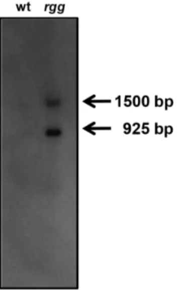
expression, northern blotting was done using RNA isolated during the post-exponential phase of growth from both the wild-typeS. pyogenesstrain NZ131 and anrggmutant. Two distinct transcripts were detected and both were more abundant in the mutant strain compared to the wild-type strain (Fig. 1), which was consistent with our previous finding that Rgg repressesspd-3expression [17]. The more abundant transcript was approximately 925 bp in length and accounted for 65% of the transcript signal, as determined by densitometry.
Mapping thespd-3 Transcriptional Start Sites
Primer extension analysis was used to determine the 59termini of the two transcripts. Extension with primer spd3PEc_96 (Table 1) showed a transcript that originated 27 bp upstream of the predictedspd-3 open reading frame (ORF) (Fig. 2). This origin, coupled with a putative transcriptional terminator 91 bp
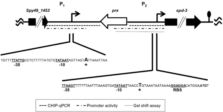
down-stream of the spd-3ORF, predicted a 919 bp transcript, which corresponded to the more abundant approximately 925 bp transcript detected by northern blotting (Fig. 1). A variety of primers were used in attempts to identify the start site of the longer transcript by using primer extension; however, we were unable to do so, possibly due to secondary structure formed within the large untranslated region. As an alternative approach, 59 RACE was used and the results showed that the 59 terminus was 594 bp upstream of thespd-3 start codon (Fig 3). The results predicted a 1,487 bp transcript, which also correlated with the size of the larger transcript identified with northern blotting (Fig. 1). In addition, the start of transcription coincided with the non-coding DNA region previously shown to be bound by Rggin vivoduring the exponential phase of growth [24]. Analyses of the DNA proximal to the transcriptional start sites revealed the presence of putative -10 and -35 RNA polymerase binding sites (Fig. 3). The two transcriptional start sites were designated P1and P2(Fig. 3).
Rgg Binds Specifically to DNA Containing the P1 and P2 Promoters
ChIP coupled with DNA genechips (ChIP-chip) and gel-shift assays previously showed that Rgg binds to P1[25]; however, we
did not previously detect binding to P2. Therefore, we re-analyzed
Rgg binding to both sites by using the more sensitive ChIP-qPCR procedure. As expected, DNA containing P1 was enriched by
approximately 9-fold in strain SA5, which encodes an Rgg-Myc fusion protein used to facilitate immunoprecipitation, compared to the control strain (Fig. 4). In addition, the P2region was enriched
nearly 75-fold (Fig. 4). The results show that Rgg binds to both the P1and P2DNA (Fig. 4). Binding near P2 was further evaluated
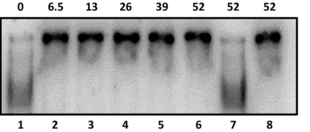
with gel-shift assays and the results confirmed that Rgg specifically binds to DNA containing P2(Fig. 5).
Rgg Directly Regulatesspd-3Expression
To characterize the two spd-3 promoters further, prophage DNA regions bound by Rgg (P1 and P2) were cloned adjacent to a promoterless firefly luciferase (luc) gene present in the shuttle plasmid pKSM720 [26]. The recombinant plasmids were used to transform both the wild-type and the rgg mutant strains to determine if expression from P1 and P2 was regulated by Rgg. The use of the plasmid based reporter system also allowed us to measure promoter activity apart from the prophage, thus avoiding confounding factors potentially associated with prophage in-duction, such as variation in gene copy number. Promoter activity was detected with P2DNA and the presence of Rgg in the
wild-type strain decreased transcription, consistent with other data indicating that Rgg represses spd-3 transcription (Table 2). Similarly, a fragment containing both spd-3 promoters (P1 and P2), showed significantly more activity compared to the fragment containing only P2 (Table 2). Again, more promoter activity was detected in thergg mutant strain, consistent with Rgg-dependent repression ofspd-3expression by direct binding to the promoter regions (Table 2).
Rgg Acts incisto regulate spd-3Promoters
Finally, we used the transcriptional reporter system and ChiP-qPCR to determine if Rgg bound incisto regulate expression of thelucgene. One PCR primer was specific tolucand a second was upstream of P1. The results showed that Rgg bound directly to the
plasmid DNA to repress expression ofluc(Fig. 6).
Figure 1. Detection ofspd-3transcripts.Northern blotting using an
spd-3specific probe and RNA isolated from the wild type (wt) and the
rggmutant (rgg) strains showed two transcripts in the sample obtained from the mutant strain. The approximate sizes were determined by using an RNA ladder (not shown).
Discussion
Temperate bacteriophage can ferry virulence-associated genes encoding a variety of toxigenic and enzymatic exoproteins among bacteria. InS. pyogenes, prophages encode superantigens, which are responsible for toxic shock syndrome; extracellular DNases, which assist in the pathogen’s escape from the innate immune response; and surface exposed proteins, which have not been characterized. Despite the importance of the gene products to human diseases caused by S. pyogenes, comparatively little is known about the regulation of their expression. Here, we examined the role of Rgg in the repression of the bacteriophage encoded extracellular DNase Spd-3. Twospd-3 transcripts were identified and the 59
termini determined. The abundance of both transcripts was elevated in an rgg mutant strain and Rgg bound to DNA containing the promoters of both transcripts, indicating that Rgg binding represses expression. To analyze regulation outside the context of the prophage, which can excise from the chromosome and thereby increasespd-3copy number, a plasmid-based reporter system was used to measurespd-3promoter activity. The results confirmed that Rgg repressed expression from both promoters. Finally we showed with ChIP-qPCR that Rgg bound to DNA incis
to effect transcription of the reporter gene. The results show that the chromosomally encoded regulator Rgg represses expression of the prophage-encoded virulence gene spd-3expression by direct binding to prophage promoters.
Rgg regulates dozens of genes in strain NZ131 in a growth-phase associated manner, including many known to contribute to virulence [16,27,28]. It does so, in part, by changing its DNA binding specificity in response to glycolytic flux via direct protein-protein interaction with LacD.1 [29]. In addition, Rgg binds to hydrophobic peptides, which modulates regulatory activity [30,31]. The genes controlled by Rgg are functionally diverse and include both cell-associated proteins and soluble exoproteins Table 1.Oligonucleotides.
Primer Sequencea(59–39) Reference or source
spd3fwd GCCAGACCCTTGCTGCTAATCCA 24
spd3rev GGTGCCTGTAAAATACGAATAAATAAGT 24
spd3fwd1 GGCGTAGCATTTAAATAAACGGAA This study
spd3rev1 GGCAAGGAGGGTAAAAATGCTAAC This study
pspd3_IRfwd GCGGATCCGTCGGACTAGTCTATGACAAA 24
pspd3_IRrev GCCTCGAGATCCATGTCCTCCTTTTATTATTTAC 24
spd3_237fwd TTTTACCCTCCTACTTATTTATTCG This study
spd3_300_BamHIfwd GCGGATCCCGGAATTAATTAAAATATTTTTGTCC 24
spd3_378_XhoIrev GCCTCGAGAAGTAGCGGGAGAAGTGCTAATGG This study
groEL2fwd GCTACTCGACGTAACATTGTG 24
groEL2rev GGAGCCTTCGTACCCAGCAT 24
spd3_cfwd GCGGATCCATGTCTAAATCAAATCGTCGTA This study
spd3_crev GCCTCGAGTTCGGTTTCTAAATTACTATCTTC This study
spd3_PEc_96 TCTGGCTGCCGTAACAGTACTTGTG This study
spd3prx36 GAAGTAGCGGGAGAAGTGCTAATGG This study
AAP-G GGCCACGCGTCGACTAGTACGGGGGGGGGGG This study
LucRev1 GCCAAGCTGGAATTCGAGCTCCCAT This study
Nested prx GGACAAAAATATTTTAATTAATTCCG This study
AUAP GGCCACGCGTCGACTAGTAC This study
a
Underlined nucleotides are restriction sites incorporated into primer. doi:10.1371/journal.pone.0061312.t001
associated with virulence [17,25]. For example, Rgg represses expression of the cell wall-associated antiphagocytic M protein in the exponential phase of growth and activates expression of a secreted cysteine protease SpeB in the post-exponential phase [16,27,32]. The results of the current study, together with results from previous studies, show that Rgg coordinates the expression of both prophage and chromosomally-encoded virulence genes in response to environmental cues. Consistent with these findings, Rgg contributes to the outcome ofS. pyogenesinfection as assessed with animal models and as observed in human epidemics of invasive disease [33] [34].
Excision of prophage from the chromosome is associated with increased gene copy number and often with simultaneous expression of prophage-encoded virulence genes. In some instances the repressor of the lytic phase also directly represses expression the prophage-encoded virulence genes. For example, expression of the prophage-encoded Shiga toxin (StxAB) in
Escherichia coliis primarily controlled by a prophage repressor that also controls lysogeny [35,36]; thusstxABexpression is dependent on prophage induction. In contrast, expression of the prophage-encoded cholera toxin (Ctx) is controlled by several chromoso-mally encoded regulators and is not dependent on induction of the
Figure 3. Schematic location of the twospd-3promoters.Transcriptional start sites are indicted by asterisks and bold face. The predicted -10 and -35 RNA polymerase binding sites of each promoter are indicated with bold type and underlined. A putative ribosome binding sites (RBS) associated with P2is similarly indicated. The Spd-3 start codon is shown in italics and bold type. Dotted lines indicate the target DNAs used with ChIP-qPCR, gel-shft assays, and transcriptional fusion assays. The lollipop symbol indicates the location of a putative transcriptional terminator. doi:10.1371/journal.pone.0061312.g003
encoding prophage [37]. Thus two paradigms have emerged from the study of the regulation of prophage encoded virulence factors. InS. pyogenes, expression of prophage encoded virulence genes, including those encoding superantigens, has been associated with the induction of prophage [4,8,9]; however, induction does not appear to be necessary for expression [4]. Moreover, expression of the phage-encoded extracellular DNase Sdn (SpyM3_1409) decreases following mitomycin C induction of prophage, despite an increase insdncopy number [4]. In this study, we showed that Rgg directly represses the promoters controllingspd-3expression, even in the absence of prophage induction, as determined by using a plasmid based reporter system. Although our investigation focused on the regulation of spd-3 expression, the results are consistent with a model in which the expression of prophage encoded virulence factors in S. pyogenes is not dependent on induction the lytic cycle and is controlled, at least in part, by chromosomally encoded regulatory loci. Given the relevance of prophage encoded superantigens and secreted DNases to human disease, additional investigation into the regulation of other prophage-encoded virulence factors is warranted.
Phenotypic variation is a hallmark of many pathogens, in-cluding S. pyogenes, and can result in heterogeneous clinical outcomes of infection. We previously showed that Rgg binds to non-coding DNA upstream of a prophage integrase/excisionase (Spy49_0746c) to repress expression [17,24]. Increased expression of the integrase/exicisionase in anrggmutant strain was associated with a decrease in the frequency of prophage excision from the chromosome [17]. The current study extends the idea that Rgg regulates specific prophage encoded genes, which alters the
phenotype of the pathogen. Given the tremendous variation in the number and composition of bacteriophage among different isolates ofS. pyogenes[7], we speculate that the direct regulation of MGEs by Rgg contributes, directly or indirectly, to the variation in the Rgg regulon observed among various isolates ofS. pyogenes[38] and potentially the clinical outcome of human infection.
Materials and Methods
Bacterial Strains and Culture Conditions
S. pyogenes strain NZ131 (serotype M49) was isolated from a patient with acute post-streptococcal glomerulonephritis (Table 3) [22]. NZ131 and its genetic derivatives includingrgg- (32) and SA5 and have been previously described (24).S. pyogenesstrains were grown with Todd-Hewitt broth (Becton Dickinson, Spark, MD) containing 0.2% (wt/vol) yeast extract at 37uC in a 5% CO2
atmosphere without agitation.E. coliDH5awas grown with
Luria-Bertani medium at 37uC with agitation. When necessary, antibiotics was added to the growth media at the following concentrations: carbenicillin at 100mg/ml forE. coli; spectinomy-cin at 100mg/ml for bothE. coliandS. pyogenes; erythromycin at 2.5mg/ml forS. pyogenes; kanamycin at 50mg/ml forE. coliand 500mg/ml forS. pyogenes.
Figure 5. Rgg binds specifically to P2.Rgg binding to the non-coding DNA upstream ofspd-3containing P2 was assessed by incubating with 0, 6.5, 26, 39 and 52 pmoles (Lanes 1–6) of purified Rgg with radiolabeled target DNA. Lane 7) Specific un-labeled competing DNA was added to the reaction. Lane 8) Labeled non-specific DNA (groEL)was added to the reaction.
doi:10.1371/journal.pone.0061312.g005
Table 2.Bothspd-3promoters (P1 and P2) are repressed by Rgg.
Promoter [Luciferase] units
wild-type rggmutant
P2a
145 (69) 35,857 (20,462)
P1 & P2a 1,486 (855) 335,979 (61,184)
aA 457 bp fragment containing the P2 promoter or a 761 bp fragment
containing both the P1 and P2 promoters was cloned adjacent to the luciferase reporter gene. The mean (standard deviation) from independent experiments is shown.
doi:10.1371/journal.pone.0061312.t002
Figure 6. Figure 6. Rgg binds to episomal P2 containing DNA. The amount of the non-coding DNA upstream ofspd-3cloned into a transcriptional reporter fusion plasmid was measured by quantitative PCR, as a negative control,groELin ChIP samples obtained from thergg
mutant and strain SA6. Experiments were conducted at least three times, and the means and standard deviations are shown.
DNA Manipulation
To isolate plasmid DNA fromE. coli, either the QIAprep Spin Miniprep Kit (Qiagen, Valencia, CA) or Maxi/Midi prep purification systems (Qiagen) was used. DNA fragments were PCR amplified with GoTaq DNA polymerase (Promega, Madi-son, WI) and the amplified DNA was separated by using agarose gel electrophoresis and purified using the SpinPrep Gel DNA kit (EMD Milliopore, Darmstadt, Germany). DNA sequencing to confirm various constructs was done at Iowa State University (Ames, IA).
Promoter Activity Assays
A shuttle plasmid (pKSM720) containing luc [26], which encodes firefly luciferase, was used to construct transcriptional fusions. Two DNA regions upstream ofspd-3(2761 to2382 and
21 to 2237) were amplified using pspd3_IRfwd and spd3_378_XhoIrev; spd3_237fwd and pspd3_IRrev primer com-binations, respectively (Table 1). The 379 bp and 237 bp DNA fragments were gel purified, digested withBamHI and XhoI, and cloned 59tolucbetweenBglII andXhoI of pKSM720 (Table 3) to create pSA27 and pSA28, respectively (Table 3). NZ131 was transformed with pKSM720, pSA27, and pSA28 by electropora-tion to create wt::luc, wt::Pspd3-379luc, and wt::Pspd3-457luc, respectively (Table 3). Similarly, the NZ131 rgg mutant was transformed with pKSM720, pSA27, and pSA28 to creatergg-::luc,
rgg-::Pspd3-379luc, and rgg-::Pspd3-457luc, respectively (Table 3). Construction of the recombinant plasmids was confirmed by PCR.
The S. pyogenes strains containing the transcriptional fusion plasmids were grown with THY broth to the exponential phase of growth (A600,0.35) and luciferase activity was measured
according to manufacturer’s instructions (Promega).
Electrophoretic Mobility Shift Assays (EMSA)
An Rgg-maltose binding protein fusion protein (Rgg-MBP) was expressed inE. coliand purified as previously described [24]. Non-coding DNA upstream ofspd-3(corresponding to nucleotides21 to2457 bp relative to the spd-3start codon) was amplified by using primers spd3fwd1 and spd3rev1 primers and NZ131 genomic DNA as a template (Table 1). The fragment was isolated by using agarose gel electrophoresis, purified, and cloned into pGEM-T-easy (Promega, Madison, WI) to create pSA29. As a non-specific control of DNA binding, a similarly sized fragment was similarly prepared by using groEL2fwd and groEL2rev primers, which are specific to the groEL ORF (Table 1). The fragments were excised from pGEM-T easy, gel purified, depho-sphorylated, and end labeled with [c32P] ATP using
poly-nucleotide kinase. Different amounts of Rgg-MBP were incubated in 25ml of binding buffer (25 mM Tris-Cl, pH 7.5, 0.1 mM EDTA, 75 mM NaCl, 1 mM dithiothreitol, 10% glycerol, and 0.5mg/ml of calf thymus DNA) at room temperature for 20 min. Competition experiments were conducted by including unlabeled DNA prior to protein addition. The reaction mixtures were separated with a 6% nondenaturing polyacrylamide gel. The gels were dried, exposed to an Amersham Biosciences storage Table 3.Bacterial strains and plasmids.
Strain or plasmid Description Source or reference
Strains
E. coli
DH5a hsdR17 recA1 gyrA endA1 relA1 Invitrogen
S. pyogenes
NZ131 M49 serotype D.R. Martin, New Zealand
rgg- NZ131rggmutant, EmR 32
SA5 NZ131rgg-complemented with pSA3, EmR, KanR 24
SA6 SA5transformed with pSA12 This study
wt::luc NZ131 transformed with pKSM720, SpecR 24
wt::Pspd3-luc NZ131 transformed with pSA6, SpecR 24
wt::Pspd3-379luc NZ131 transformed with pSA27, SpecR This study
wt::Pspd3-457luc NZ131 transformed with pSA28, SpecR This study
rgg-::luc NZ131rggmutant transformed with pKSM720, SpecR 24
rgg-:: Pspd3-luc NZ131rggmutant transformed with pSA12, SpecR 24
rgg-::Pspd3-379luc NZ131 transformed with pSA27, SpecR This study
rgg-::Pspd3-457luc NZ131 transformed with pSA28, SpecR This study
Plasmids
pGEM-T-Easy
pKSM720 GAS replicating plasmid with firefly luciferase and RBS, SpecR 26
pSA12 Non-coding region upstream ofspd-3was cloned into pKSM720, SpecR 24
pSA27 Non-coding region between -761 and -382 upstream ofspd-3was cloned
into pKSM720, SpecR This study
pSA28 Non-coding region between -1 and -457 upstream ofspd-3was cloned into pKSM720, SpecR
This study
pSA29 Non-coding region between -225 and -472 upstream ofspd-3was cloned
into pGEM-T-easy vector, AmpR This study
phosphor screen and imaged with a Typhoon 9400 instrument (GE Healthcare, Piscataway, N.J.).
RNA Isolation and Northern Blotting
Overnight cultures of NZ131 and the rgg mutant were inoculated into 40-ml THY broth to anA600of 0.08. The cultures
were grown to the post-exponential phase of growth (A600,0.6).
Total RNA was isolated as described previously [17]. The concentration and quality of RNA was assessed with an Agilent 2100 Bioanalyzer (Agilent, Palo Alto, CA) using an RNA 6000 Nano LabChip kit (Agilent). Fifteen micrograms of total RNA from each strain was separated with a 1.5% agarose-0.66 M formaldehyde gel in morpholinepropanesulfonic acid (MOPS) running buffer (20 mM MOPS, 10 mM sodium acetate, 2 mM EDTA; pH 7.0). RNA was blotted onto Hybond N+
membranes (GE Healthcare Biosciences, Pittsburgh, PA.) with the Turbo-blotter alkaline transfer system (Schleicher & Schuell, Keene, N.H.), according to the manufacturer’s instructions. RNA was fixed to the membrane by baking at 80uC for 30 min. Thespd-3
coding DNA was amplified using spd3_cfwd and spd3_crev primers (Table 1), separated by agarose gel electrophoresis, the fragment excised from the gel, and purified as described above. Using the purifiedspd-3coding DNA as a template, [a-32P]dCTP
radiolabelled probes were synthesized by the random-primed method (Ready-To-Go Labeling Kit; Pharmacia). Membranes were hybridized under aqueous conditions at 65uC with the radiolabelled probes. The blots were washed and exposed to an Amersham Biosciences storage phosphor screen and imaged with a Typhoon instrument.
Primer Extension Analysis
The 59end of thespd-3transcript was determined with 15mg of RNA isolated from NZ131 and the rgg mutant using an AMV reverse transcriptase primer extension kit (Promega) according to manufacturer’s instructions. The spd3PEc_96 primer (Table 1) was end-labeled with [c32P] ATP using polynucleotide kinase and the extension products were separated with a 6% polyacrylamide-urea sequencing gel. The 59end was mapped by comparison to a sequencing reaction generated with a SequiTherm EXCELTMII DNA Sequencing Kit (Epicentre Biotechnologes, Madison, Wisconsin) with the end-labeled spd3PEc_96 and a DNA template that contained the entire non-coding DNA upstream ofspd-3.
59Rapid Amplification of c-DNA Ends (59-RACE)
The 59end of the largerspd-3transcript was determined using the 59 RACE System for Rapid Amplification of cDNA Ends, Version 2.0 (Invitrogen, Carlsbad, California) according to
manufacturer’s instructions. First strand cDNA was synthesized using 15mg of total RNA, the gene specific primer spd3prx36, and Superscript III Reverse Transcriptase (Invitrogen). After the first strand cDNA synthesis, the mRNA template was removed with the RNase cocktail (Ambion, Austin, Texas). Single stranded cDNA was purified using a DNA Clean and Concentrator-5 kit (Zymo Research Irvine, California). A homopolymeric tail was added to the 39-end of the cDNA using recombinant TdT (Invitrogen) and dCTP. Five mL of dC-tailed single stranded c-DNA was amplified using abridged anchor primer (AAP-G) and nested prx primers (Table 1). The amplified product was diluted 100-fold and 5mL was used in a second-round of PCR amplification with abridged universal amplification primer (AUAP) and nested prx primers. The PCR products were separated by electrophoresis on a 1% agarose gel, purified, and sequenced at the Iowa State Sequencing facility.
ChIP-qPCR
A ChIP assay was performed using the experimental protocol previously described [24]. Briefly, cultures ofS. pyogenesstrainsrgg
-[32] and SA5 (encoding an Rgg-myc fusion protein [24]) were grown to anA600of approximately 0.6, which corresponds to the
post-exponential phase of growth. The cultures were treated with 1% formaldehyde (w/v). DNA bound to Rgg-Myc was immuno-precipitated with a monoclonal antibody to Myc (Invitrogen, Carlsbad, CA). Control samples were similarly prepared from an NZ131rggmutant strain. Immunoprecipitated DNA was purified and specific regions quantitated with PCR using ABsolute SYBR Green ROX Mix (ABgene House, Surrey, United Kingdom). Primer pairs pspd3_IRfwd, spd3_378_XhoIrev and Spd3_237fwd, pspd3_IRrev were used to quantitate DNA containing P1 and P2, respectively (Table 2). Primers pspd3_IRfwd and LucRev1 were used to measure DNA cloned upstream of thelucgene (Table 2). For the control region, primers groEL2fwd and groEL2rev were used (Table 2). Enrichment was normalized to the amount of non-specific groEL DNA in pre-cipitated samples, as previously described [24].
Acknowledgments
We thank Dr. Adhar Manna for technical assistance.
Author Contributions
Conceived and designed the experiments: MSC SA. Performed the experiments: SA. Analyzed the data: MSC SA. Contributed reagents/ materials/analysis tools: SA MSC. Wrote the paper: SA MSC.
References
1. Cunningham MW (2000) Pathogenesis of group A streptococcal infections. Clin Microbiol Rev 13: 470–511.
2. Stevens DL (1992) Invasive group A streptococcus infections. Clin Infect Dis 14: 2–11.
3. Musser JM, Shelburne SA, 3rd (2009) A decade of molecular pathogenomic analysis of group A Streptococcus. J Clin Invest 119: 2455–2463.
4. Banks DJ, Lei B, Musser JM (2003) Prophage induction and expression of prophage-encoded virulence factors in group A Streptococcus serotype M3 strain MGAS315. Infect Immun 71: 7079–7086.
5. Medini D, Donati C, Tettelin H, Masignani V, Rappuoli R (2005) The microbial pan-genome. Curr Opin Genet Dev 15: 589–594.
6. Ferretti JJ, Ajdic D, McShan WM (2004) Comparative genomics of streptococcal species. Indian J Med Res 119 Suppl: 1–6.
7. Banks DJ, Beres SB, Musser JM (2002) The fundamental contribution of phages to GAS evolution, genome diversification and strain emergence. Trends Microbiol 10: 515–521.
8. Zabriskie JB (1964) The Role of Temperate Bacteriophage in the Production of Erythrogenic Toxin by Group a Streptococci. J Exp Med 119: 761–780.
9. Broudy TB, Pancholi V, Fischetti VA (2002) The in vitro interaction of Streptococcus pyogenes with human pharyngeal cells induces a phage-encoded extracellular DNase. Infect Immun 70: 2805–2811.
10. Ferretti JJ, McShan WM, Ajdic D, Savic DJ, Savic G, et al. (2001) Complete genome sequence of an M1 strain of Streptococcus pyogenes. Proc Natl Acad Sci U S A 98: 4658–4663.
11. Aziz RK, Kotb M (2008) Rise and persistence of global M1T1 clone of Streptococcus pyogenes. Emerg Infect Dis 14: 1511–1517.
12. Beres SB, Carroll RK, Shea PR, Sitkiewicz I, Martinez-Gutierrez JC, et al. (2010) Molecular complexity of successive bacterial epidemics deconvoluted by comparative pathogenomics. Proc Natl Acad Sci U S A 107: 4371–4376. 13. Beres SB, Sylva GL, Barbian KD, Lei B, Hoff JS, et al. (2002) Genome sequence
of a serotype M3 strain of group A Streptococcus: phage-encoded toxins, the high-virulence phenotype, and clone emergence. Proc Natl Acad Sci U S A 99: 10078–10083.
15. Winter JE, Bernheimer AW (1964) The Deoxyribonucleases of Streptococcus Pyogens. J Biol Chem 239: 215–221.
16. Lyon WR, Gibson CM, Caparon MG (1998) A role for trigger factor and an rgg-like regulator in the transcription, secretion and processing of the cysteine proteinase of Streptococcus pyogenes. Embo J 17: 6263–6275.
17. Dmitriev AV, McDowell EJ, Kappeler KV, Chaussee MA, Rieck LD, et al. (2006) The Rgg regulator of Streptococcus pyogenes influences utilization of nonglucose carbohydrates, prophage induction, and expression of the NAD-glycohydrolase virulence operon. J Bacteriol 188: 7230–7241.
18. Buchanan JT, Simpson AJ, Aziz RK, Liu GY, Kristian SA, et al. (2006) DNase expression allows the pathogen group A Streptococcus to escape killing in neutrophil extracellular traps. Curr Biol 16: 396–400.
19. Amulic B, Hayes G (2011) Neutrophil extracellular traps. Curr Biol 21: R297– 298.
20. Uchiyama S, Andreoni F, Schuepbach RA, Nizet V, Zinkernagel AS (2012) DNase Sda1 allows invasive M1T1 Group A Streptococcus to prevent TLR9-dependent recognition. PLoS Pathog 8: e1002736.
21. Sumby P, Barbian KD, Gardner DJ, Whitney AR, Welty DM, et al. (2005) Extracellular deoxyribonuclease made by group A Streptococcus assists pathogenesis by enhancing evasion of the innate immune response. Proc Natl Acad Sci U S A 102: 1679–1684.
22. McShan WM, Ferretti JJ, Karasawa T, Suvorov AN, Lin S, et al. (2008) Genome sequence of a nephritogenic and highly transformable M49 strain of Streptococcus pyogenes. J Bacteriol 190: 7773–7785.
23. Proft T, Moffatt SL, Berkahn CJ, Fraser JD (1999) Identification and characterization of novel superantigens from Streptococcus pyogenes. J Exp Med 189: 89–102.
24. Anbalagan S, McShan WM, Dunman PM, Chaussee MS (2011) Identification of Rgg binding sites in the Streptococcus pyogenes chromosome. J Bacteriol 193: 4933–4942.
25. Anbalagan S, Dmitriev A, McShan WM, Dunman PM, Chaussee MS (2012) Growth phase-dependent modulation of Rgg binding specificity in Streptococcus pyogenes. J Bacteriol 194: 3961–3971.
26. Kinkel TL, McIver KS (2008) CcpA-mediated repression of streptolysin S expression and virulence in the group A streptococcus. Infect Immun 76: 3451– 3463.
27. Chaussee MS, Somerville GA, Reitzer L, Musser JM (2003) Rgg Coordinates Virulence Factor Synthesis and Metabolism in Streptococcus pyogenes. J Bacteriol 185: 6016–6024.
28. Chaussee MS, Watson RO, Smoot JC, Musser JM (2001) Identification of Rgg-regulated exoproteins of Streptococcus pyogenes. Infect Immun 69: 822–831. 29. Loughman JA, Caparon MG (2006) A novel adaptation of aldolase regulates
virulence in Streptococcus pyogenes. Embo J 25: 5414–5422.
30. Shelburne SA, 3rd, Olsen RJ, Makthal N, Brown NG, Sahasrabhojane P, et al. (2011) An amino-terminal signal peptide of Vfr protein negatively influences RopB-dependent SpeB expression and attenuates virulence in Streptococcus pyogenes. Mol Microbiol 82: 1481–1495.
31. Federle M (2012) Pathogenic streptococci speak, but what are they saying? Virulence 3: 92–94.
32. Chaussee MS, Ajdic D, Ferretti JJ (1999) The rgg gene of Streptococcus pyogenes NZ131 positively influences extracellular SPE B production. Infect Immun 67: 1715–1722.
33. Olsen RJ, Laucirica DR, Watkins ME, Feske ML, Garcia-Bustillos JR, et al. (2012) Polymorphisms in regulator of protease B (RopB) alter disease phenotype and strain virulence of serotype M3 group A Streptococcus. J Infect Dis 205: 1719–1729.
34. Carroll RK, Shelburne SA, 3rd, Olsen RJ, Suber B, Sahasrabhojane P, et al. (2011) Naturally occurring single amino acid replacements in a regulatory protein alter streptococcal gene expression and virulence in mice. J Clin Invest 121: 1956–1968.
35. Wagner PL, Neely MN, Zhang X, Acheson DW, Waldor MK, et al. (2001) Role for a phage promoter in Shiga toxin 2 expression from a pathogenic Escherichia coli strain. J Bacteriol 183: 2081–2085.
36. Neely MN, Friedman DI (1998) Functional and genetic analysis of regulatory regions of coliphage H-19B: location of shiga-like toxin and lysis genes suggest a role for phage functions in toxin release. Mol Microbiol 28: 1255–1267. 37. Waldor MK, Friedman DI (2005) Phage regulatory circuits and virulence gene
expression. Curr Opin Microbiol 8: 459–465.