SEBBEN AL ETAL.
18 REV ASSOC MED BRAS 2017; 63(1):18-20
IMAGE IN MEDICINE
High cervical spine spondylodiscitis management and
literature review
ANDRÉ LUIS SEBBEN1,2*, XAVIER SOLER GRAELLS1,2, MARCEL LUIZ BENATO2, PEDRO GREIN DEL SANTORO2, ÁLYNSON LAROCCA KULCHESKI2
1Orthopedics and Traumatology Service, Hospital de Clínicas, Universidade Federal do Paraná (UFPR), Curitiba, PR, Brazil 2Orthopedics and Traumatology Service, Hospital do Trabalhador, UFPR, Curitiba, PR, Brazil
S
UMMARYStudy conducted at Hospital do Trabalhador, Universidade Federal do Paraná (UFPR), Curitiba, PR, Brazil
Article received: 2/3/2016 Accepted for publication: 5/2/2016
*Correspondence: Address: Rua General Carneiro, 181
Curitiba, PR – Brazil CEP 80060-000 [email protected]
Research project approved by the Research Ethics Committee of Hospital do Trabalhador, CAAE no.: 48619315.5.0000.5225
http://dx.doi.org/10.1590/1806-9282.63.01.18
Spondylodiscitis affecting the cervical spine is the most unusual type. Disease progression can be dramatic, even causing quadriplegia and death. We present an unusual case that progressed with osteolytic lesions between C2 and C3, causing cord compression and epidural abscess. The patient was treated surgically by a double approach and improved without neurological deicits and with better inlammatory markers. We reviewed the current literature on the subject.
Keywords: spinal disease, neck pain, discitis.
I
NTRODUCTIONPyogenic spinal infections are rare and affect 1 to 7% of all cases of osteomyelitis. However, its incidence has been increasing, mainly due to the increased longevity of the population and a higher incidence of comorbidities that cause immunosuppression.1 Discitis predominantly
oc-curs in the lumbar spine, followed by the thoracic spine and, to a lesser extent, the cervical spine.2 The literature
regarding cervical spondylodiscitis is scarce. Its presenta-tion may be more dramatic and with rapid evolupresenta-tion, causing early neurological deicits. Emergency treatment is mandatory, since it can progress to fulminant sepsis and neurological complications.3
We are reporting an unusual case of cervical pyo-genic spondylodiscitis. The literature was revised in order to better understand the subject.
C
ASE REPORTMale patient, 59 years old, farmer. The initial complaint was intense neck pain for 2 months with progressive wors-ening. This was associated with constitutional symptoms including weight loss of 10 kg in 45 days, loss of appetite, fever, adynamia, and night sweats. Upon physical exami-nation, the patient was emaciated, febrile (38.3ºC), pros-trate, and presented intense pain upon anterior cervical
palpation with an antalgic posture in semilexion. Passive and active cervical mobilization was painful. Muscle strength, deep tendon relexes, and sensitivity in the limbs were preserved.
Laboratory tests on admission showed 8,200 leuko-cytes with 2% Auer rods, erythrocyte sedimentation rate (ESR) of 100 mm/h and c-reactive protein (CRP) of 35 mg/L. Blood culture from two samples showed no growth of microorganisms.
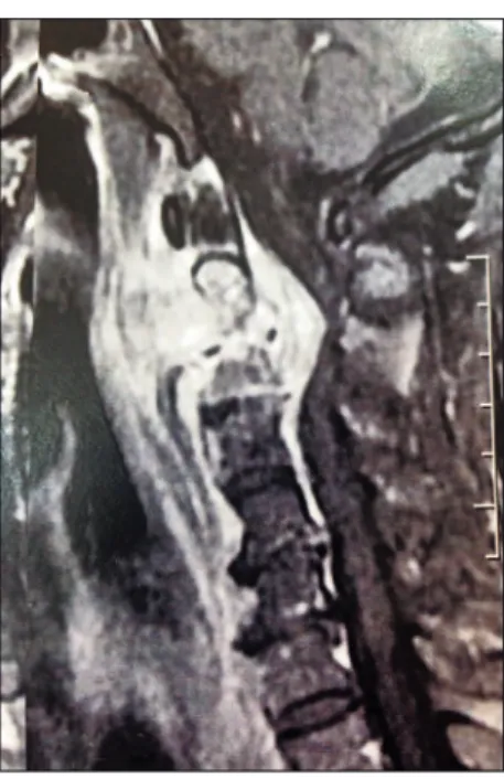
X-ray and computerized axial tomography of the cervi-cal spine showed lytic lesions between C2 and C3. The nuclear magnetic resonance imaging (NMR) showed signs suggestive of spondylodiscitis in C2-C3 associated with the presence of a massive epidural abscess compressing the ventral surface of the spinal cord, as well as involvement of paravertebral and prevertebral soft tissues (Figure 1). Bone scintigraphy ruled out an expansive tumor process and conirmed the NMR indings.
HIGHCERVICALSPINESPONDYLODISCITISMANAGEMENTANDLITERATUREREVIEW
REV ASSOC MED BRAS 2017; 63(1):18-20 19
growth of multisensitive Staphylococcus aureus and anato-mopathological examination conirmed inlammation.
In accordance with the sensitivity spectrum of the germ, oxacillin 500 mg, intravenous, every 4 hours was given for 3 weeks, followed by cefalexin 500 mg, peroral, every 6 hours for another 5 weeks, with a total of 8 weeks of antibiotic therapy.
The patient progressed satisfactorily with improvement of the pain, as assessed using a visual pain scale, which was 10 in the preoperative period and fell to 2 on the 5th day
after surgery. The Oswestry 2.0 questionnaires were also applied before and after surgery, with 49/50 points in the preoperative period, ranking as an invalid, and 4/50 in the postoperative period, showing an excellent post-surgical outcome. The patient was discharged after three weeks without complaints and with laboratory examinations that showed a decrease in inlammatory markers. Six weeks after surgery, the patient was still using a Philadelphia cervical collar, and already showed signs of osseointegration of the graft. The patient remained asymptomatic.
D
ISCUSSIONCervical spondylodiscitis is rare, given that most vertebral abscess and cases of discitis occur in the thoracic and lumbar spine. The annual incidence varies from 0.5 to 2.5 cases per 100,000 inhabitants. Spondylodiscitis is the primary mani-festation of hematogenous osteomyelitis in patients over 50 years of age, representing 3 to 5% of all cases of osteomyelitis.4
Pathogens can affect the spine by three routes: hema-togenous, external inoculation or contiguity.
The arterial hematogenous route is the predominant one, enabling the infection to be disseminated from dis-tant sites.5
Although a broad spectrum of microorganisms have been identiied (bacteria, mycobacteria, fungi and parasites), a monobacterial etiology predominates, with S. aureus be-ing the most common.6 The present case reiterates the
higher prevalence of this germ in the literature.
The main risk factors include use of intravenous drugs and comorbidities such as diabetes and terminal chronic renal failure.6 We found no risk factors in the case
re-ported, which makes it even more atypical.
As shown in the literature,5 NMR is the most sensitive
(93-96%) and speciic (92.5-97%) test for the early detection of spondylodiscitis. In most cases, it can differentiate between pyogenic infections, neoplasms, and tuberculosis.
FIGURE 1 Cervical NMR image showing signs of spondylodiscitis
in C2-C3 with spinal cord compression and epidural abscess.
FIGURE 2 Transoperative image of posterior ixation with the
iliac graft.
SEBBEN AL ETAL.
20 REV ASSOC MED BRAS 2017; 63(1):18-20
Furthermore, it can deine the paravertebral and epidural spaces better.7 This complementary examination was used
to diagnose a case that progressed slowly and was funda-mental for the diagnostic deinition, treatment and reso-lution of the case.
In order to direct the antibiotic therapy, a percutaneous biopsy, which is a safe and minimally invasive procedure, may bey performed. If the irst sample is negative, some experts recommend taking another.8 Friedman reported
50% positivity in cultures produced from percutaneous biopsies. Surgical debridement is reserved for patients who present abscesses and neural compression and need to have their spine stabilized, a fact that occurred in our case. Oth-er indications include debridement of devitalized tissue and removal of infected implant material. Speciic antibi-otics should be administered for 8 to 12 weeks after surgery, according to the results of the culture. Infections in most of the patients are resolved using this approach.9
Classically, the standard treatment was corpectomy and placement of structured grafts without the use of the implant material. Currently, most surgeons have preferred techniques that provide greater stability to the targeted site using implant material in patients treated for spondylo-discitis. There is preference for the posterior route in the cervical spine in order to avoid the main complications, which include graft migration, failure of the synthesis ma-terial and esophageal istula. Several authors have reported no complications related to the use of these implants.10
Our patient was treated according to this protocol and recovered satisfactorily.
C
ONCLUSIONAlthough cervical spondylodiscitis is a rare disease, it is a diagnosis that should not be overlooked in patients who have indolent neck pain associated with constitu-tional symptoms. Early diagnosis and initiation of ther-apy are the only means of avoiding disease progression, thus preventing patients from having sequelae that are often irreversible.
R
ESUMOEspondilodiscite da coluna cervical alta: manejo e revisão da literatura
A espondilodiscite, que acomete a coluna cervical, é a de localização mais rara. Pode ter uma evolução dramática, inclusive causando tetraplegia e óbito. Apresentamos um caso atípico que evoluiu com lesões osteolíticas entre C2 e C3, causando compressão medular e abscesso epidural. O paciente foi submetido a tratamento cirúrgico por dupla abordagem e evoluiu bem, sem déicits neurológi-cos e com melhora dos marcadores inlamatórios. Revi-samos a literatura vigente sobre o assunto.
Palavras-chave: doenças da coluna vertebral, cervical-gia, discite.
R
EFERENCES1. Kulcheski AL, Graells XS, Benato ML, Santoro PG, Sebben AL. Espondilodiscite fúngica por Candida albicans: um caso atípico e revisão da literatura. Rev Bras Ortop. 2015; 50(6):739-42.
2. Baker AS, Ojemann RG, Swartz MN, Richardson EP. Spinal epidural abscess. N Engl J Med. 1975; 293(10):463-8.
3. Schimmer RC, Jeanneret C, Nunley PD, Jeanneret B. Osteomyelitis of the cervical spine: a potentially dramatic disease. J Spinal Disord Tech. 2002; 15(2):110-7.
4. Jensen AG, Espersen F, Skinhøj P, Rosdahl VT, Frimodt-Møller N. Increasing frequency of vertebral osteomyelitis following Staphylococcus aureus bacteraemia in Denmark 1980-1990. J Infect. 1997; 34(2):113-8. 5. Cottle L, Riordan T. Infectious spondylodiscitis. J Infect. 2008; 56(6):401-2. 6. Kulowski J. Pyogenic osteomyelitis of the spine: an analysis and discussion
of 102 cases. J Bone Joint Surg. 1936; 18(2):343-64.
7. Hopkinson N, Stevenson J, Benjamin S. A case ascertainment study of septic discitis: clinical, microbiological and radiological features. QJM. 2001; 94(9):465-70.
8. Rankine JJ, Barron DA, Robinson P, Millner PA, Dickson RA. Therapeutic impact of percutaneous spinal biopsy in spinal infection. Postgrad Med J. 2004; 80(948):607-9.
9. Cebrián Parra JL, Saez-Arenillas Martín A, Urda Martinéz-Aedo AL, Soler Ivañez A, Agreda E, Lopez-Duran Stern LL. Management of infectious discitis. Outcome in one hundred and eight patients in a university hospital. Int Orthop. 2012; 36(2):239-44.