Authors
Nordeval Cavalcante
Araújo1
Lilimar da Silveira Rioja2
Maria Alice Puga
Rebelo3
1Nephrology Service of Hospital Universitário Pedro Ernesto of Universidade do Estado do Rio de Janeiro - UERJ
2Pathology Service of Hospital Universitário Pedro Ernesto da UERJ
3Medical School of UERJ
Submitted on: 8/28/2009 Accepted on: 11/27/2009
Corresponding author:
Nordeval Cavalcante Araújo Rua São Salvador, 14 / 1404 Flamengo
CEP: 22231-130 Rio de Janeiro – RJ
We declare no confl ict of interest.
A clinical predictor index for renal survival
A
BSTRACTIntroduction: A clinical index that discrimi-nates disease progression independent of his-topathologic features may be valuable in the best timing of biopsy. Objective: This study addresses the question if a clinical index ba-sed on cortical echogenicity, renal length to body height ratio (KL/H), and serum creati-nine levels predicts renal survival. Methods:
The study enrolled 154 patients. Biopsy spe-cimens were graded for chronic (glomerular obsolescence, segmental glomerular sclero-sis, tubular atrophy and interstitial fibrosis) and acute (mesangial proliferation, leucocyte permeation, crescent and fibrinoid necrosis and interstital infiltrate) index by the sum of scored lesions. A chronic clinical index was created by the sum of scored cortical echo-genicity relative to liver or spleen, creatini-ne serum levels and KL/H. The study end point was start on dialisis. Results: Higher grade of chronic clinical and biopsy indices were associated with poorer long-term renal survival. Five out of six patients with serum creatinine levels > 2.5mg/dL, highest corti-cal echogenicity and KL/H < 0.60, before biopsy, startet on dialysis and one increased creatinine levels up to 4.5 mg/dL. The nic clinical index correlates well with chro-nic biopsy index. Conclusions: The chronic clinical index could be useful to predict a clinical setting in which a renal biopsy will show advanced chronic and irreversible le-sion. In patients with highest grade of clini-cal parameters renal biopsy can be obviate. As a chronicity of illness index for groups of patients with renal medical diseases, the system could be useful in outcome compari-sons and evaluation of therapeutic efficacy.
Keywords: needle biopsy, chronic kidney failure, interventional ultrasonography, me-asures of association, exposure, risk or ou-tcome, severity of illness index.
[J Bras Nefrol 2010;32(1):27-32]©Elsevier Editora Ltda.
I
NTRODUCTIONRenal biopsy is, nowadays, a widely used approach to patients with renal disease. If the kidneys are small the risk factor is increased and the histological diagnosis is thought to be negligible to the patient management.1,2 Therefore, in this situation
patients should not be referred for renal biopsy. However, there is no consensus on renal length value below which the biop-sy should not be performed.3,4,5 Since the
kidney length to body height ratio better represents kidney size than absolute renal length6, its use in a biopsy series would be
valuable.
Serum creatinine is the most commonly used measure of renal function in clinical practice today, however, isolated serum creatinine could not differentiate acute from chronic kidney failure. A clinical pa-rameter that could help us to differentiate acute from chronic kidney failure at early stage is lacking.
Ultrasonography is now a well-known non-invasive method of evaluating kid-neys with various medical disorders. The assessment of relative hepatorenal echoge-nicity has been used as an important pa-rameter in renal parenchymal evaluation. In addition to its value in parenchymal assessment, sonography is the method of choice to determine location of the punc-ture site in renal biopsy.
Many reports tried to find a rela-tionship between sonographic features and the histopathological lesions in kid-ney biopsy.4,7,8,9,10,11 Some prospective7,11,12
and retrospective4,8,9,10,13 reports found
a good correlation between renal corti-cal echogenicity and tubulointerstitial abnormality seen in biopsy specimens. Moreover, it was considered of prog-nostic value for cortical echogenicity in
childhood nephrotic syndrome13 and
In accordance with chronicity and activity indices for lupus nephritis lesions, MP, LP, CNF and mononu-clear II were classified as acute lesions and GO, SGS, TA and IF as chronic16. Patients were assigned a
biop-sy chronicity index based on the the sum of grade of GO, SGS, TA, and IF. Likewise, a biopsy acute index was assigned based on the sum of the grade of MP, LP, CFN and II. A clinical index of renal chronicity was created by scoring cortical echogenicity grade (grade 0: 0; grade 1: 1; grade 2: 2), serum creatinine levels (≤ 1.4 mg/dL: 0; > 1.4 < 2.5 mg/dL: 1; ≥ 2.5 mg/dL: 2) and right kidney length to height ratio (KL/H) (< 0.60: 1; ≥ 0.60: 0).
The chronic clinical index, chronic and acute biop-sy indices were then further grouped to facilitate sta-tistical analysis as follows: I, for sum ranging from 0 to 2; II, for sum ranging from 3 to 5 and III, for sum equal or more than 6 to create both chronic and acute biopsy indices. If serum creatinine level was equal to or less than 1.4 mg/dL the chronic clinical index was 0. If serum creatinine level was greater than 1.4 mg/dL, the value of the chronic clinical index was the sum of the assigned weights for all measurements recorded: I, for sum ranging from 0 to 1; II, for sum ranging from 2 to 3 and III, for sum ranging from 4 to 5.
The pathological and sonographic data were analyzed blindly, respective to sonogram and histo-logical findings, and independently by two different corresponding specialists. Both had knowledge of cli-nical information.
The study end point was start of dialysis. For pa-tients who did not reach the outcome the assessment of renal function was made at least one year later.
The decision to treat with steroids or immunosu-ppressive drugs was based solely on patient physi-cian discretion.
The association between indices was done using Spearman correlation test.
Comparison of survival was done by Kaplan-Meier method, with differences in survival curves evaluated by log rank sum testing.
The influence of the indices on the risk of a speci-fic outcome was investigated by the Cox proportional hazards regression model. The level of significance p < 0.05 was used to indicate a statistical difference.
R
ESULTSBesides kidney size clinical and laboratory data are shown in Table 1. The mean serum creatinine level was 2.09 mg/dL (ranging from 0.5 to 11.7 mg/dL). 71 patients had impaired renal function (serum creatinine > 1.4 mg/dL). Ten biopsies out of 154 yielded less than five glomeruli and were ruled out of the study. Table 2 lists the histological diagnosis in the 144 patients based on the results of light mi-croscopy and immunofluorescence.
Curiously, in the medical literature,15 the decision
to perform a biopsy does not take into account renal cortical echogenicity.
Renal biopsy indications based on current criteria have yielded many reports that found an incidence of predominantly chronic lesions up to 17.2 % of the biopsies1. Therefore, a clinical index that
discrimina-tes disease progression independent of histopatholo-gical features may be valuable in the best timing of renal biopsy.
This prospective study was designed to address the feasibility of a clinical index based on cortical echogenicity, renal length to body height ratio, and serum creatinine levels as a predictor factor for renal survival.
M
ATERIAL ANDM
ETHODSThis study is a prospective clinicopathological analy-sis of 154 patients who were submitted to renal sono-graphic examination before percutaneous biopsy. The patients ranged in age from 13 to 88 years at the time of biopsy (mean age 37.59 ± 15.97 years); there were 82 females and 72 males.
The variables included in the evaluation of the ultrasound images were kidney length (KL) and cor-tical echogenicity. Because of the well-known indivi-dual variation in kidney size according to the height, sex and age of subjects we calculated the relative kidney length by dividing the absolute renal length (in milimeters) by the subject body height (in centi-meters) (KL/H).6
Sonograms were graded by degree of cortical echogenicity relative to liver/spleen parenchyma in the following manner: echogenicity of the cortex of the kidney less than that of the liver or spleen (grade 0), echogenicity of the cortex of the kidney equal to that of the liver or spleen (grade 1), echogenicity of the cortex of the kidney greater than that of the liver or spleen (grade 2). The echogenicity of the cortex of the right kidney was preferred in all cases but in cases of highly echogenic liver, with or without distal attenuation, the echogenicity of the left kidney was considered. Cortical echogenicity grade 0 was assu-med as normal.
Table 1 DISTRIBUTIONOFCONTINUOUSCLINICALAND LABORATORYDATAANDKIDNEYSIZE.
Age Disease Creatinine Albumin Proteinuria Hematocrit KL KL/H duration
years years mg/dL g/dL mg/24h % mm
mean 37.59 3.52 2.09 2.97 5972 35.85 109.7 0.67
SD 15.97 10.22 1.94 1.07 5673 7.44 10.56 0.06
SD - standard deviation; KL - kidney length; KL/H - kidney length/ height.
Table 2 HISTOLOGICALDIAGNOSIS
FSG 26
SLE 22
MGN 13
Minimal change disease 12
IgAN 12
MPGN 8
Sclerosing GN 6
Hypertension 6
Collapsing FSG 4
Amyloidosis 4
HIV 4
TI Nephritis 3
DM 3
Postinfectious GN 3
Proliferative GN 2
Indeterminate 2
IgMN 2
HCV 2
Thrombotic microangiopathy 2
Miscelaneous 8
FSG – focal segmental glomerulosclerosis; MGN – membra-nous glomerulonephritis; IgAN – immunoglobulin A nephropa-thy; IgMN – immunoglobulin M nephropanephropa-thy; MPGN – mem-brano-proliferative glomerulonephritis; TI – tubulointerstitial; SLE – systemic lupus erythematosus; DM – diabetes mellitus; HCV – hepatitis C virus.
Cortical echogenicity was graded 0 in 53 pa-tients (37.6%), 1 in 52 papa-tients (36.9%), and 2 in 36 patients (25.5%). In three patients with both he-patic and splenic disease the cortical echogenicity
was not evaluated. KL/H was equal to or more than 0.60 in 124 patients and less than 0.60 in 19. In one bedridden patient the height could not be measured.
Curiously, 40.6% of the patients with normal serum creatinine and 34.0% with normal cortical echogenicity had mild chronic histological lesions. Moderate to severe chronic lesions figures were 7.8% to normal creatinine patients and 8.5% to normal cortical echogenicity patients.
Out of 154 patients 134 were followed-up for a mean of 40.5 months after biopsy. The duration of the follow-up from the time of biopsy was sig-nificantly shorter in patients who started on dialy-sis. Forty-one patients reached the outcome after a mean follow-up of 24.3 months and 93 did not require dialysis during a mean follow-up of 47.6 months. By the end of follow-up we recorded 9 deaths without start dialysis and 11 cases who abandoned treatment or did not complete one ye-ar after biopsy.
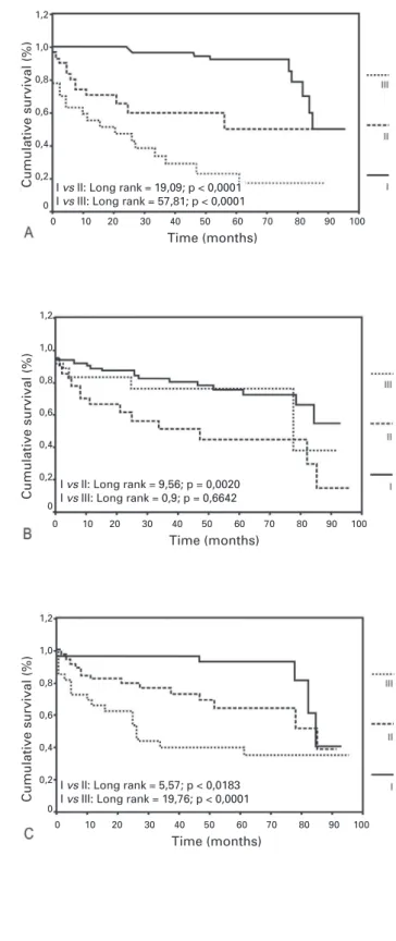
During the overall follow-up the outcome de-veloped at some time in 12.2 % of the patients of the grade I, 38.7 % of the patients of the gra-de II (I vs II: p < 0.0002) and 74.1 % of the pa-tients of the grade III (I vs III: p < 0.0001) of the chronic clinical index (p < 0.0001). Outcome was reached in 23.8 % of the patients of grade I, 55.6 % of the patients of grade II (I vs II: p = 0.0024) and 27.8 % of the patients of grade III (I vs III: p = 0.6616) of acute biopsy index (ns) and 12.2 % of grade I, 34.1 % of grade II (I vs II: p = 0.0546) and 56.3 % of grade III (I vs III: p = 0.0014) of the chronic biopsy index. Before biopsy six grade III chronic clinical index patients shared the same features: serum creatinine levels higher than 2.5 mg/dL, renal cortical echogenicity grade II and kidney length to height ratio less then 0.60. At the end of the follow-up five out of six started on dialysis and one increased serum creatinine levels to 4.5 mg/dL.
Figure 1. Renal survival according to chronic clinical index (A), acute biopsy index (B) and chronic biopsy index (C)..
Cumulative survival (%)
Cumulative survival (%)
Cumulative survival (%)
0 10 20 30 40 50 60 70 80 90 100
Time (months) I vs II: Long rank = 19,09; p < 0,0001 I vs III: Long rank = 57,81; p < 0,0001
1,2
1,0
0,8
0,6 0,4
0,2 0
0 10 20 30 40 50 60 70 80 90 100
Time (months) I vs II: Long rank = 9,56; p = 0,0020 I vs III: Long rank = 0,9; p = 0,6642
1,2
1,0
0,8 0,6
0,4
0,2 0
0 10 20 30 40 50 60 70 80 90 100
Time (months) I vs II: Long rank = 5,57; p < 0,0183 I vs III: Long rank = 19,76; p < 0,0001
1,2
1,0 0,8
0,6
0,4
0,2 0
The chronic clinical index correlated well wi-th chronic biopsy index (Spearman correlation = 0.484; p < 0.001) but not with acute biopsy index (Spearman correlation = 0.137; p = 0.131)
The Cox proportional hazards regression mo-del showed that, independent of biopsy indices, higher chronic clinical index was associated with poorer long-term renal survival (Table 3).
D
ISCUSSIONThe inability of physicians to adequately measure the chronicity of kidney medical diseases has led to many unnecessary renal biopsies.1
Although serum creatinine is a major risk factor for renal survival, it does not fully account for the risk. Clinical markers commonly used for renal di-sease evaluation, including hypertension, hematuria, proteinuria, serum albumin, serum cholesterol and serum triglycerides failed to predict clinical outcome. Therefore, other risk factors must be identified in or-der to better evaluate the need for renal biopsy.
Ultrasound is able to outline macroscopic kidney anatomy features like location, size, contours, mobili-ty, and to detect tumors, calculi and hydronephrosis. For renal medical disease purposes the assessment of the echogenicity of the renal compartments (cortex and medulla) are also important. Nowadays the so-nographic evaluation of the kidney includes cortical echogenicity, corticomedullary definition and pyra-mid proeminence.
The search for a relationship between sonogra-phic appearance of the kidney and histopathological findings was the aim of many reports in recent ye-ars. Indeed, increased cortical echogenicity correlates well with severity and chronicity of tubulointerstitial lesions.7 From laboratory data increased serum
crea-tinine levels correlated with enhanced cortical echo-genicity.7 Since creatinine plays a very important
role when we are dealing with renal diseases, the above association strongly supports the value of renal sonogram as a sensible marker of parenchy-mal renal disease. Based on our findings we would like to extend the value of kidney sonography in renal medical disease.
In adjunction to other clinical and laboratory parameters cortical echogenicity could be useful to predict a clinical setting in which a renal biopsy will show advanced chronic and irreversible lesion. The chronic clinical index is reliable in predicting outcome in groups of patients and indicates chro-nicity of renal disease at the time of admission.
0.60 reached the outcome and one patient increa-sed serum creatinine to levels higher than 4.0 mg/ dL, despite treatment. For this group of patients the renal biopsy did not improve medical management, therefore biopsy can be obviated in these cases.
The strong association between biopsy chroni-city and echographic-laboratory indices allows us to anticipate the chronic lesions and obviate the biopsy. These results indicate that high-risk pa-tients can be identified by noninvasive measure-ments of cortical echogenicity and renal size asso-ciated with serum creatinine.
The relatively better outcome of patients with high biopsy acute index in contrast to high biopsy chronic index suggests that acute lesions, although usually dramatic, are often responsive to therapy. The chronic clinical index uses routinely availa-ble, objective data, is quickly computed, and could be useful to obviate renal biopsy in some patients. Preliminary evaluation in this study demonstrated
Table 3 RISK OFOUTCOMEACCORDINGTOGRADEOFINDICES
Chronic Clinical Index
Outcome I II III
Yes 9 (12.2%) 14 (38.7%) 20 (74.1%)
No 60 (87.8%) 24 (61.3%) 7 (25.9%)
OR - 5.44 10.60
CI 95% - 2.21 - 13.35 4.24 - 26.54
p - <0.001 <0.001
Acute Biopsy Index
Outcome I II III
Yes 19 (23.8%) 15 (55.6%) 5 (27.8%)
No 61 (76.3%) 12 (44.4%) 13 (72.2%)
OR - 3.18 1.28
CI 95% - 1.51 - 6.70 0.42 - 3.87
p - 0.002 0.661
Chronic Biopsy Index
Outcome I II III
Yes 6 (12.2%) 15 (34.1%) 18 (56.3%)
No 43 (87.8%) 29 (65.9%) 14 (43.8%)
OR - 2.60 4.89
CI 95% - 0.98 - 6.87 1.84 - 12.94
p - 0.055 0.001
OR – odds ratio; CI – confi dence interval
a strong correlation between chronic clinical index and renal survival. As a chronicity index of disease for groups of patients with renal medical diseases, the system could be useful in outcome comparisons and evaluation of therapeutic efficacy. Before it is widely used, however, multi-institutional valida-tion is needed.
R
EFERÊNCIAS1. Roger SD, Beale AM, Cattell WR, Webb AW. What is the value of measuring renal parenchymal thickness before renal biopsy? Clin Radiol 1994;49:45-9. 2. Di Nardo R, Ianicelli E, Leonardi D, Manganaro L, Mormile
F, Pecci G. Aspetti ecografici delle nefropatie mediche e con-fronti com agobiopsia. Ann Ital Med Int 1989;4:207-12. 3. Jerassi R, Kusteva R, Kiperova B. Indications for
4. Hricak H, Cruz C, Romanski R, Uniewski MH, Levin NW, Madrazo BL, et al. Renal parenchymal dise-ase: sonographic-histologic correlation. Radiology 1982;144:141-7.
5. Stanley JH, Cornella R, Loevinger E, Schabel SI, Curry NS. Sonography of systemic lupus nephritis. Am J Roentgen 1984;142:1165-8.
6. Miletié D, Fuckar Z, Sustié A, Mozetié V, Stimac D, Zauhar G. Sonographic measurement of abso-lute and relative length in adults. J Clin Ultrasound 1998;26:185-9.
7. Araújo NC, Rioja LS, Rebelo MAP. Doença parenqui-matosa renal: correlação histológico-sonográfica. Rev Assoc Med Bras 2008;54:48-54.
8. Brenbridge NA, Chevalier RL, Kaiser DL. Increased re-nal echogenicity in pediatric rere-nal disease: histopatho-logic correlations. J Clin Ultrasound 1986;14:595-600. 9. Hayden Jr. CK, Santa-Cruz FR, Amparo EC, Brouhard B, Swischuk LE, Ahrendt DK. Ultrasonographic evalua-tion of the renal parenchyma in infancy and childhood. Radiology 1984;152:413-7.
10. Patel JP. Renal parenchymal disease: histopathologic-sonographic correlation. Urol int 1986;41:289-91.
11. Rosenfield AT, Siegel NJ. Renal parenchymal dise-ase: Histopathologic-Sonographic correlation. Am J Roentgen 1981;137:793-8.
12. Page JE, Morgan SH, Eastwood JB, Smith SA, Webb DJ, Dilly SA, et al. Ultrasound findings in renal paren-chymal disease: comparison with histological appea-rances. Clinical Radiology 1994;49:867-70.
13. Gershen RS, Brody AS, Duffy LC, Springate JE. Prognostic value of sonography in childhood nephrotic syndrome. Pediatr Nephrol 1994;8:76-8.
14. Choyke PL, Grant EG, Hoffer FA, Tina L, Korec S. Cortical echogenicity in the hemolytic uremic syndro-me: clinical correlation. J Ultrasound Med 1988;7:439-42.
15. Silkensen JR, Kasiske BL. Laboratory assessment of kid-ney disease: clearance, urinalysis, and kidkid-ney biopsy. In: Brenner BM and Rector FC, editors. The Kidney. 16th ed. Philadelphia: WB Saunders; 2004. p 1079-1106. 16. Golbus J. Lupus nephritis. Classification, prognosis,