S
CIENTIFICA
RTICLES Revista Brasileira de FisioterapiaEffects of an active eccentric stretching program
for the knee flexor muscles on range
of motion and torque
Efeitos do alongamento ativo excêntrico dos músculos flexores do joelho
na amplitude de movimento e torque
Batista LH1, Camargo PR1, Oishi J2, Salvini TF1
Abstract
Objective: To evaluate the changes in knee range of motion (ROM) and torque of knee flexor and extensor muscles after an active eccentric stretching program for the knee flexor muscles. Methods:Thirty-four volunteers (23 women and 11 men), aged 34.42±9.3 years, performed an active eccentric stretching program for the knee flexor muscles in the standing posture, consisting of seven repetitions of one minute each, with 30 seconds of resting between them. The stretching program was performed twice a week, for four weeks. Knee extension ROM and the torque of the knee flexor and extensor muscles were evaluated using an isokinetic dynamometer before and after the stretching program. The torque was evaluated in the isometric, isokinetic concentric and eccentric modes at 30°/s and 60°/s. Results: There was an increase in ROM from 53.7±13° to 30.1±16° (p=0.0001), in isometric torque of the flexors from 89±32Nm to 93±33Nm (p=0.01) and of the extensors from 178±67Nm to 187±73Nm (p=0.006). The concentric and eccentric torque of the flexors at 30°/s increased from 90±31Nm to 96±31Nm (p=0.001) and from 100±34Nm to 105±35Nm (p=0.01), respectively. The concentric torque of the extensors at 60°/s increased from 144±51Nm to 151±58Nm (p=0.02), and the eccentric torque at 30°/s increased from 175±71Nm to 189±73Nm (p=0.01). Conclusions: The stretching program proposed was effective for increasing the flexibility of the stretched muscles and the torque of the agonist (stretched) muscle groups and their antagonists.
Key words: active eccentric stretching; flexibility; torque; ROM; knee flexors.
Resumo
Objetivo: Avaliar a amplitude de movimento (ADM) e o torque flexor e extensor do joelho após a realização de um programa de alongamento ativo excêntrico dos músculos flexores do joelho. Materiais e métodos:Trinta e quatro voluntários (23 mulheres e 11 homens), 34,42±9,3 anos, realizaram um programa de alongamento ativo excêntrico dos músculos flexores do joelho na postura em pé, que consistiu de sete repetições de um minuto com 30 segundos de descanso entre as repetições. O programa de alongamento foi realizado duas vezes por semana, durante quatro semanas. A ADM de extensão e o torque flexor e extensor do joelho foram avaliados no dinamômetro isocinético pré e pós-programa de alongamento. O torque foi avaliado nos modos isométrico e isocinético concêntrico e excêntrico a 30°/s e 60°/s. Resultados: Houve aumento na ADM de 53,7±13° para 30,1±16° (p=0,0001), no torque isométrico flexor de 89±32Nm para 93±33Nm (p=0,01) e extensor de 178±67Nm para 187±73Nm (p=0,006). O torque flexor concêntrico e excêntrico a 30°/s aumentou de 90±31Nm para 96±31Nm (p=0,001) e de 100±34Nm para 105±35Nm (p=0,01), respectivamente. O torque extensor concêntrico a 60°/s aumentou de 144±51Nm para 151±58Nm (p=0,02) e o excêntrico a 30°/s de 175±71Nm para 189±73Nm (p=0,01).
Conclusões: O programa de alongamento proposto foi efetivo para aumentar a flexibilidade dos músculos alongados e torque dos grupos musculares agonistas (alongados) e seus antagonistas.
Palavras-chave: alongamento ativo excêntrico; flexibilidade; torque; ADM; flexores do joelho.
Received: 27/03/2007 – Revised: 11/09/07 – Accepted: 14/03/2008
1Muscle Plasticity Unity of the Neurosciences Laboratory, Physical Therapy Department, Universidade Federal de São Carlos (UFSCar) – São Carlos (SP), Brazil 2Estatistics Department
Financial Support: This work was funded by the Fundação de Amparo à Pesquisa do Estado de São Paulo (Fapesp) and by the Conselho Nacional de Desenvolvimento Científico e Tecnológico (CNPq).
Correspondence to: Tania Fátima Salvini, Departamento de Fisioterapia, Universidade Federal de São Carlos, Rodovia Washington Luís, km 235, CEP 13565-905, São Carlos (SP), Brazil, e-mail: [email protected]
Introduction
he immobilization of muscles in a stretched position pro-motes an increase of their length because of increments in the number of sarcomeres in series of the muscular ibrils1. his adaptation happens in an attempt to re-establish the ideal physiological superposition of actin and myosin ilaments, that make the muscle able to generate higher levels of strength in this new functional length2. herefore, stretching can increase muscular lexibility, and in addition, could cause changes in the development of maximum strength.
In the measurement of the range of motion of knee exten-sion and hip lexion, many studies have reported increases of knee lexor muscle lexibility, subsequent to the application of various stretching programs3-5. However, few studies considered the correlations between changes in lexibility and muscular torque. Many of these studies analysed acute post-stretching6-8 muscle responses, even though, there is a lack of data related to long-term stretching efects9,10.
Worrell, Smith and Winegardner9 submitted knee lexor muscles to two kinds of stretching: static and proprioceptive neuromuscular facilitation (PNF). he authors veriied that the amplitude of knee extension did not sufer any changes af-ter stretching, although an increase occurred of the eccentric and concentric torque of the muscular group that were stre-tched. Hortobágy et al.10 noticed an increase in the lexibility of the knee lexor agonist muscles subsequent to a static stre-tching program performed with this muscle group. Although the performance of this muscle group was not analysed, the authors demonstrated improvements in the performance of the knee extensor muscles in the antagonist group. Conse-quently, according to the authors, it is possible that the stre-tched agonist muscles could have inluenced the mechanical properties of the antagonist group. Similarly, Winters et al.11 defend the view that active stretching of the agonist muscular group can improve the performance of its antagonist group. Even so, the authors suggest that other studies should investi-gate these correlations more closely. he results of the studies regarding the changes in lexibility of the stretched muscles and their antagonists are still a controversial subject in the scientiic literature.
Analysing the above mentioned studies, some characteris-tics were veriied: a) the various kinds of knee lexors stretched, in the standing position, were not performed with unloading of the weight on the member that was stretched, but this was su-pericially supported4,5; b) the improvement of knee lexor mus-cle lexibility was greater when they remained under tension during stretching3,4, as is characterized by active stretching.
It is important to point out that the majority of authors who carried out research on humans showed the eiciency of
static, passive, dynamic or PNF stretching techniques5, howe-ver, active static stretching has not been frequently studied11. Published studies which have shown, the majority performed with animals, demonstrated that active eccentric stretching is highly suggested for promoting muscular stretching, since it stimulates faster adaptations in length of the muscles, and consequently, increases muscle lexibility, that could also cause changes in the levels of strength development12.
hus, greater knowledge of the changes in lexibility and muscular torque of agonist and antagonist muscle groups, performed in a clinic or during the practice of sports, could provide scientiic evidence of the eiciency of active eccentric stretching applied in humans. For this reason, this study aimed to evaluate the efects of an active eccentric stretching program applied to knee lexors, performed in a standing position, with unloading of weight on the stretched member, with lexibility evaluated by measuring the amplitude of the knee extension, lexor torque and knee extension.
Materials and methods
Subjects
hirty-four subjects participated in this study (23 wo-men and 11 wo-men) aged 34.42±9.3 years old. he subjects should demonstrate the following characteristics: a) have a sedentary lifestyle; b) and a minimum of 20° amplitude of the dominant member knee extension13, which was measured using an isokinetic dynamometer and c) be in good health. In this case, subjects who participated in the study showed no inlammatory or osteomioarticular disturbances, as well as, no cognitive or cardiovascular problems of the lower limbs and/or spine which could prevent them from performing the study procedures.
All subjects were informed of the objectives and the pro-cedures of the study and signed an informed consent form in accordance with the resolution 196/96 of the National Health Council. he study was approved by the Ethics Committee of the Universidade Federal de São Carlos (UFSCar), number 179/2007, and was in accordance with the demands of the Hel-sinki declaration for studies performed with humans.
Procedures
178
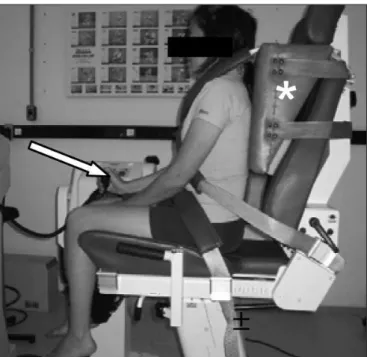
Figure 1. Positioning of the volunteer in the seat of the dynamometer: device (arrow) that was used to start or stop the passive evaluation, and accessory (∗) to keep the hip joint at 90° of flexion, respectively.
of motion and torque were evaluated using isokinetic dy-namometer (Biodex Multi-joint System 3), using only the dominant member.
Evaluations
a) Measurement of the amplitude of knee extension
Position on the equipment: a quilt accessory was created to be placed on the back of the dynamometer chair (Figure 1) to maintain the subject’s hips inlected approximately 90°. his procedure maintained the pelvis closest to the neutral position during the evaluations. Afterwards, the subject was positioned and stabilized on the dynamo-meter chair using diagonal and pelvic belts and the dynamometer’s axis was aligned with the lateral condyle of the femur. he subjects were advised to close their eyes and remain relaxed during the test;
Measurements: the subject could operate the dynamome-ter using a device (Figure 1), so that the resistance arm of the equipment would passively start to extend the knee up to 90° of knee lexion, at a speed of 2°/s. hey were also advised to stop the dynamometer resistance arm using the device as soon as they felt the beginning of knee lexor muscular tension in stretching, which occurred normally between 90° and 20° of knee extension for the subjects who were selected, could not perform total knee exten-sion (0°). he measurements were taken three times and the mean value was used for the statistical analysis14. No
warm-up was performed before the measurement of the amplitude of movement.
b) Measurement of the isometric torque of the knee lexor and extensor muscles: after the range of motion was mea-sured, the subject would warm-up on a stationary bicycle for ive minutes at a speed of 20km/h and performed self-stretching of the knee lexor and extensor muscles. Afterwards, they would once again be positioned on the dynamometer chair, this time without the support of the quilt. he maximum isometric torque of the knee exten-sion was evaluated using maximum voluntary isometric contraction (MVIC) at 80° of knee lexion, as suggested by Marginson and Eston15. hree MVICs were performed and the highest peak torque reached was maintained. Each contraction was maintained for ive seconds, with a rest interval of 90 seconds. he same procedure was used for the evaluation of the knee lexor torque, except that the lexor torque was evaluated with knee lexed at 30°, in agreement with Murray et al.16.
c) Measurement of the isokinetic torque of the knee le-xor and extensor muscles:the concentric and eccentric isokinetic torque of the knee lexor and extensor muscles were evaluated using an range of motion of 60º17, starting from 90º of knee lexion. It is important to indicate that the concentric-eccentric program was used to analyse the maximum contractions of the knee extensor muscles, whereas the eccentric-concentric program was used to evaluate the knee lexor muscles. hese evaluations were performed at a speed of 30º/s and 60º/s for both muscular groups. Five consecutive movements of knee extension and lexion were performed at each speed, with a rest interval of two minutes. he evaluation always started at a speed of 30°/s. It is also important to state that ive sub-maximum contractions were performed before the maximum tests in order for the subjects to get used to the equipment. All tests were carried out by the same therapist.
Stretching
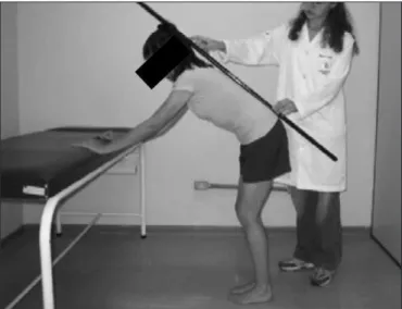
Figure 2. Posture used for the active eccentric stretching of the knee flexor muscles. Note the knee and trunk flexion, alignment of the spine and support of the hands on the stretcher.
Table 1. Knee extension range of motion (ROM) and torque of the flexors and extensors of the knee evaluated in the isometric mode at 80 and 30º of flexion, respectively, and in the concentric and eccentric modes at 30°/s and 60°/s, pre- and post-intervention.
Pre-intervention Post-intervention
Extension ROM (°) 53.7±13.0 30.1±16.0*
Extensor torque (Nm)
Isometric 178.6±67.8 187.5±73.5*
30°/s
Eccentric 175.4±71.6 189.9±73.8 *
Concentric 155.3±59.5 161.6±65.1
60°/s
Eccentric 177.8±74.1 183.1±68.0
Concentric 144.4±51.6 151.57±58.2*
Flexor torque (Nm)
Isometric 89.6±32.62 93.8±33.1*
30°/s
Eccentric 100.3±34.2 105.6±35 *
Concentric 90.7±31.7 96.7±31.8*
60°/s
Eccentric 102.9±35.2 103.9±33.4
Concentric 99.4±34.9 100.7±31.5
Results are mean±standard deviation. *p<0.05 when compared to the pre-intervention period. they stated that they felt maximum supportable knee lexor
stretching, without pain. As soon as this maximum tension was achieved, the stretching should be maintained for one minute. At the end, the subjects returned to the initial po-sition and remained still for 30 seconds. hen they would repeat the procedure.
he same procedure was performed seven times, two times a week (with an interval of two days in between each session), for a period of four weeks. It is important to in-dicate that a bar remained over the subject’s spine during all of stretching time in an attempt to prevent possible compensations.
Statistical analyses
Paired Student t-tests and the Wilcoxon test were used to evaluate the knee extension amplitude and knee lexors and extensors torque, before and after the stretching program. he irst was used to evaluate parametric data and the later was used to evaluate non-parametric data. A signiicance level of p<0.05 was considered at the conclusion of the analysis.
Results
• Evaluation of the amplitude of movement: there was an
increase of approximately 23.6° in the knee extension am-plitude (p=0.0001t), when comparing the previous data and after the stretching program (Table 1).
• Knee extensor concentric and eccentric isometric and
isokinetic torque: the knee extensor torque peak increa-sed after the stretching program in all evaluated modules:
isometric (p=0.006); concentric isokinetic at the speed of 60°/s (p=0.02) and eccentric at the speed of 30°/s (p=0.01), as shown in Table 1.
• Knee lexor concentric and eccentric isometric and isoki
-netic torque:as speciied in Table 1, the peak knee lexor torque also increased after the stretching program in the evaluated modules: isometric (p=0.01); concentric isokine-tic at the speed of 30°/s (p=0.001, Wilcoxon) and eccentric at the speed of 30°/s (p=0.01, Wilcoxon).
Discussion
he results of this study showed that the knee lexor stre-tching program, eccentrically and actively performed, with unloading of the body weight over the stretched member, was efective in increasing knee extension range of motion and in increasing the knee extensors and lexors torque.
180
the range of motion increased 23.6°. his indicated that the procedure used in the present study showed better advantages for gains in the range of motion.
Nelson and Bandy20 did not observe any diferences in gains of amplitude of knee extension after eccentric active and passive stretching. In both stretching programs, the ave-rage of gains in range of motion was 12°. his diference was perhaps due to better positioning of the pelvis, which was maintained in ante-version position during the performance of the stretching of the knee extensors and lexors that was not mentioned in other studies5. Although Sullivan, Dejulia and Worrell4 found an increase of just 11° in the amplitude of the knee extension, after completing the stretching programs, they have indicated the relevance of pelvis positioning during the performance of stretching of the knee lexors. hus, the maintenance of the pelvic positioning guaranteed muscular tension of the knee lexors during the performance of the exercises. Taylor, Brooks and Ryan21, carrying out research using animals, noticed that the combination of stretching and contraction in an exercise program can be more efective than a program just consisted of stretching, since the applica-tion of higher levels of tension on the musculotendinous unit causes more viscoelastic stress. he results of this study point to a necessity of future studies in which diferent stretching postures should be taken, unloading the weight on the stre-tched leg, demanding simultaneous contractions of diferent groups of muscles to better stabilize the knee3.
he gains in the knee extension amplitude might have been caused by changes in muscular length due to increases in the number of sarcomeres in series1,2. However, it is im-portant to point out that many of these changes were not observed with human muscles22. Studies which have been carried out on animals23 and humans24 show that increases in muscular length can also occur due to changes in the con-junctive tissues.
Peak knee extensor and lexor isometric torque increased after the performance of the stretching program. Since the subjects showed shortening of the knee lexor muscles, they could not inlect the trunk and extend the knees at the same time during the performance of the stretching. Because of this, the exercises were performed with semi-lexed knees. his increased the activity of the quadriceps and the knee lexors by co-contraction, to maintain the posture25. Considering the principle of speciicity26,27, a training program produces physio-logical adaptations in the muscles, which are being trained in response to the stimuli of the exercise that has been performed. Accordingly, the speciicity of a stretching program is related to increases of the knee extensor and lexor isometric torques.
he peak concentric and eccentric isokinetic extensor torques also increased after the performance of the stretching
program. Hortobágyi et al.10 defend the viewpoint that incre-ases in the lexibility of the knee lexors may inluence the intrinsic extensor mechanical properties. hey showed that there were increases in the strength of the knee extensors after passive lexor stretching. As previously stated and in accordance with Winters et al.11, when a muscular group is stretched, the antagonists contract. herefore, the standing active stretching performed might have caused neural adap-tations which controlled the level of tension of the muscle, the number of active motor units, the frequency and synchrony of activation between them26. he peak isokinetic lexor tor-que increased likewise during the eccentric and concentric contractions, at a speed of 30º/s. One of the reasons for this increase in the torque of the lexors would be a reduction of neural stability levels. According to Hamill and Knutzen28, active stretching induces a more pronounced response of the Golgi tendon organs and can attenuate their response by per-mitting higher levels of tension of the muscles that received active stretching.
Worrell, Smith and Winegardner9, also observed increa-ses in concentric knee flexor torque after the performance of stretching programs. They attributed these increases to a greater ability of the stretched muscle to store elastic po-tential energy absorbed during the eccentric contractions. This would improve the later strength of the concentric contractions. The phenomena of the improvements in strength of the concentric contractions subsequent to the eccentric pre-stretching contractions of the same muscles is well accepted in the scientific community. Most of the improvements in strength after stretching is due to the passive components as well as the contractible and active components of the muscles29. Taylor, Brooks and Ryan21 re-lated that, after stretching, some viscoelastic properties of the muscular conjunctive tissue change, the resistance ten-sion diminishes and the muscle becomes more complacent. Thus, it would be able to store more potential elastic energy during eccentric contractions. Analysing this present data, the increases in peak knee flexor concentric torque that was observed in this study, might have been caused by increases of the elastic components, since there were increases in the flexibility of the knee flexors.
1. Tabary JC, Tabary C, Tardieu G, Goldspink G. Physiological and structural changes in the cat’s soleus muscle due to immobilization at different lengths by plaster casts. J Physiol. 1972;224(1):231-44.
2. Williams PE, Goldspink G. Changes in sarcomere length and physiological properties in immobilized muscle. J Anat. 1978;127(Pt 3):459-68.
3. Moore MA, Hutton RS. Electromyographic investigation of stretching techniques. Med Sci Sports Exerc. 1980;12(5):322-9.
4. Sullivan MK, Dejulia JJ, Worrell TW. Effect of pelvic position and stretching method on hamstring muscle flexibility. Med Sci Sports Exerc. 1992;24(12):1383-9.
5. Bandy WD, Irion JM, Briggler M. The effect of time and frequency of static stretching on flexibility of the hamstring muscles. Phys Ther. 1997;77(10):1090-6.
6. Cramer JT, Housh TJ, Johnson GO, Miller JM, Coburn JW, Beck TW. Acute effects of static stretching on peak torque in women. J Strenght Cond Res. 2004;18(2):236-41.
7. Power K, Behm D, Cahill F, Carroll M, Young W. An acute bout of static stretching: effects on force and jumping performance. Med Sci Sports Exerc. 2004;36(8):1389-96.
8. Marek SM, Cramer JT, Fincher AL, Massey LL, Dangelmaier SM, Purkayastha S, et al. Acute effects of static and proprioceptive neuromuscular facilitation stretching on muscle strength and power output. J Athl Train. 2005;40(2):94-103.
9. Worrell TW, Smith TL, Winegardner J. Effect of hamstring stretching on hamstring muscle desempenho. J Orth Phys Ther. 1994;20(3):154-9.
10. Hortobágyi TJ, Faludi J, Tihanyi J, Merkely B. Effects of intense “stretching”- flexibility training on the mechanical profile of the knee extensors and on the range of motion of the hip joint. Int J Sports Med. 1985;6(6):317-21.
11. Winters MV, Blake CG, Trost JS, Marcello-Brinker TB, Lowe L, Garber MB, et al. Passive versos active stretching of hip flexor muscles in subjects with limited hip extension: a randomized clinical trial. Phys Ther. 2004;84(9):800-7.
12. Goldspink G, Scutt A, Loughna PT, Wells DJ, Jaenicke T, Gerlach GF. Gene expression in skeletal muscle in response to stretch and force generation. Am J Physiol. 1992;262(3 Pt 2):R356-63.
13. Davis DS, Ashby PE, McCale KL, McQuain JA, Wine JM. The effectiveness of 3 stretching techniques on hamstring flexibility using consistent stretching parameters. J Strength Cond Res. 2005;19(1):27-32.
14. Batista LH, Camargo PR, Aiello GV, Oishi J, Salvini TF. Avaliação da amplitude articular do joelho: correlação entre as medidas realizadas com o goniômetro universal e no dinamômetro isocinético. Rev Bras Fisioter. 2006;10(2):193-8.
15. Marginson V, Eston R. The relationship between torque and joint angle during knee extension in boys and men. J Sports Sci. 2001;19(11):875-80.
16. Murray MP, Gardner GM, Mollinger LA, Sepic SB. Strength of isometric and isokinetic contractions: knee muscles of men aged 20 to 86. Phys Ther. 1980;60(4):412-9.
17. Wallin D, Ekblom B, Grahn R, Nordenborg T. Improvement of muscle flexibility. A comparison between two techniques. Am J Sports Med. 1985;13(4):263-8.
18. Reid DA, McNair PJ. Passive force, angle, and stiffness changes after stretching of hamstring muscles. Med Sci Sports Exerc. 2004;36(11):1944-8.
19. Decoster LC, Scanlon RL, Horn KD, Cleland J. Standing and supine hamstrings stretching are equally effective. J Athl Train. 2004;39(4):330-4.
20. Nelson RT, Bandy WD. Eccentric training and static stretching improve hamstring flexibility of high school males. J Athl Train. 2004;39(3):254-8.
by the above mentioned authors, only the concentric test re-sulted in higher peak torque of the knee lexors at the lowest speed. he results found in the present study difered from the results in the scientiic literature, except for the results found for concentric contractions of the knee lexors. hat indicates that more studies should be carried out to elucidate the chan-ges in torque for the various muscular groups (agonists and antagonists) when submitted to eccentric active stretching programs. hese studies might be more useful if they used faster speeds than the speeds used in this study. It is also im-portant to state that the large diferences found in the results of the isokinetic tests, in this study, may have occurred be-cause of the diiculty the subjects showed when performing them. he subjects showed some diiculty especially to start the eccentric tests of both muscular groups. hese diiculties
might had been eased if the knee lexor eccentric test began with a lower angle of knee lexion (between 80 and 70°), and in the meantime, the stretching would be performed using an amplitude of 60°. In addition, the subjects could have been more familiarized with the equipment, which might have in-luenced the knee extensor tests.
Conclusion
he results of this study show that the stretching program for the knee lexor muscles, actively and eccentrically perfor-med with unloading of body weight, were efective in increa-sing the amplitude of knee extension, as well as torque of the knee lexors and extensors.
182
21. Taylor DC, Brooks DE, Ryan JB. Viscoelastic characteristics of muscle: passive stretching versos muscular contractions. Med Sci Sports Exerc. 1997;29(12):1619-24.
22. Gajdosik RL. Passive extensibility of skeletal muscle: review of the literature with clinical implications. Clin Biomech (Bristol, Avon). 2001;16(2):87-101.
23. Taylor DC, Dalton JD Jr, Seaber AV, Garrett WE Jr. Viscoelastic properties of muscle-tendon units. The biomechanical effects of stretching. Am J Sports Med. 1990;18(3):300-9.
24. Magnusson SP, Aagaard P, Nielson JJ. Passive energy return after repeated stretches of the hamstring muscle-tendon unit. Med Sci Sports Exerc. 2000;32(6):1160-4.
25. Perry J, Antonelli MS, Ford W. Analyses of knee joint forces during flexed-knee stance. J Bone Joint Surg. 1975;57(7):54-61.
26. McCafferty WB, Horvath SM. Specificity of exercise and specificity of training: a subcellular review. Research Quarterly. 1977;48(2):358-71.
27. Aagaard P. Training-induced changes in neural function. Exerc Sport Sci Rev. 2003;31(2):61-7.
28. Hamill J, Knutzen KM. Biomechanical basis of human movement. Baltimore: Lippincott Williams & Wilkins; 1999.
29. Herzog W, Leonard TR. Force enhancement following stretching of skeletal muscle: a new mechanism. J Exp Biol. 2002;205(Pt 9):1275-83.