S
CIENTIFICA
RTICLES Revista Brasileira de FisioterapiaSkeletal muscle weakness and exercise
intolerance in patients with chronic
obstructive pulmonary disease
Fraqueza muscular esquelética e intolerância ao exercício em pacientes
com doença pulmonar obstrutiva crônica
Silva KR1, Marrara KT1, Marino DM2, Di Lorenzo VAP1,3, Jamami M1,3
Abstract
The aim of this study was to evaluate the functional capacity and the performance of respiratory and quadriceps muscles in patients with chronic obstructive pulmonary disease (COPD) and relate them to nutritional status and forced expiratory volume in the first second (FEV1). Methods: Twelve patients with moderate COPD (70±7 years, FEV1 52±17% predicted, body mass index (BMI) 23±4kg/m2) and seven
healthy volunteers (69±8 years, FEV1 127±12% predicted, BMI 27±3kg/m2) were evaluated. All of them underwent body composition
analysis, measurement of respiratory muscle strength (maximum inspiratory pressure, MIP, and maximum expiratory pressure, MEP), cardiorespiratory exercise test (CET) and evaluation of palm grip strength, peak torque and total work or endurance of the quadriceps femoris. Results: The patients with COPD had lower values for the free-fat mass (LBM) index (18±1 versus 21±1kg/m2, p≤0.05), maximum
load attained in the CET (60±20 versus 102±18 watts, p≤0.01), MIP (58±19 versus 87±21cmH2O, p≤0.05), palm grip strength (38±6
versus 47±5kg, p≤0.05), peak torque (103±21 versus 138±18Nm, p≤0.05) and total work of the quadriceps femoris (1570±395 versus
2333±568J, p≤0.05) when compared with the control group (independent Student’s t test). There was no correlation between FEV1 and the variables studied, while the LBM correlated with the total work of the quadriceps (Pearson, r=0.6290, p≤0.05). Conclusions: These results indicate that patients with COPD show weakness of the inspiratory and quadriceps muscles and lower functional capacity, when compared with a healthy group. Moreover, they suggest that the degree of airflow obstruction is not a good predictor for quantifying the nutritional and muscle impairments in patients with COPD.
Key words: exercise intolerance; muscle performance; functional capacity; COPD.
Resumo
O objetivo deste estudo foi avaliar a capacidade funcional e o desempenho da musculatura respiratória e periférica e relacioná-los com o estado nutricional e volume expiratório forçado no primeiro segundo (VEF1). Materiais e métodos: Foram avaliados 12 pacientes com doença pulmonar obstrutiva crônica (DPOC) moderada a grave (70±7 anos, VEF1 de 52±17% previsto, índice de massa corpórea (IMC) de 23±4kg/m2)
e sete indivíduos saudáveis (69±8 anos, VEF1 de 127±12% previsto, IMC de 27±3kg/m2). Todos realizaram análise da composição corporal,
medida da força muscular respiratória (pressão inspiratória máxima, PImax, e pressão expiratória máxima, PEmax), teste de exercício cardiorrespiratório (CRET), avaliação da força de preensão palmar, pico de torque e trabalho total ou endurance do quadríceps femoral. Resultados: Os pacientes com DPOC tiveram valores reduzidos do índice de massa magra corpórea (LBMI) (18±1 versus 21±1kg/m2,
p≤0,05), da carga máxima atingida no CRET (60±20 versus 102±18watts, p≤0,01), da PImax (58±19 versus 87±21cmH2O, p≤0,05), da força de preensão palmar (38±6 versus 47±5kg, p≤0,05), do pico de torque (103±21 versus 138±18Nm, p≤0,05) e do trabalho total do quadríceps femoral (1570±395 versus 2333±568J, p≤0,05) quando comparado com o grupo controle (teste t de Student não pareado). Não houve correlação entre VEF1 e as variáveis estudadas; o LBMI correlacionou-se com o trabalho total do quadríceps (Pearson, r=0,6290, p≤0,05). Conclusões: Estes resultados indicam que os pacientes com DPOC apresentam fraqueza muscular inspiratória e periférica e menor capacidade funcional, quando comparados com o grupo saudável. Além disso, sugere que o grau de obstrução ao fluxo aéreo não é um bom preditor para quantificar as debilidades nutricionais e musculares dos pacientes com DPOC.
Palavras-chave: intolerância ao exercício; desempenho muscular; capacidade funcional; DPOC.
Received: 24/01/2007 – Revised: 09/07/2007 – Accepted: 22/02/2008
1Graduate program in Physical Therapy, Universidade Federal de São Carlos (UFSCar) – São Carlos (SP), Brazil 2Physical Therapist
3Physical Therapy Department
Correspondence to: Karina Rabelo da Silva, Unidade Saúde Escola (USE), UFSCar, Rodovia Washington Luís, km 235, São Carlos (SP), Brazil, e-mail: [email protected]
Introduction
he chronic obtrusive pulmonary disease (COPD) is a re-spiratory disorder characterized by the chronic blocking to the air low which is not totally reversible and is associated with an inlammatory response of the lungs to harmful particles or gases, caused mainly by smoking1. Besides the chronic inlam-mation of the airways, the presence of active inlammatory cells and the increase of the plasmatic levels of pro-inlammatory cytokines in the systemic circulation were detected, which along with oxidative stress contribute to nutritional changes and skeletal muscular disorders in these patients2, both con-tributing to the low endurance for physical exercise, especially among those individuals with a moderate to serious degree of obstruction in air low3.
Among the mechanisms involved in the development of the peripheral muscular disorder of COPD patients, the fol-lowing stand out: deconditioning by disuse; pro-inlammatory cytokines (TNF-α, interleucines-6 and 8, for example), reduced anabolic hormones (testosterone), hypoxemia and/or hyper-capnia, malnutrition, and long-term use of corticoids1,4,5.
Studies have demonstrated that COPD patients often show signiicant mass loss6, respiratory muscle weakness7, upper-limb strength reduction, and obvious diminishments in strength8,9 and endurance10 of the quadriceps muscles when compared to healthy control subjects. All of these factors ac-count for increases in the mortality rates among these patients, not to mention their low quality of life4.
he aim of the present study was to point out that a more comprehensive evaluation, one that might detect the various impairments to which the bearer of such a disease may be susceptible, will contribute to better planning of elaborate pulmonary rehabilitation in a customized fashion Besides this, there are to date very few studies that make use of the isoki-netic dynamometer to evaluate the enduranceof the femoral quadriceps in the COPD.
hus, this investigation intends to test the patients capacity to exercise, as well as the strength of their respiratory muscles and the performance of the palm pressure, strength and endur-ance of the femoral quadriceps, and to correlate these variables with their nutritional condition’s and degree of airway obtru-sion. It also aimed at comparisons of these results with those of sedentary individuals who did not sufer from respiratory disorders.
Materials and methods
his study was carried out between February and October of 2006, and focused on patients with a COPD diagnosis sent
for treatment at the Functional Respiratory Reeducation Pro-gram. It was approved by the Committee for Ethics in Research with Human Beings of the Universidade Federal de São Carlos (UFSCar), under protocol n° 235/2006 and all of the partici-pants (COPD group and control group) signed a term of free agreement for participation form.
Criteria for inclusion and exclusion
Male patients with a clinical diagnosis for COPD, ranging from moderate to serious, according to GOLD’s1 classiication were included, as well as ex-smokers, with a long smoking his-tory (48±17 packets/year for 44±16 years), who were clinically stable and without orthopedic, neurological, or cardiovascular impairments, and/or cognitive changes that might compro-mise their participation in the suggested protocol. he hypoxe-mic patients (peripheral oxygen saturation (SpO2) <88% at rest) and/or those who used oxygen at home were excluded.
COPD group
Ten individuals with a moderate degree of the disease, and eight serious degree patients started the evaluations. Two se-riously impaired individuals were sent to the hospital during the data collection. Out of the subjects with a moderate degree, one did not adapt to the cycloergometer cyclage, one gave up claiming to having personal problems, and two were excluded for being absent more than three times on the scheduled days. Twelve subjects with COPD concluded the tests (six moderate and six serious). hree patients (25%) had used inhalatory cor-ticoids for at least six months prior to the study.
Control group
Ten male volunteers composed the control group, all aged 58 or older, sedentary or with a low level of physical activity (a two hour weekly walk, at most), non-smokers, without any osteomuscular, neurological, cardiovascular or pulmonary pathologies that might have prevented them from undergoing the tests. hree volunteers were excluded, one of them for interrupting the cardiorespiratory test (CRT) too soon due to joint pain in the left knee, one for showing hypertension be-fore the CRT, and another one for having undergone prostate micro-surgery during the evaluations. hus, the inal sample was made up of seven individuals.
Procedures
All the healthy patients and volunteers were submitted to a pulmonary function test using the Vitalograph Spirotrac IV
syprometer, duly calibrated, following the guidelines of the Bra-zilian Society of Pneumology and Tisiology11. he forced vital capacity (FVC) and the forced expiratory volume in the irst second (FEV1) measurements were taken, and Tifenau’s index (VEF1/CVF) was calculated.
he complete evaluation occurred over the course of four diferent days, in no longer than two weeks, consisting of:
• anthropometric evaluations and body mass index (BMI): the
measurements were taken on a Filizola anthropometric scale, with the subject bare foot and semi-nude. hese were the total body mass (BM) in kg, and the height in m, and then the BMI (BMI=mass/height2) was calculated, according to the following classiication: BMI<20kg/m2 as low mass, BMI between 20 and 24.9kg/m2 as normal mass, BMI from 25 to 29.9kg/m2 as overmass and BMI≥30 kg/m2, obese12;
• evaluation of the body composition: the evaluation of the
body composition was carried out by means of the bioelec-tric impendence analysis (BIA) at 50kHz on digital scales (Tanita, model UltimateScale) following the recommenda-tions of the manufacturer. All of the subjects underwent the analysis after fasting for at least ten hours to standardize li-quid uptake. Stemming from the fat obtained, the Lean Body Mass (LBM) was calculated with the formula
100 BM x Fat % LBM = BM
-13
.
Next, the lean body mass index (LBMI) was determined, which is the relation between the LBM and the height squared (kg/ m2)14,15. he classiication of malnutrition for men through the LBMI varied from LBMI≤16kg/m2 16 to 17.4kg/m2 15;
• functional capacity evaluation: the incremental test was
performed on a cyclo-ergometer itted with an electronic brake system (Ergo-FIT, model Ergo 167 Cycle). he pro-tocol was as follows: initial phase (two minutes) seated, without activity, on the bike’s saddle; loading incremental phase (10watts at every two minutes), starting at 15watts until the individual got exhausted; period of active recovery (15watt load for two minutes); recovery phase (six minutes) without activity on the bike’s saddle. During the last 30 se-conds of each phase described above and before each stage of the incremental loading phase, some measures were taken, namely blood pressure (BP), heart rate (HR), SpO2, and the score of Borg’s CR10 Scale17, which was obtained upon questioning the subjects about fatigue or dyspnea and pain or fatigue in the lower limbs. Criteria for interrup-ting the test: reaching the maximum HR given by the for-mula: 220-age, systolic above 220mmHg or its non-increase with a greater workload and/or variation of the diastolic BP greater than 15mmHg, SpO2 reduction under 80% or at the individual’s request due to pain or thoracic burn, dizziness,
cephalea, intense dyspnea, pain in the lower limbs and/or fatigue;
• muscular performance evaluation (at least 72 hours after CRET):
- the evaluation of the respiratory muscular strength was obtained through maximal inspiratory pressure (MIP) and maximal expiratory pressure (MEP) using a manovacuometer (Ger-Ar) scaled between 300 and +300cmH2O, equipped with an adapter for the mouth-piece and containing a escape valve through an oriice of approximately 2mm in diameter, to dissipate the pressures generated by the muscles of the face and of the oropharynx18. Both the MIP and the MEP were per-formed at least three times for each individual, in the orthostatic position, and with the use of a nose clip. he MIP was obtained by means of the residual volume, whereas the MEP was from the total pulmonary capa-city. he patient received due verbal encouragement and in case there was a diference of more than 10% be-tween the measurements, a new maneuver was perfor-med. In the interest of statistical analyses, the greatest value was maintained.
- the evaluation of the isometric force developed by the muscles of the forearm and of the hand was made through palm pressure with a hand-grip style dyna-mometer (Takei Physical Fitness Test, model TKK 5401 Grip-D). he protocol that was used was with the subject in an orthostatic position, with the upper limb (UL) stretched out down the body. Five maximal voluntary contractions (MVC) were required for each limb with a 30-second rest between each repetition19. For the statistical analyses, the greatest value obtained in the hand-grip was considered regardless of the UL where it originated.
- strength and endurance measurements of the dominant quadriceps were taken through the evaluation of the peripheral muscles performance ( femoral quadriceps) of the dominant lower limb (LL) was carried out using an isokinetic dynamometer (Biodex, model Biodex Multi-Joint System 2, Biodex Medical System Inc.) by means of concentric MVCs following a reciprocal protocol exten-sion and lexion in two distinct situations (strength and endurance). For the evaluation of muscle strength, the subject performed ive MVCs in a row at an angular speed of 60°/s, using the greatest torque peak of the extensor muscle for strength analysis. After a ive-minute rest, the endurance measurement began by means of three accu-mulated series of 15 MVCs each at 150°/s19, with a four-minute rest between them, and the total work developed by the quadriceps muscle was calculated. he individuals
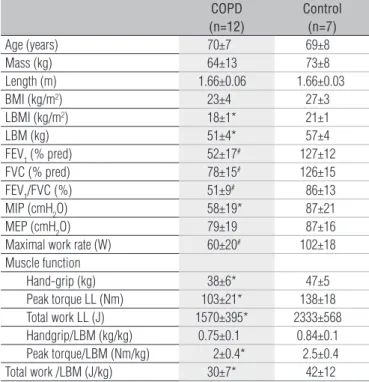
Data are presented as mean±sd, *p≤0.05, #p≤0.01. BMI=body mass index;
LBMI=free-fat mass index; LBM=free-LBMI=free-fat mass; FEV1=forced expiratory volume in one second; FVC=forced vital capacity; FEV1/FVC=Tiffeneau index; MIP=maximal inspiratory pressure; MEP=maximal expiratory pressure; LL=lower limbs.
Table 1. Subject characteristics and studied variables.
COPD (n=12)
Control (n=7)
Age (years) 70±7 69±8
Mass (kg) 64±13 73±8
Length (m) 1.66±0.06 1.66±0.03
BMI (kg/m2) 23±4 27±3
LBMI (kg/m2) 18±1* 21±1
LBM (kg) 51±4* 57±4
FEV1 (% pred) 52±17# 127±12
FVC (% pred) 78±15# 126±15
FEV1/FVC (%) 51±9# 86±13
MIP (cmH2O) 58±19* 87±21
MEP (cmH2O) 79±19 87±16
Maximal work rate (W) 60±20# 102±18 Muscle function
Hand-grip (kg) 38±6* 47±5
Peak torque LL (Nm) 103±21* 138±18 Total work LL (J) 1570±395* 2333±568 Handgrip/LBM (kg/kg) 0.75±0.1 0.84±0.1 Peak torque/LBM (Nm/kg) 2±0.4* 2.5±0.4
Total work /LBM (J/kg) 30±7* 42±12
had their HR, SpO2 and BP monitored before and soon after each one of the aforementioned tests.
he tests with the isokinetic dynamometer were carried out by a single trained examiner. he equipment was duly calibrated, following the manufacturer’s instructions. he in-dividual was seated, and the suitable adjustments for the ideal alignment of the knee joint to the center of the dynamometer were maintained and the attachments to the member tested and to the trunk. Gravity-correction procedures were taken prior to the tests, as well as warming-up (three submaximal contractions before each speed applied), as well as verbal en-couragement during the tests.
Statistical analyses
he variables studied are shown as their mean values±standard deviation (sd). Considering the normal beha-vior of the variables, given by Kolmogorov and Smirnov’s test, the non-paired t-Student test was used for comparisons be-tween the groups, and paired for intra-group comparisons. For the study of the correlations between the variables, the Pearson’s correlation coeicient was used with the level of signiicance set at p≤0.05. For the sample composition of the study, α=0.05 and power greater than or equal to 80% were used to detect signii-cant diferences in the variables under scrutiny by means of the StatMate 2 Program (GraphPad Software, Inc., v. 2.0).
Results
he subjects’ characteristics are shown in Table 1. here were no signiicant diferences in the anthropometric and demographic data, such as age, mass, height, and BMI. Never-theless, the LBMI was quite lower in the COPD group than in the control group. hose patients demonstrated considerably lower spyrometric values than those obtained by the control group. he FEV1did not demonstrate any signiicant correla-tions between the selected variables (p>0.05). Of the 12 patients evaluated, three (25%) showed energetic-protein malnutrition for having BMI<20kg/m2 and LBMI<17.4kg/m2.
Functional capacity
he maximal load achieved by the COPD group was subs-tantially lower than the one reached by the control group (Ta-ble 1), and there was a positive correlation (Pearson, r=0.8038, p≤0.01) between the maximal load attained and the torque peak of the dominant lower limb (Figure 1). Of the 12 patients evaluated, four (33.3%) interrupted the CRET due to pain in their lower limbs, one (8.3%) because of pain in his lower limbs and dyspnea, four (33.3%) due to dyspnea, two (17%) because of general fatigue, and one (8.3%) for reaching the maximal HR for his age group.
Muscular performance
For the MIP (58±19 versus 87±21cmH
2O), the palm pres-sure was (38±6 versus 47±5kg), torque peak (103±21 versus 138±18Nm), and the femoral quadriceps endurance (1570±395 versus 2333±568J) were signiicantly smaller in COPD patients
when compared to the control group (Table 1). he femoral quadriceps endurance showed a positive correlation (Pearson, r=0.6290, p≤0.05), with the LBMI (Figure 1).
Expressed as a percentage of the mean value of the heal-thy subjects9,the COPD group showed palm pressure strength of 82±12%, torque peak 75±15%, and quadriceps enduranceof 67±17%. Upon comparing the percentages of the palm pressure strength developed in relation to the torque peak and to the qua-driceps endurance, in the COPD group, there was a noteworthy diference between the limbs (paired t-Student test, p≤0.05), which revealed less strength of the lower limb (in relation to the upper limb). In the control group, these diferences were not observed. Additionally, the muscular function of the dominant quadriceps, both strength and endurance, expressed by the LBM (Table 1) were signiicantly lower in the COPD group if compa-red to the control group. However, the strength developed by the upper limbs using the hand-grip as shown by the LBM, did not show signiicant diferences between the groups (Table 1).
Figure 1. Correlations COPD group (A) Correlation between the peak torque (Nm) quadríceps and maximal work rate in CRET (W). (B): Correlations between LBMI (kg/m2) and quadriceps endurance (J).
A
20 30 40 50 60 70 80 90 100
60 70 80 90 100 110 120 130 140 150
B
600 800 1000 1200 1400 1600 1800 2000 2200
Endurance (J) 16.0
16.5 17.0 17.5 18.0 18.5 19.0 19.5 20.0 20.5 21.0
r =0.6290, p≤0.05 r =0.8038, p≤0.01
Max. load (W)
T
o
rq
u
e
(
N
.M
)
L
B
M
I
(k
g
/m
²)
Discussion
his study showed that the nutritional condition evaluated by the LBMI, as well as the functional capacity and the mus-cular performance in absolute terms were reduced in patients with COPD when compared to healthy individuals having similar anthropometric characteristics. No correlations were observed between the nutritional condition, functional capac-ity and muscular performance and the FEV1.
he causes of intolerance to exercise in the COPD patients are traditionally focused on the limitations of the ventilatory system and gas exchange20. Nonetheless, some studies3,21 have demonstrated that these are not the only reasons for low capacity for exercise. A relevant factor causing physi-cal limitations is peripheral muscle dysfunction, marked by structural abnormalities, by lowering of the muscular mass and mitocondria relations, changes in the type and size of the muscle ibers and reduction of the oxidative enzymes, reduction of functional strength and resistance, or related to the muscular bioenergetics, such as reductions of oxygen uptake and pH, and increases in the levels of lactates4. Pe-ripheral muscle disorders compromise COPD patients’ ca-pacities to exercise5 since the reduction of peripheral muscle strength is related to physical capacity and to the intensity of symptoms during the incremental exercise test, regardless of the pulmonary function21,22.
In the course of this investigation, we found a strength re-duction of the femoral quadriceps muscle in the COPD group, when compared to the control group, a inding that is corrob-orated by other reports8,23. We also detected positive correla-tions between femoral quadriceps strength evaluated by peak torque, and the maximum load achieved in the CRET, which suggests that the less strength this muscle develops, the less it is capable of performing dynamic exercises for this muscle group. Aside from the peripheral muscle weaknesses, there were also a signiicant correlations between the ventilatory parameters (minute ventilation, current volume, and respira-tory frequency) and the tolerance to exercise, although with a low correlation with the disease’s seriousness as evaluated by the FEV1
24. No signiicant correlations were found between
the maximal exercise load or the muscular performance and the FEV1. It was evident that the airway obtrusion was not a good predictor to estimate the physical and muscular capac-ity of individuals with COPD.
Among the participants of the COPD group, the interrup-tions in the CRET were caused, most of the times, either by dyspnea or by pain in the lower limbs implying that, for some patients, the limiting symptom of the efort is caused by de-iciencies of the peripheral muscles, as suggested by some authors5,25, and not only to ventilatory changes. he weaknesses
of the peripheral muscles of the lower limb in patients with COPD have often been attributed to atrophy by disuse or to physical deconditioning10,23, with an obvious reduction of the transverse section of the thigh area evaluated by a computed tomography8,9 as well as increases in the proportion and atro-phy of type IIA and IIX ibers26.
In the COPD group, the palm pressure strength corre-sponded to 82% of the value scored by the control group, and the strength and endurance of the quadriceps corresponded respectively to 75 and 67% of the values obtained by the con-trol group. hese results suggested that the muscular debility in the COPD particularly afected the lower limb muscles when compared to the upper limbs, a inding conirmed by other researchers5,8. One possible explanation for that would be the reduction of activities which involved walking, in attempt to avoid the sensations of dyspnea, the predominance of daily life activities with the use of the upper limb, and the use of
The Global Initiative for Chronic Obstructive Lung Disease (GOLD) – 1.
workshop report, global strategy for diagnosis, management, and prevention of COPD, 2005 [cited 2006 Jan 20]. Available from: <http://goldcopd.com/>
Agustí AG, Noguera A, Sauleda J, Sala E, Pons J, Busquets X. Systemic 2.
effects of chronic obstructive pulmonary disease. Eur Respir J. 2003;21(2):347-60.
the scapular girdle muscles during accessory breathing. his reduced the impairment of the upper limbs through disuse.
Another inding of this investigation was the reduction of the femoral quadriceps muscle endurance, which can be ex-plained by the putative change of the iber-type proportions. his might favor the reduction of the oxidative capacity, thus heightening the disposition to fatigue and diminishing the resistance of the peripheral muscles, as remarked by other researchers28,29. Other factors, already mentioned, that may have also contributed to muscle loss are the use of oral corti-coids8 and mass loss6.
Concerning the use of oral systemic corticoids, none of the patients in the COPD group used this medicine and three (25%) used it for at least six months prior to the study (the association of β2 agonist/inhalatory corticoid ( formoterol/ budesonida). Nevertheless, the inhalatory corticoids (budes-onida, for example) have milder adverse systemic efects when compared to the systemic corticoids (prednosolona, for example)30.
COPD patients often lose mass and/or have LBM reduc-tion6. Depletion of the lean mass may occur even in patients whose mass is within normal standards, and is commonly ac-companied by BMI or total body mass reduction16,31. he lean mass is regarded as an indirect measure of muscle mass, and its reduction afects both the peripheral muscles, respiratory functions, the capacity to exercise, and increases the risk of mortality6,12. herefore, nutritional depletion exerts a consider-able impact on COPD.
his paper considered the prevalence of nutritional deple-tion in reladeple-tion to funcdeple-tional capacity, muscle performance, and airway obtrusion in a group of COPD patients. Twenty-ive per cent of subjects sufered from energy-protein malnutrition, which was close to the expected percentages among stable out-patient clinical individuals, which is usually around 27%16. Although several studies relate LBM and/or LBMI reductions to a reduction of the respiratory and peripheral muscular func-tions, as well as tolerance to exercise31, this research showed that the LBMI only had signiicant correlations with the quad-riceps endurance, which demonstrated that the smaller the LBM, the less resistance the muscles will have to remain in dynamic activity.
Engelen et al.9 showed that the absolute strength of the MS and of the femoral quadriceps were lower in COPD pa-tients when compared to healthy subjects, but the ratio
between peripheral strength with the LBM or the LBM of the extremities was not signiicant between the groups when tested separately. his suggests that the lowest peripheral strength developed by the patients with COPD was caused by the reduction of the muscular mass, with no associations between the muscular function and the FEV1. he present results are in accordance with these indings, which attested that the UL and LL muscular performance of patients with COPD was reduced when compared to healthy individuals, and that there was no connection with the FEV1.Besides, the relation between palm pressure strength and the LBM, did not signiicantly difer between both groups Yet, the ratio of the quadriceps strength and the LBM contrasted with the indings of Engelen et al.9, since a diference between those groups was detected. It is suggested that the muscular weak-ness of the upper limbs, as measured indirectly by means of the hand-grip in COPD patients may be attributed to the reduction of lean mass. Still, the weakness of the upper limbs did not result in the same responses as the upper limb mus-cles. However, one of the drawbacks of this study was the lack of predicting correction formulae and/or of regression equa-tions15 to obtain the LBM by using the BIA. he equipment used did not provide the tissue resistance values, essential for the use of predictive formulae. hus, a more simple model was used to verify the body composition as a reference point, by using only two components ( fat and lean tissues) which could generate less reliable LBM results32.
he physical training improved the both the muscular functions and tolerance to exercise4 and induced an anabolic response, with an increase of the LBM in normal mass COPD patients19. On the other hand, with individuals who sufered from malnutrition, the training was related to the increase of the protein catabolism, causing the thin mass depletion condi-tion to worsen33.
It was concluded that, besides pulmonary impairment, COPD brought about systemic manifestations that triggered intolerance to exercise and muscular weakness, not to men-tion probable nutrimen-tional changes. Based on the indings of this study, Physical herapy evaluations must contemplate the various aspects related to COPD, thus contributing to the elaboration of a custom-made pulmonary rehabilitation program that focuses on the evident weaknesses, and provid-ing special attention and guidance to those patients suferprovid-ing from malnutrition.
174
Palange P, Forte S, Onorati P, Paravati V, Manfredi F, Serra P, et al. Effect 3.
of reduced body mass on muscle aerobic capacity in patients with COPD. Chest.1998;114(1):12-8.
American Thoracic Society, European Respiratory Society. Skeletal muscle 4.
dysfunction in chronic obstructive pulmonary disease. A statement of the American Thoracic Society and European Respiratory Society. Am J Respir Crit Care Med.1999;159(4 Pt 2):S1-40.
Mador MJ, Bozkanat E. Skeletal muscle dysfunction in chronic obstructive 5.
pulmonary disease. Respir Res. 2001;2(4):216-24.
Wouters EFM. Nutrition and metabolism in COPD. Chest. 2000;117(5 6.
Suppl 1):274-80S.
Orozco-Levi M. Structure and function of the respiratory muscles 7.
in patients with COPD: impairment or adaptation? Eur Respir J. 2003;46:41s-51s.
Bernard S, Leblanc P, Whitton F, Carrier G, Maltais F. Peripheral muscle 8.
weakness in patients with chronic obstructive pulmonary rehabilitation. Am J Respir Crit Care Med. 1998;158:629-39.
Engelen MP, Schols AMWJ, Does JD, Wouters EFM. Skeletal muscle 9.
weakness is associated with wasting of extremity lean mass but not with airflow obstruction in patients with chronic obstructive pulmonary disease. Am J Clin Nutr. 2000;71:733-8.
Serres I, Gautier V, Varray A, Préfaut C. Impaired skeletal muscle endurance 10.
related to physical inactivity and altered lung function in COPD patients. Chest. 1998;113(4):900-5.
Sociedade Brasileira de Pneumologia e Tisiologia. Diretrizes para testes 11.
de função pulmonar. J Pneumol. 2002;28(Suppl 3):S1-238.
Prescott E, Almdal T, Mikkelsenz KL, Tofteng CL, Vestbo J, Lange P. 12.
Prognostic value of mass change in chronic obstructive pulmonary disease: results from the Copenhagen City Heart Study. Eur Respir J. 2002;20(3):539-44.
McArdle WD, Katch FI, Katch VL. Avaliação da composição corporal. 13.
In: McArdle WD, Katch FI, Katch VL, editores. Fisiologia do exercício: energia, nutrição e desempenho humano. 5a ed. Rio de Janeiro: Guanabara Koogan; 2003. p. 772-814.
VanItallie TB, Yang MU, Heymsfield SB, Funk RC, Boileau RA. 14.
Height-normalized indices of the body’s lean mass and fat mass: potentially useful indicators of nutritional status. Am J Clin Nutr. 1990;52(6):953-9.
Kyle UG, Janssensb JP, Rochat T, Raguso CA, Pichard C. Body composition 15.
in patients with chronic hypercapnic respiratory failure. Respir Med. 2006;100(2):244-52.
Vermeeren MA, Creutzberg EC, Schols AM, Postma DS, Pieters WR, 16.
Roldaan AC, et al. Prevalence of nutritional depletion in a large out-patient population of patients with COPD. Respir Med. 2006;100(8):1349-55.
Borg G. Borg’s perceived exertion and pain scales. In: Human kinetics. 17.
United States: Champaign; 1998.
Supinski G. Determination and interpretation of inspiratory and expiratory 18.
pressure measurements. Clin Pulm Med. 1999;6:118-25.
Neder JA, Nery LE. Protocolos. In: Neder JA, Nery LE, editores. Fisiologia 19.
clínica do exercício: teoria e prática. São Paulo: Artes Médicas; 2003. p. 176-82.
Maltais F, Jobin J, Sullivan MJ, Bernard S, Whittom F, Killian KJ, et al. 20.
Metabolic and hemodynamic responses of lower limb during exercise in patients with COPD. J Appl Physiol. 1998;84(5):1573-80.
Hamilton AL, Killian KJ, Summers E, Jones NL. Muscle strength, symptom 21.
intensity and exercise capacity in patients with cardiorespiratory disorders. Am J Respir Crit Care Med.1995;152(6 Pt 1):2021-31.
Gosselink R, Troosters T, Decramer M. Peripheral muscle weakness 22.
contributes to exercise limitation in COPD. Am J Respir Crit Care Med. 1996;153(3):976-80.
Man WD, Soliman MGG, Nikoletou D, Harris ML, Rafferty GF, Mustfa N, et 23.
al. Non-volitional assessment of skeletal muscle strength in patients with chronic obstructive pulmonary disease. Thorax. 2003;58(8):665-9.
Bauerle O, Chrusch CA, Younes M. Mechanisms by which COPD affects 24.
exercise tolerance. Am J Respir Crit Care Med. 1998;157(1):57-68.
Jeffery Mador M, Kufel TJ, Pineda L. Quadriceps fatigue after cycle 25.
exercise in patients with chronic obstructive pulmonary disease. Am J Respir Crit Care Med. 2000;161(2 Pt 1):447-53.
Gosker HR, Engelen MPKJ, van Mameren HV, van Dijk PJV, van der Vusse 26.
GVDJ, Wouters EF, et al. Muscle fiber type IIX atrophy is involved in the loss of lean mass in chronic obstructive pulmonary disease. Am J Clin Nutr. 2002;76(1):113-9.
Dourado VZ, Tanni SE, Vale AS, Faganello MM, Sanchez FF, Godoy I. 27.
Manifestações sistêmicas na doença pulmonar obstrutiva crônica. J Bras Pneumol; 2006;32(2):161-71.
Coronell C, Orozco-Levi M, Méndez R, Ramírez-Sarmiento A, Gáldiz JB, 28.
Gea J. Relevance of assessing quadriceps endurance in patients with COPD. Eur Respir J. 2004;24(1):129-36.
Janaudis-Ferreira T, Wadell K, Sundelin G, Lindström B. Thigh muscle 29.
strength and endurance in patients with COPD compared with healthy controls. Respiratory Medicine. 2006;100(8):1451-57.
Maltais F, Ostinelli J, Bourbeau J, Tonnel AB, Jacquemet N, Haddon J, 30.
et al. Comparison of nebulized budesonide and oral prednisolone with placebo in the treatment of acute exacerbations of chronic obstructive pulmonary disease: a randomized controlled trial. Am J Respir Crit Care Med. 2002;165(5):698-703.
Franssen FME, Broekhuizen R, Janssen PP, Wouters EFM, Schols 31.
AMWJ. Effects of whole-body exercise training on body composition and functional capacity in normal-mass patients with COPD. Chest. 2004;125(6):2021-8.
Sun SS, Chumlea CW, Heymsfield SB, Lukaski HC, Schoeller D, Friedl K, 32.
et al. Development of bioelectrical impedance analysis prediction equations for body composition with the use of a multi component model for use in epidemiologic surveys. Am J Clin Nutr. 2003;77(2):331-40.
Engelen MP, Wouters EF, Deutz NE, Menheere PP, Schols AM. Factors 33.
contributing to alterations in skeletal muscle and plasma amino acid profile in patients with chronic obstructive pulmonary disease. Am J Clin Nutr. 2000;72(6):1480-7.