S
CIENTIFICA
RTICLES Revista Brasileira de FisioterapiaCardiovascular responses in the seated
posture of the Global Postural
Reeducation (GPR) method
Respostas cardiovasculares durante a postura sentada da Reeducação
Postural Global (RPG)
Mota YL, Barreto SL, Bin PR, Simões HG, Campbell CSG
Abstract
Objective: To evaluate heart rate (HR), systolic arterial pressure (SAP), diastolic arterial pressure (DAP), mean arterial pressure (MAP) and double product (DP) responses in the seated posture of the Global Postural Reeducation (GPR) method. Methods: Nine healthy female volunteers (23±2.1 years; 56.4±7.8kg; 1.61±0.05m, 21.6±2.4kg/m2), without experience of the GPR, method underwent a treatment
session in the seated posture. It was a three-step experiment: pre-posture resting, posture maintenance and post-posture recovery. In both the resting and the recovery step, the volunteers remained seated for 20 minutes and arterial pressure and HR were measured every five minutes. The posture maintenance step lasted for three minutes and was implemented three times with one-minute intervals between implementations. Arterial pressure and HR were measured every 1.5 minutes, while the posture was being maintained. Results: The SAP, DAP, MAP and DP values were significantly greater (p<0.05) from the third to the ninth minute of maintaining the posture (154±14, 107±11, 122±9mmHg and 16,478±2,802mmHg/min) in comparison with the pre-posture resting values (109±10, 74±7, 85±8mmHg and 9,374±1,687mmHg/min) and the post-posture recovery values. However, these values returned to the resting values within the first five minutes of post-posture recovery. The HR while maintaining the posture was not statistically different from the pre-posture resting HR.
Conclusions: Significant increases in SAP, DAP, MAP and DP were observed while maintaining the seated posture of the GPR method that was used in this study, but these values returned to the resting values within the first five minutes of post-posture recovery.
Key words: arterial pressure; double product; heart rate; isometric exercise; global postural reeducation.
Resumo
Objetivo: Avaliar as respostas da freqüência cardíaca (FC), pressão arterial sistólica (PAS), diastólica (PAD), média (PAM) e duplo produto (DPr), durante a postura sentada do método de Reeducação Postural Global (RPG). Materiais e métodos: Nove voluntárias saudáveis (23±2,1 anos, 56,4±7,8kg, 1,61±0,05m, 21,6±2,4kg.m2-1), inexperientes na prática do método RPG, foram submetidas
a uma sessão de RPG na postura sentada, realizada em três fases: repouso pré-postura, execução da postura e recuperação pós-postura. No repouso e na recuperação, as voluntárias permaneceram sentadas por 20 minutos, sendo PA e FC verificadas a cada cinco minutos. A fase de execução da postura foi realizada em três séries e mantida por três minutos cada, com intervalo de um minuto entre elas. A verificação da PA e da FC foi realizada a cada um minuto e 30 segundos de execução da postura.
Resultados: Os valores de PAS, PAD, PAM e DPr foram significativamente maiores (p<0,05) do terceiro ao nono minuto da execução
da postura (154±14, 107±11, 122±9mmHg e 16.478±2.802mmHg.min-1) quando comparados aos valores de repouso pré-postura
(109±10, 74±7, 85±8mmHg e 9.374±1.687mmHg.min-1) e aos valores de recuperação pós-postura. Porém, estes valores retornaram
aos valores de repouso nos primeiros cinco minutos de recuperação pós-postura. Durante a execução da postura, a FC não foi estatisticamente diferente da FC de repouso pré-postura.Conclusões: Elevações significativas da PAS, PAD, PAM e DPr foram observadas durante a execução da postura sentada da RPG empregada nesse estudo, mas retornaram aos valores de repouso nos primeiros cinco minutos de recuperação pós-postura.
Palavras-chave:pressão arterial; duplo produto; freqüência cardíaca; exercício isométrico, reeducação postural global.
Received: 11/09/2006 – Revised: 12/12/2006 – Accepted: 08/04/2008
161
Universidade Católica de Brasília (UCB) – Brasília (DF), Brazil.
Introduction
The rehabilitation of patients with postural misalig-nment and pain involves the use of methods that include different types of exercise. Global Postural Reeducation (GPR) is one of the methods that include exercises involving isometric muscular contraction1. The GPR method was ori-ginally developed by Phillipe Emmanuel Souchard in 1987, based on the theory of muscular chains2, which are formed by the organization of static muscles that occurs through muscle overlaps and connections by means of aponeuroses. Thus, the tension of a given muscle group is transmitted to the entire chain3. The GPR method includes five muscular chains: dorsal chain, brachial chain, anterior chain of the neck, antero-lumbar chain and respiratory chain4.The use of these postures aims to correct the existing retractions in the different muscular chains2.
he GPR postures were described as follows: “frog on the loor with open and folded arms; frog in the air with open and folded arms, standing against the wall, standing in the middle, seated and standing postures with anterior incline”4. he pur-pose of the seated position is to strengthen the paravertebral muscles, and to stretch the muscles of the dorsal chain, such as the gastrocnemius, soleus, lexor digitorum longus, lexor digitorum brevis and hamstring muscles1.
Prior physical evaluation of the morphological alterations makes it possible to decide which postures are most suitable to correct the patient’s postural misalignments during treatment. Two diferent postures can be used in the same session, taking into consideration the patient’s ability to sustain that posture. A GPR session lasts one hour on average and usually takes place once a week5.
GPR uses active muscle stretching postures in which the stretching is made possible by the patients’ participation in their own corrections and isometric contractions in the incre-asingly eccentric positions of the shortened muscles5. he iso-metric contractions cause a progressive rise in blood pressure (BP) and heart rate (HR), and the rise in BP during isometric contraction varies according to the muscular tension that oc-curs during the exercise6-8.
BP is the product of cardiac output (CO) times peripheral vascular resistance (PVR). In isometric exercise, there is a minor increase in CO, particularly due to HR increase and to expressive PVR elevation, which contributes to a substantial increase in systolic blood pressure (SBP) and diastolic blood pressure (DBP)6-8. he boost in CO is primarily due to the HR increase. he rise in PVR may be inluenced by the accumu-lation of local metabolites that stimulate muscular chemi-cal receptors, by aferent discharges from muscular nerve endings to the cardiovascular center, and by an increase in
intrathoracic pressure caused by Valsalva’s maneuver, which often accompanies the isometric exercises9.
The sustained contraction of isometric exercise lowers blood and oxygen supply to the muscular tissue, thus HR ri-ses linearly with the increase in the need for oxygen uptake9. Double product (DP) is one of the indicators of myocardial oxygen uptake. It can be obtained indirectly by multiplying the HR by the SBP and has been regarded as an important indicator of cardiac work load10.Isometric exercises cause significant modifications in HR and BP6-8, and these va-riations depend on the percentage of maximal voluntary contraction, on the time of contraction and on the muscle group involved. However, there are no scientific reports on the cardiovascular responses triggered by the GPR. Thus, because GPR involves the isometric contraction of large muscle groups for a long period of time, there is the concern that this method might cause the patient’s BP, HR, and DP to soar. The purpose of this study was to analyze the HR, BP, and DP responses during the “seated” posture of the GPR method in young, healthy individuals.
Materials and methods
Subjects
his was a cross-sectional investigation involving nine female subjects enrolled in the Physical herapy under-graduate course (23±2.1 years old; 56.4±7.8kg; 1.61±0.05m; 21.6±2.4kg.m2-1). he procedures of the present study were approved by the Ethics Committee in Human Research of Universidade Católica de Brasília (UCB), under protocol nº 082/2005. After undergoing a clinical evaluation and illing out a questionnaire, subjects were selected to take part in the study. Selection criteria were: no high BP (SBP<140mmHg and PAD<90mmHg)11,12, no smoking habit, obesity, or cardiores-piratory disease, and no regular exercise regime or previous experience with the GPR method.
Subjects were advised to have a good night’s rest and avoid cofee, alcohol, foods with high-sodium content and physical exercise in the 48 hours prior to the tests. Before executing the posture, subjects were informed about the ex-perimental protocol and the purpose of the study by means of a consent form, and then had their height and total body mass measured.
Procedures
163
and of a sphygmomanometer (Missouri São Paulo, SP, Brazil), which consists of an inlatable cuf connected to a calibrated mercury manometer. he BP measurement was obtained through arterial occlusion by inlating the cuf and correlating the auscultation of the heart beats to the values being displayed in the mercury column. he sounds perceived during the BP measurement are called Korotkof sounds, and are classiied into ive phases, in which phase 1 ( irst sound) corresponds to the SBP value, and phase 5 (sound muling or disappearing) corresponds to the DBP value13. A HR monitor (Polar Coded, Finland) was used, and the weight and height were measured using a scale (Filizola Personal Line, Campo Grande, MS, Bra-zil) with an attached stadiometer. he posture was performed on a stretcher (ISP, Instituto São Paulo, Cascavel, PR, Brazil). Data collection took place at the Cardiopulmonary Rehabilita-tion Laboratory of UCB.
Experimental protocol
he subjects had no previous experience performing the postures of the GPR method, therefore they attended a prac-tice session to learn about monitoring techniques, evolution of seated posture, correct breathing, and adaptation to the labo-ratory environment. he subjects returned to the labolabo-ratory for data collection on a diferent day.
he cuf was placed irmly about 2 to 3cm from the an-tecubital fossa, and the rubber bulb was centralized on the left brachial artery. he watch containing the heart monitor screen was put on the right wrist, and the sensor on the subject’s thorax.
he experiment was carried out in three stages: pre-posture rest, posture performance, and post-posture recovery. In the pre-posture rest, subjects remained seated on the stretcher for 20 minutes, in a comfortable position, with no support on the trunk and with their knees in semi-lexion, with a posture and position similar to the GPR posture used in the study. BP and HR were measured every ive minutes for 20 minutes, and the mean was used to compare the values during and after the posture.
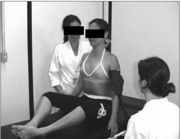
The performance of the seated posture began with the subject in the correct posture: hip flexion with the spine following the physiological kypholordotic curves when viewed in the sagittal plane (Figure 1). In the frontal plane, no lateral or rotational misalignment should be detected. The weight of the body was evenly distributed on each ischium, with the knees starting in semi-flexion facing ou-tward and feet together5.
Each curve of the spine was corrected by axial manual traction in all necessary planes starting with lumbar traction, then the thoracic traction, until it ofered a support to the
occipital bone7.he corrections were maintained manually by the therapist and by the abdominal and paravertebral mus-cle activity of the individual. he progression of posture was performed by maintaining the trunk position with an axial traction and extending with internal knee rotation and plan-tar dorsilexion.
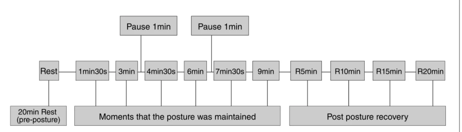
The posture was carried out in three series of three minu-tes each with one-minute intervals during which the subject remained in the same posture, though relaxed, totaling nine minutes of posture performance. BP and HR were checked every one minute and 30 seconds, id est at one minute and 30 seconds and three minutes after the beginning of each series, known as posture moment: 1min30s, 3min, 4min30s, 6min, 7min30s, and 9min, as detailed in Figure 2. During posture performance, the therapist made use of verbal com-mands and manual contact, requesting the maintenance of the alignment and the necessary postural corrections, in order to optimize stretching and avoid compensations. The subject inhaled softly, and let out prolonged, maximal exha-lations, lowering the ribs and contracting the abdomen as much as possible to stretch the respiratory muscular chain, while the therapist helped to maintain the axial elongated position. In post-postural recovery, the individual remained in the same posture as the pre-posture rest for 20 minutes, and HR and BP were measured every five minutes. As a criterion for post-posture hypotension, we considered the significant BP reduction in the recovery values when com-pared to the mean of the four measurements taken during the pre-posture rest.
he measurements of both BP and HR were taken by a sin-gle researcher, and the performance of the GPR method was conducted by a physical therapist certiied by the GPR course taught by Phillipe Emmanuel Souchard.
Figure 1. Representation of the Global Postural Reeducation (GPR)
Results
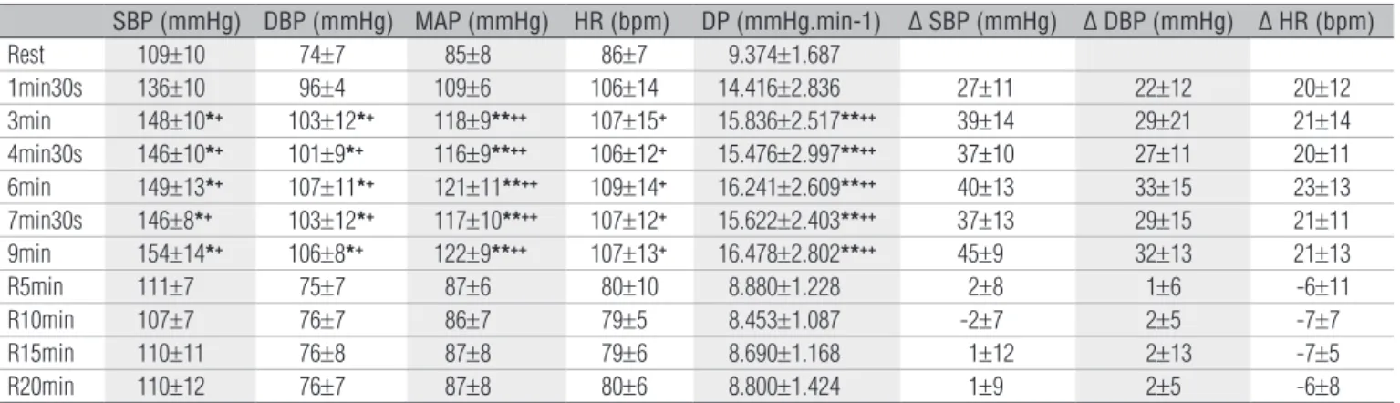
The SBP, DBP, MBP, and DP values (Table 1 and Fi-gure 3) during posture performance were significantly higher (p<0.05) between the third and ninth minute of the task, reaching values of up to 154±14; 107±11; 122±9mmHg and 16,478±2,802mmHg.min-1 when compared to the pre-posture rest values (109±10; 74±7; 85±8mmHg and 9,374±1,687mmHg.min-1) for SBP, DBP, MBP and DP, respec-tively. At these moments, there was a mean increase of up to 45.0±9; 33.0±15; 37.0±10mmHg; and even 7,104.0mmHg.min-1 above the pre-posture rest values for SBP, DBP, MBP and DP, respectively (Table 1).
As for the SBP and DBP values for the post-posture reco-very (R5min to R20 min, Table 1 and Figure 3), they did not statistically difer from those obtained in the pre-posture rest.
With regard to the HR values, in spite of the mean incre-ase between 20±11 and 23±13bpm from one minute and 30 seconds to nine minutes of posture (Table 1), the diferences did not reach statistical signiicance when compared to the pre-posture rest values.
Discussion
he signiicant increase of the variables studied from the third minute of posture performance is a common physiologi-cal response to isometric exercise. As the posture is carried out, the patient performs the stretch determined by the therapist, maintaining isometric contractions particularly in the paraver-tebral muscles, which is the probable cause of the cardiovascu-lar alterations found in this study. In this kind of exercise, there is a mechanical compression of the arterial system, which brings about a persistent reduction in muscular perfusion and subsequent increase of the total PVR. In this case, sympathetic
Rest 1min30s 3min 4min30s 6min 7min30s 9min R5min R10min R15min R20min
Pause 1min Pause 1min
Moments that the posture was maintained
20min Rest
(pre-posture) Post posture recovery
Figure 2. Schematic representation of the periods when the blood pressure and the heart rate were measured (Rest – Pre-posture resting; periods
of posture execution from 1min30s to 9min; Post-posture recovery – R5min to R20min).
Statistical analysis
he results are presented with their mean values and res-pective standard deviations. In order to test the normality of the data, we used Kolmogorov Smirnov’s method. We also compared the HR, SBP, DBP, MBP, and DP values taken for pre-posture rest, pre-posture performance and post-pre-posture recovery. Because the DP and MBP values had normal distribution, the variance analysis (ANOVA) with Tukey’s post hoc was applied. To compare the results for HR, SBP, and DBP for pre-posture rest, posture performance and post-posture recovery, we ap-plied Friedman’s non-parametric test for repeated measures, followed by Dunn’s multiple-comparison techniques. he level of signiicance was p<0.05.
Figure 3. Mean results of the systolic blood pressure (SBP), diastolic
blood pressure (DBP) and mean arterial pressure (MAP), in the moments of pre-posture resting (Rest), execution of the posture (1min30s to 9min) and of post-posture recovery (R5min R20min).
SBP DBP MAP
BP (mmHg)
70 80 90 100 110 120 130 140 150 160
RES
T
1min30s 4min30s 7min30s
3min 6min 9min
R5min
R1
0min
R1
5min
R20min
+ *
+ * +*
+ * +
* + * +
* + * +*+*
+ * + * +
* + *
+ * + * +
* + * +*
+ *
*p <0.05 in relation to the rest pre-posture for SBP and DBP; +p<0.05 in relation to the
165
nervous system (SNS) activity is boosted, thus increasing CO and mean BP in an attempt to reestablish and/or increase muscular blood low14-16.Also, in the beginning of the isometric exercise, there is an inhibition of cardiac vagal activity (reduc-tion of parasympathetic modula(reduc-tion) followed by an activa(reduc-tion of the sympathetic response6,7,17-19,as well as an interaction be-tween the central command and the mechanoreceptors found in the muscles. his potencializes the SNS responses, causing the BP to rise7,19-21.
he study revealed that between the third and ninth mi-nute of posture performance there were signiicant increments in the SBP, DBP, MBP, and DP values. he magnitude of these responses may be directly related to the duration of the efort combined with the diiculty maintaining the muscle contrac-tion and the size of the muscle mass involved in this type of isometric exercise14,15,22.In the study developed by Polito and Farinatti22, BP responses increased in proportion to the num-ber of repetitions (time of performance of the movements) in a resisted dynamic exercise. he present study used three series of isometric contractions lasting three minutes each and found that, as the posture performance time and the interval dis-tance progressed (three, six, and nine minutes), the values for SBP, DBP, MBP, and DP also increased. Gandevia and Hobbs8 studied individuals performing intense isometric contractions (handgrip) to the point of fatigue and suggested that the che-moreceptors of the muscles were activated in direct proportion to the duration of isometric contractions, thus contributing to BP increase during contractions.
In the inal moments of the test, the subjects reported the inability to maintain posture due to muscle fatigue. Polito and Farinatti22 observed that the repetitions to the point of fatigue (muscle failure to sustain a certain force) caused gre-ater elevations in BP.
he DBP during isometric exercise, caused by the mainte-nance of the posture, had a diferent response from that obser-ved by other authors. As the physical exercise is performed, the DBP remains constant or oscillates slightly, about 10mmHg, in relation to the values at rest23.In the present study, there was an elevation in the DBP between 22 and 33mmHg during posture performance when compared to pre-posture rest, and this signiicant increase was observed from the third minute of posture maintenance onwards, as with the SBP. his provides us with an important practical application, id est that this GPR posture should not be sustained for more than three minutes by high-risk patients, such as those who sufer from sympto-matic and asymptosympto-matic cardiovascular diseases, especially high BP. However, further research is needed to conirm this claim. In addition, there was a tendency for the SBP, DBP, and MBP to fall in relation to the previous value in the stages of the posture performance that followed the one-minute intervals (4min30s and 7min30s), which suggests that the use of longer pauses may be helpful for this type of patient (Figure 3), or even for patients of other age groups whose BP tends to be higher.
In a study by Williams6, the researcher detected a SBP/ DBP rise from 138/82 at rest to 224/156mmHg (+86/74mmHg) during a sustained contraction of the lower limb at 70% of the maximal voluntary contraction. At that point, the patient was overcome by fatigue and could no longer sustain the muscular contraction. In our study, we perceived an elevation from 109/74 at rest to 154/106mmHg (+45/32mmHg) in nine minutes of posture. It is important to consider that the intensity and dura-tion of the efort to the point of fatigue in the aforemendura-tioned study were diferent from those employed in the present study, as was the method of BP measurement. he intra-arterial me-thod, though invasive, produces the most realistic BP values. he BP values obtained in these studies show that isometric
SBP (mmHg) DBP (mmHg) MAP (mmHg) HR (bpm) DP (mmHg.min-1) ∆ SBP (mmHg) ∆ DBP (mmHg) ∆ HR (bpm)
Rest 109±10 74±7 85±8 86±7 9.374±1.687
1min30s 136±10 96±4 109±6 106±14 14.416±2.836 27±11 22±12 20±12 3min 148±10*+ 103±12*+ 118±9**++ 107±15+ 15.836±2.517**++ 39±14 29±21 21±14
4min30s 146±10*+ 101±9*+ 116±9**++ 106±12+ 15.476±2.997**++ 37±10 27±11 20±11
6min 149±13*+ 107±11*+ 121±11**++ 109±14+ 16.241±2.609**++ 40±13 33±15 23±13
7min30s 146±8*+ 103±12*+ 117±10**++ 107±12+ 15.622±2.403**++ 37±13 29±15 21±11
9min 154±14*+ 106±8*+ 122±9**++ 107±13+ 16.478±2.802**++ 45±9 32±13 21±13
R5min 111±7 75±7 87±6 80±10 8.880±1.228 2±8 1±6 -6±11 R10min 107±7 76±7 86±7 79±5 8.453±1.087 -2±7 2±5 -7±7 R15min 110±11 76±8 87±8 79±6 8.690±1.168 1±12 2±13 -7±5 R20min 110±12 76±7 87±8 80±6 8.800±1.424 1±9 2±5 -6±8
Table 1. Mean+sd results of systolic blood pressure (SBP), diastolic blood pressure (DBP), mean arterial pressure (MAP), heart rate (HR), double
product (DP) and delta variation (∆) of the SBP, DBP and HR during the pre-posture resting (Rest), moments of posture execution (1min30s; 3min; 4min30s; 6min; 7min30s and 9min) and during the 5th, 10th, 15th, and 20th min of post-posture recovery (R5min, R10min, R15min and R20min) from the seated posture of the GPR technique.
exercise causes excessive BP increase, which varies according to the intensity and duration of the muscle contraction.
his signiicant increase both in SBP and DBP in the present study may also be caused by the increase in the intrathoracic pressure due to the additional contraction of the exhalation muscles as a consequence of Valsalva’s maneuver16,24.In this maneuver, the person tries to exhale while the glottis is closed, thus the increase in intrathoracic pressure causes a decrease in venous return due to the collapse in the veins that cross the thorax, elevating BP16. Narloch and Brandstater25 investigated the inluence of this breathing technique on the BP of young athletes during weight-bearing exercise and found an increase in the SBP/DBP at rest with and without Valsalva’s maneuver, from 127/82 to 180/155mmHg, during the performance of ive repetitions of the exercises at 85% of maximal repetitions from 178/156 to 267/239mmHg using the intra-arterial technique of BP measurement. his study allows us to assert that Valsalva’s maneuver contributes to an even greater BP rise than that in-duced by the physical exercise, including excessive BP elevation even when the maneuver is performed at rest. Stretching du-ring GPR is carried out in association with deep and prolonged exhalation5 and the patient may, despite advice to the contrary, perform Valsalva’s maneuver at given moments in which the maintenance of the posture becomes uncomfortable, thus increasing BP. In addition to Valsalva’s maneuver, the seated position of the GPR posture itself may have contributed to the reduction in venous return and therefore to the elevation of BP. Post-exercise hypotension (PEH) is characterized by a re-duction in BP during the post-exercise recovery phase, when the pressure values are signiicantly lower than those obtained in the pre-exercise stage. In this study, no statistical diference was found between BP at rest and post-posture recovery BP in the course of the 20 minutes during which it was taken, and the BP returned to its rest values in the irst ive and ten minutes of recovery. According to Brum et al.26 the characteristic, type, in-tensity, and duration of the exercise determine the magnitude of PEH. hus, the model put forward in this study in terms of duration, posture, pause, and type of muscular contraction did not result in post-posture hypotension in the course of the 20 minutes during which BP was measured. he efects of other variations, such as duration of posture performance and of post-posture recovery, as well as the posture used on recovery BP must be investigated. In a study developed by Simões27, the PEH of SBP was only observed after 30 minutes of recovery following resisted exercise performed in a 20-minute circuit, whereas the PEH of DBP was not observed. It is possible that after the 20 minutes of post-posture recovery there was PEH induced by the nine minutes of performance of the seated posture of the GPR method, though this was not investigated in the present study.
he DP is a parameter obtained by multiplying HR by SBP and represents cardiovascular stress with a high correlation (r=0.88) to myocardial O2 consumption and is an important indicator of cardiac workload during the physical exertion10. he present study found a signiicant increase in DP (75.8%), which went from 9,374 at rest to 16,478mmHg.min-1 at nine minutes of posture, showing that the GPR method employed in the study caused a considerable cardiovascular overload. he-refore, the risk/beneit must be taken into account when using this method on patients with high BP and heart disease.
In the study by Andrade, Barbosa Júnior and Pulcenelli28, the DP increase was a result of the signiicant elevation of SBP and HR in response to the number, intensity, and length of repetitions performed in resistance exercise. he DP can vary from around 6,000 at rest to 4,000mmHg.min-1 (an increase of approximately 567%) or more, depending on the intensity and mode of exercise17.
We observed that the increase in DP during posture perfor-mance was below the value of the cutof point for angina pec-toris ( from 30,000mmHg.min-1). he young, healthy subjects of this study achieved approximately 55% (16,478mmHg.min-1) of this threshold. Nevertheless, it is necessary to investigate the hemodynamic responses during the application of this method in high-risk populations. It is important to know the cardiac overload imposed by diferent exercises and therapies, as it helps in the selection of therapies that ofer the least impact, especially when dealing with populations at risk.
With regard to MBP, we observed a signiicant increase between the third and ninth minute of posture performance when compared to the pre-posture rest values. MBP represents the mean force exerted by the blood against the arterial walls during the entire cardiac cycle. For this calculation, the dura-tion of the diastole is considered 1/3 greater than that of the systole24. he increase in MBP while posture is sustained may be related to the signiicant DBP increase16 in the present study. According to Polito and Farinatti16, the DBP response during the exercise, not exceeding the normal response, is considered the facilitator of myocardial perfusion. herefore DBP and the MBP values should be monitored during posture performance to reduce ischemic risk.
167
increase in CO. However, a limitation that might have contri-buted to results in the present study was the fact that HR was not continually recorded, but only when the other variables were registered (1min30s; 3min; 4min30s; 6min; 7min30s and 9min of posture). For this reason, future studies should monitor HR on a beat-by-beat basis.
A few limitations of the study must be highlighted. BP was measured using the auscultatory method. It is known that the use of invasive methods such as the intra-arterial catheteriza-tion would yield more precise, reliable, and valid results, des-pite being invasive and onerous. However, it was not possible to employ these procedures in the present study. In addition, all the hemodynamic variables investigated in the present study were elevated during the posture performance stage of the GPR posture in relation to the pre-posture rest values. Ho-wever, all of them returned to values close to those observed in the pre-posture rest in the irst ive minutes of post-posture recovery. In spite of these limitations, the present study inten-ded to encourage physical therapists to monitor the cardio-vascular responses of patients who have been recommended
GPR treatment, particularly those at risk, such as individuals with symptomatic and asymptomatic cardiovascular disease, especially high BP. It is evident that the clinical practice of GPR has had beneicial results in terms of improving lexibility, but before it can be ofered to patients as an alternative, risk-free treatment, it must be scientiically validated.
Conclusions
References
Souchard PE. Reeducação postural global: método do campo fechado. 4ª 1.
ed. São Paulo: Ícone; 2001.
Teodori RM, Moreno MA, Fiore Júnior JF, Oliveira ACS. Alongamento da 2.
musculatura inspiratória por intermédio da reeducação postural global (RPG). Rev Bras Fisioter. 2003;7(1):25-30.
Yokohama TV. A prática do Iso-stretching na melhora da expansibilidade 3.
toracopulmonar, verificada através da Espirometria e da Cirtometria [Monografias do curso de Fisioterapia]. Cascavel (PR): Unioeste; 2004.
Souchard PE. O stretching global ativo: a reeducação postural global a 4.
serviço do esporte. 2ª ed. São Paulo: Manole; 1996.
Souchard PE, Ollier M. As escolioses: seu tratamento fisioterapêutico e 5.
ortopédico. São Paulo: Realizações; 2001.
Williams CA. Effect of muscle mass on the pressor response in man 6.
during isometric contractions. J Physiol. 1991;435:573-84.
Friedman DB, Peel C, Mitchell JH. Cardiovascular responses to 7.
voluntary and nonvoluntary static exercise in humans. J Appl Physiol. 1992;73(5):1982-5.
Gandevia SC, Hobbs SF. Cardiovascular responses to static exercise in 8.
man: central and reflexes contributions. J Physiol. 1990;430:105-17.
Ghorayeb N, Barros Neto TL. O exercício: preparação fisiológica, avaliação 9.
médica, aspectos especiais e preventivos. São Paulo: Atheneu; 1999.
Leite TC, Farinatti PTV. Estudo da freqüência cardíaca, pressão arterial 10.
e duplo produto em exercícios diversos para grupamentos musculares semelhantes. Rev Bras de Fisio do Exercício. 2003;2(1):29-49.
Chobanian AV, Bakris GL, Black HR, Cushman WC, Green LA, Izzo JL Jr, 11.
et al. The Seventh Report of the Joint National Committee on Prevention, Detection, Evaluation, and Treatment of High Blood Pressure: the JNC 7 report. JAMA. 2003;289:2560-72.
Sociedade Brasileira de Hipertensão, Sociedade Brasileira de Cardiologia, 12.
Sociedade Brasileira de Nefrologia. V Diretrizes Brasileiras de Hipertensão Arterial. São Paulo; 2006, 1-50.
Chobanian AV, Bakris GL, Black HR, Cushman WC, Green LA, Izzo JL 13.
Jr, et al. Seventh report of the Joint National Committee on Prevention, Detection, Evaluation, and Treatment of High Blood Pressure. Hypertension. 2003;42(6):1206-52.
Mcardle WD, Katch FI, Katch VL. Fisiologia do exercício: Energia, nutrição 14.
e desempenho humano. 4ª ed. Rio de Janeiro: Guanabara Koogan; 1998.
Nóbrega ACL. Fisiologia do exercício. Revista SOCERJ. 2000; 15.
XIII(4):19-23.
Polito MD, Farinatti PTV. Respostas de freqüência cardíaca, pressão 16.
arterial e duplo-produto ao exercício contra-resistência: uma revisão da literatura. Rev Port Ciências Desp. 2003b;3(1):79-91.
Power S, Howley E. Fisiologia do exercício – Teoria e aplicação ao 17.
condicionamento e ao desempenho. 3ª ed. São Paulo: Manole; 2000.
González-Camarena R, Carrasco-Sosa S, Román-Ramos R, Gaitán-18.
González MJ, Medina-Bañuelos V, Azpiroz-Leehan J. Effect of static and dynamic exercise on heart rate and blood pressure variabilities. Med Sci Sports Exerc. 2000;32(10):1719-28.
Ray CA, Rea RF, Clary MP, Mark AL. Muscle sympathetic nerve responses 19.
to static leg exercise. J Appl Physiol. 1992;73(4):1523-9.
Iellano F, Massaro M, Raimond G, Peruzzi G, Legramante JM. Role of 20.
muscular factors in cardiorespiratory responses to static exercise: contribuition of reflex mechanisms. J Appl Physiol. 1999;86(1):174-80.
Smolander J, Aminoff T, Korhonen I, Tervo M, Shen N, Korhonen O, et al. 21.
Heart rate and blood pressure responses to isometric exercise in young and older men. Eur J Appl Physiol Occup Physiol. 1998;77(5):439-44.
Polito MD, Farinatti PTV. Considerações sobre a medida da pressão arterial 22.
em exercícios contra-resistência. Rev Bras Med Esporte. 2003a;9(1):25-31.
Conselho Nacional de Ergometria. Indicações e contra-indicações dos 23.
testes ergométricos. Arq Bras Cardiol. 1995;65(2):191-211.
Robergs RA, Roberts SO, Silva AC. Princípios
24. fundamentaisdefisiologia doexercício para aptidão, desempenho e saúde. São Paulo: Phorte; 2002.
Narloch JA, Brandstater ME. Influence of breathing technique on arterial 25.
blood pressure during heavy weight lifting. Arch Phys Med Rehabil. 1995;76(5):457-62.
Brum
26. P, Forjaz C, Tinucci T, Negrão CE. Adaptações agudas e crônicas do exercício físico no sistema cardiovascular. Revista Paulista Educ Física. 2004;18:21-31.
Simões GC. Efeitos de diferentes intensidades de exercício resistido 27.
sobre as respostas hemodinâmicas em indivíduos diabéticos tipo 2 e não diabéticos [dissertação de mestrado em Educação Física e Saúde]. Brasília: UCB, 2006.
Andrade FM, Barbosa Júnior OA, Pulcenelli AJ. Estudo comparativo do 28.
duplo produto no treinamento de força em séries piramidais crescente e decrescente. Revista Digital Vida & Saúde. 2002;1(3).
Marin-Neto JA, Maciel BC, Gallo Júnior L, Junqueira Junior LF, Amorin 29.