S
CIENTIFICA
RTICLES ©Revista Brasileira de FisioterapiaEffect of gallium-aluminum-arsenide laser
therapy (660Nm) on recovery of the sciatic
nerve in rats following neurotmesis lesion and
epineural anastomosis: functional analysis
Efeito da terapia com laser de arsenieto de gálio e alumínio (660Nm) sobre a
recuperação do nervo ciático de ratos após lesão por neurotmese seguida de
anastomose epineural: análise funcional
Reis FA1, Belchior ACG1, Nicolau RA2, Fonseca TS1, Carvalho PTC1
Abstract
Context: Peripheralnerve injuries result in sensory and motor losses in the innervated area and can hinder individuals’ daily activities.
Objective: The objective was to analyze the influence of applying gallium-aluminum-arsenide (GaAlAs) laser (660Nm) on the functional recovery of the sciatic nerve in rats. Methods: The sciatic nerve of 12 Wistar rats was subjected to injury consisting of neurotmesis and epineural anastomosis. The rats were divided into two groups: control and laser therapy. After the injury, a GaAlAs laser was used (660Nm, 4J/cm², 26.3mW and 0.63cm² beam) at three equidistant points on the injury, for 20 days. Footprint impressions were obtained from the animals before and seven, 14 and 21 days after the surgical procedure and the sciatic functional index (SFI) was calculated.
Results: Comparison of the SFI did not show any significant difference (p>0.05) between the two groups. Conclusions: The parameters and methods used for the laser therapy did not produce any effect on the SFI over the period evaluated.
Key words: nerve regeneration; low-level laser therapy; sciatic nerve.
Resumo
Contextualização:As lesões nervosas periféricas podem comprometer atividades diárias de um indivíduo e resultam em perda da sensibilidade e motricidade do território inervado. Objetivo:Com o intuito de acelerar os processos regenerativos, objetivou-se analisar a influência da aplicação do laser de arsenieto de gálio e alumínio (AsGaAl, 660Nm) sobre a recuperação funcional do nervo ciático de ratos. Materiais e métodos:O nervo ciático de 12 ratos Wistar foi submetido à lesão por neurotmese e anastomose epineural e divididos em dois grupos: controle e laserterapia. Após a lesão, utilizou-se o laser de GaAlAs, 660Nm, 4J/cm2, 26,3mW, feixe de 0,63cm2, em
três pontos eqüidistantes sobre a lesão, por 20 dias. As impressões das pegadas dos animais foram obtidas antes e após (sete, 14 e 21 dias pós-operatórios) o procedimento cirúrgico e calculou-se o índice funcional do ciático (IFC). Resultados: A comparação do IFC não resultou em diferença significante (p>0,05) entre os grupos. Conclusões: Conclui-se que os parâmetros e métodos empregados na laserterapia demonstram resultados nulos sobre o IFC no período avaliado.
Palavras-chave: regeneração nervosa; terapia a laser de baixa intensidade; nervo ciático.
Received: 17/07/2007 – Revised: 12/11/2007 – Accepted: 14/03/2008
1 Physical Therapy Program, Universidade para o Desenvolvimento do Estado e da Região do Pantanal (Uniderp) – Campo Grande (MS), Brazil. 2 Tissue Biomodulation Laboratory, Research and Development Institute, Universidade do Vale do Paraíba (Univap) – São José dos Campos (SP), Brazil Correspondence to: Filipe Abdalla dos Reis, Rua Goiás, 1.709, Vila Célia, CEP 79022-355, Campo Grande (MS), Brazil, e-mail: [email protected]
216
Introduction
Peripheral nerves are often exposed to injuries from trau-matic origins, such as crushing impact and total transections, resulting in decreases or complete loss of sensory and motor capabilities in the corresponding area of innervation. he seve-rity of these deicits will depend on the involvement of various structures. he impairment on the daily activities of patients with peripheral nerve injuries is a determining factor to esta-blish the necessity of early recovery1.
The functional recovery following peripheral nerve in-jury has intrigued researchers for a long time, and it has generated a great number of scientific studies about the several intervention techniques used, the phenomena in-volved in regeneration and the evaluation methods for the results. Although there is always some recovery in most nerve injuries, they occur slowly and, many times, in an in-complete manner2. It is estimated that in some countries, the incidence of traumatic injuries is greater than 500.000 of new cases annually1, where 2.8% of the patients acquire permanent incapacities due to the length of time required for the nerve regeneration, of around two months3. This fact explains the elaboration of different therapies to reduce the levels of injury and incapacity.
To evaluate the levels of nerve injuries in experimental situ-ations, the functional assessment of gait has been shown to be a reliable and reproducible method4,5. In 1982, De Medinacelli, Freed and Wyatt5 proposed the use of a method of assessment named the Sciatic Functional Index (SFI) based upon measu-rements of the rear feet of rats. his method was used with a normal control group and in experimental groups, after the sectioning and crushing of the sciatic nerve. he experiment consisted of obtaining images of the animals’ footprints, while they walked on a track speciically built for this purpose and the images were recorded and analyzed.
he use of therapeutic resources with a regenerative pur-pose is a common practice in Physical herapy. For peripheral nerve injuries, the electric stimulation6, ultrasound therapy7 and low-intensity laser therapy (LILT)8 have been used to acce-lerate the regenerative processes, aiming at the early recovery of the patients’ functional status. LILT was used on the regene-rative processes and the functional recovery from peripheral nerve injuries in the 1970’s, and there were various reports and disagreements over the obtained results9.
By the analyses of this study, it was found that the He-Ne laser, with an emission in the red region of the electromagnetic spectrum, is the most studied wave length in the biomodu-lation of biological responses in the healing process. At this very moment, new wave lengths are now being developed and researched, like lasers in the 650 to 830Nm region. It can be
observed that, in many studies, the descriptions of parame-ters for irradiation like: dosages, average power, lengths and methods of application are widely varied, which makes the methodological understanding diicult for reproducing results and for other comparisons.
Considering the poor reproducibility studies cited in the literature and the previously observed unsatisfactory metho-dological presentation of the data, this study aimed to evaluate the efects of low intensity laser therapy on the repairing pro-cesses of the sciatic nerve. he investigation of an experimental model related to the analysis of functional recovery could pro-vide relevant data as the basis of future clinical applications in the treatment of nerve injuries.
Materials and methods
Animals
Twelve male adult rats three month old were used, from the Wistar breed, with a body weight ranging from 300 to 350g, brought from the Central Bioterium of the Universidade para o Desenvolvimento do Estado e da Região do Pantanal (Uniderp), kept under controlled conditions of luminosity and temperature, with six animals per cage, and regular feeding and water ad libitum.
he rat was chosen as the experimental animal for its ease of acquisition, handling and also for the low operating costs. Besides these factors, the similarities to the human distribu-tion of nerve trunks and the proper anatomic characteristics for surgery procedures facilitated the choice of this animal1.
All experimental procedures were done according to the rules from the Brazilian College of Animal Experimentation (Cobea), and approved by the Ethics Committee of Research from the Universidade do Vale do Paraíba (Univap), under the record number L185/2005/CEP.
Animals were randomly assigned for two experimental groups, according to the procedure to be performed:
∙ the control group (n=6): where the animals were submitted to unilateral injury by neurotmesis of the sciatic nerve, with epineural anastomosis and without irradiation;
∙ the laser therapy group (n=6): with the animals submitted to unilateral injury by neurotmesis of the sciatic nerve, with epineural anastomosis and subsequent irradiation with la-ser on the injured area for 20 consecutive days.
Surgery procedures
217 ITS
IT
PL
TS TS
A
PL
B Figure 1. Representation of the values utilized in calculating the SFI after obtaining the animals’ footprints, where ITS is the space between the second and fourth toes, TS is the space between the first and fifth toes and PL is the space between the heel and the third toe. Normal paw (A) is shown on the left and experimental paw (B) on the right.
2mg/kg) associated with acepromazine (Acepran, 1mg/ kg), both in single intramuscular doses. After 15 minutes, zolazepam and tiletamine (Zoletil 50, 40mg/kg) were ad-ministered. Once anesthetized, the animal was positioned in the ventral decubitus, with its front and back paws in ab-duction. This was done with the alcohol-iodized anti-septic , trichotomy and incisions on the side face of the right thigh, from the height of greater trochanter to the knee. The scia-tic nerve was addressed, and with the aid of a magnifying glass, about 3mm distal from its appearance, a nerve injury by complete transection of the sciatic nerve was carried out. After the injury, an epineural anastomosis of the scia-tic nerve was done with three simple stitches using nylon monofilament (Mononylon 10-0, Polysuture, Minas Gerais, Brasil). After completion of the injury, the soft tissues were sutured with simple stitches, using nylon monofilament (Mononylon 5/0, Ethicon). Following the surgery, each animal received a single dose of Fentanyl by intraperito-neal via (0.032mg/kg) aimed at infection prophylaxis and analgesic promotion, respectively. Over the next two con-secutive days, analgesic was administered every 12 hours. The injury by complete transection was preferred in this study rather than by a crushing injury, since the first one preserves the structure that supports the nerve, increasing the axonal extension, as the neural tubes are in continuity, thus facilitating the regeneration.
Laser therapy
he gallium-aluminum arsenide (GaAlAs) diode laser from the brand KLD (Amparo, São Paulo, Brasil), model En-dophoton, with the wave length of 660Nm, power of 26.3mW, beam of light area of 0.63cm2, was used in the continuous mode. he application was done by the transcutaneous punctual and by contact method (to reduce relection), with an energy density of 4J/cm2, a power density of 0.0413W/cm2 and a duration of 96.7 seconds. he mean power of the equi-pment was veriied prior to the trial with the aid of a power meter (2-Watt Broadband Power/Energy Meter, Model 13 PEM 001/J, Holland). hree points were irradiated over the surgical incision: one point in each extremity and another point in the centre. he laser therapy began during the irst post-surgery day and was applied over the following 20 days. he animals in the control group were submitted to the same procedures, however, with the laser of.
Functional analyses
Animal footprints were obtained before and after (se-ven, 14 and 21 days post-surgery) the surgery procedure,
through the use of millimetered paper strips that were engaged in a gait track built according to Dijkstra et al.3 proposal. After the initial gait training of five minutes, the animals’ paws were painted with nanjing ink and their foo-tprints were recorded for the analysis of the sciatic func-tional index (SFI).
The collected measurements were: the distances be-tween the second and fourth distal phalanx, bebe-tween the first and fifth distal phalanx and between the proximal edge of the foot and the third distal phalanx. After these variables values were obtained, they were introduced into a mathema-tical equation where the results represented the percentage of the deficit in the harmed side (S=studied paw), compared to the normal side (N=normal paw). The normal function, or the absence of injury was indicated by an index of 0% while -100% represented the complete loss of function and total nerve injury (Figure 1)10.
Data analyses
he SFI data were submitted to ANOVA analyses with post-tests of Bonferroni between the control group and the laser therapy group on the days seven, 14, and 21 of post-surgery, with signiicance level of 5% (p<0.05).
Results
218
Pre-Lesion 0.0
-20.0
-40.0
-60.0
-80.0
-100.0
-120.0
7 days
Control group Laser group
14 days 21 days
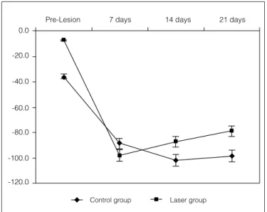
Figure 2. Graph representing means and standard deviation on the different SFI evaluation dates, for both groups.
Table 1. The different SFI evaluation dates, for both groups, with their means and standard deviation, respectively. Note comparative analyze
between the groups, where: axb<0.05; axc, axd<0.01; exf, exg, exh<0.001 and other comparisons didn’t demonstrate significant difference.
Rats
Sciatic functional index
Control Laser
Pre lesiona 7daysb 14daysc 21daysd Pre lesione 7daysf 14daysg 21daysh
R1 -6.60 -79.72 -93.30 -93.30 -9.53 -125.48 -93.30 -76.73
R2 -18.38 -93.30 -124.53 -62.53 -25.88 -79.59 -75.30 -90.81
R3 -24.91 -128.91 -141.94 -135.81 -1.58 -93.30 -93.30 -83.75
R4 -133.07 -77.90 -84.94 -91.53 23.16 -116.28 -93.30 -60.13
R5 -19.48 -77.90 -73.49 -65.19 -27.18 -93.30 -93.30 -87.91
R6 -12.71 -93.30 -93.02 -143.38 -2.97 -81.17 -75.91 -74.59
Mean -35.9 -88.8 -101.9 -98.6 -7.3 -98.2 -87.4 -79.0
SD 48.0 23.2 25.9 34.3 18.6 18.7 9.1 11.2
day; and of -98.3±34.3 on the 21st post-surgery day. For the laser therapy group, a mean value of the collected data was obtained of -7.3±18.3 on the pre-injury moment; -98.2±18.7 on the seventh post-surgery day; -87.4±9.1 on the 14th post-surgery day; and -79.0±11.2 on the 21st post-surgery day. The ANOVAs of the control group, for the different days of the SFI collection, showed that only the pre-injury values were sta-tistically significant (p<0.001) when compared to the other days (Table 1). For the laser therapy group, the pre-injury values compared to the seventh, 14th, 21st days post-surgery showed statistically significant differences (p<0.001). Howe-ver, there were no difference between the seventh, 14th, 21st days post-surgery (p>0.05).
In comparison between the two experimental groups (con-trol versus laser) it was noticed that there were no signiicant
diferences in the values between the four periods of evaluation (p>0.05) as show in Figure 2.
Discussion
There are evidences, in experimental and clinical rese-arches11 that one of the laser’s effects is to improve nerve function, to avoid wound development12, to elevate neurons metabolism and to increase the capacity to produce mye-lin. Since laser therapy is not invasive, the ability to radiate injured nerves without surgical interventions is beneficial. Endo13 claimed that the injury by neurotmesis introduces a number of variables hard to control and to standardize, however we did not find many difficulties in the present study, since the injury was standardized in all animals of both experimental groups and the surgical technique was executed by an experienced professional with specialized training. Another important fact for choosing this type of injury is because of the shortage of studies that use laser in the injury by transaction.
A variety of surgical procedures has been used in the re-pair of peripheral nerves, including epineural and perineural repair, autogenic grafts, vein grafts and entubulation, with or without association of neurotrophic factors14. he simple epi-neural anastomosis was the adopted technique because of its easy execution and because it has shown high biomechanical resistance to traction, according to Temple et al.15.
219
intensity laser therapy (LILT) employs doses of 1 to 4J/cm2, associated to an output power between 10 and 90mW, being widely used in various musculoskeletal injuries, in addition to the algic and inlammatory processes12. Based on this fact the use of a density of 4J/cm2 in this study was justiied. It is important to highlight that this parameter is widely variable in the researches using laser therapy on nerve regeneration. he use of LILT as a therapeutic method still has contradic-tions and its biomodulator efect over the peripheral nerves is obscure yet, since some studies show positive results18,21,22 while others indicate that the laser has no inluence over the peripheral nerves regeneration19.
The use of the gait track is a very common method of assessment23 that has a wide applicability on experimental researches for its easy execution and low cost of the me-thod. Some researches are trying to modernize this data collection with the use of digital cameras making it possible a more dynamic evaluation3. However, the purpose of this research better fits in the use of the conventional method with a track of wood and the use of ink nanjing. The values obtained in this study, related to the SFI, showed that, af-ter the nerve injury by complete transection, there was a severe functional loss in both experimental groups on the seventh day post-surgery, however, in the control group the functional index reduced even more on the 14th and 21st day while, in the same period, the laser therapy group showed functional improvement compared to the seventh day, although the statistical analysis did not show significant results between groups.
A probable explanation for the low SFI of the irradiated group on the seventh day post-surgery was the fact that, on the irst hours after the axon rupture, the cellular body starts to present alterations (cromatolisis), histologically charac-terized by cell ingurgitation, Nissil substance degeneration (neuron rough endoplasmic reticulum) and nucleus migra-tion from the center to the periphery. hese alteramigra-tions, in spite of the neurotransmitters production, aim at increasing protein synthesis (actin and tubulin), which are related to the axon cytoskeleton regeneration, and afect intracellular transportation and growth cone movement24. Probably, the seven days period after injury is marked by these events, but the use of laser therapy within the 24 hours after injury, could reduce the immediate functional loss, conirming the Dahlin25 claim. After the seventhday post-surgery, group 2 (laser the-rapy) presented a line of upward trend regarding to functional improvement, and this line was stabilized and upward only in group 1 (control) after the 14th day. On the 21st post-surgery day it was observed that there were no diferences between the SFI of laser and control groups. Nevertheless, when the evolution of the curves on Figure 1 is analyzed, it is observed
that the mean values were diferent, showing a probable con-tinuity of the laser action over the sciatic nerve.
De Medinacelli26 found that after one month and a half to two months after a crushing injury, functional recovery reaches its plateau, although there is no morfometrically signiicant change. An important fact to be mentioned is the functional level previous to the injury, noting that in both groups, in the evaluation period, they did not present any signiicant statistical diference, thus showing the uniformity of this research sample.
Although there is no statistical significance between the experimental groups, when the SFI is evaluated, it can be observed that on group 2 (laser) there were no signs of infections and points of suture dehiscence, which was observed in some animals from the control group (dehis-cences). Another observed fact was that the animals from this group often used the injured limb for support during feeding, standing up on two members and even to scratch themselves, while in the control group there was dehis-cence formation, making it clear the difficulty to heal the injury of adjacent tissues.
The molecular base that could justify the laser the-rapy effectiveness over the nerve regeneration is not clear. Karu27 verified that the irradiation of isolated mitochon-dria induced positive changes over the cell homeostasis. He suggested that some components of the respiratory chain (cytochromes, flavins and dehydrogenase) are able to absorb the light from a certain wavelength. Thus, this absorption results in an increase of ATP synthesis, affecting the cell levels of hydrogen, activating the ionic gradient (so-dium, potassium, calcium).
220
References
1. Rodríguez FJ, Valero-Cabré A, Navarro X. Regeneration and functional recovery following peripheral nerve injury. Drug Discovery Today: Disease Models. 2004;1(2):177-85.
2. Noble J, Munro CA, Prasad VS, Midha R. Analysis of upper and lower extremity peripheral nerve injuries in a population of patients with multiple injuries. J Trauma. 1998;45(1):116-22.
3. Dijkstra JR, Meek MF, Robinson PH, Gramsbergen A. Methods to evaluate functional nerve recovery in adult rats: walking track analysis, video analysis and the withdrawal reflex. J Neurosci Methods. 2000;96(2):89-96.
4. Varejão AS, Meek MF, Ferreira AJA, Patrício JAB, Cabrita AMS. Functional evaluation of peripheral nerve regeneration in the rat: walking track analysis. J Neurosci Methods. 2001;108(1):1-9.
5. de Medinaceli L, Freed WJ, Waytt RJ. An index of the functional conduction of the rat sciatic nerve based on measurements made from walking tracks. Experimental Neurol. 1982;77(3):634-43.
6. Mendonça AC, Barbieri CH, Mazzer N. Directly applied low intensity direct electric current enhances peripheral nerve regeneration in rats. J Neurosci Methods. 2003;129(2):183-90.
7. Raso VV, Barbieri CH, Mazzer N, Fasan VS. Can therapeutic ultrasound influence the regeneration of the peripheral nerves? J Neurosci Methods. 2005;142(2):185-92.
8. Gigo-Benato D, Geuna S, Rochkind S. Phototherapy for enhancing peripheral nerve repair: a review of the literature. Muscle Nerve. 2005;31(6):694-701.
9. Basford JR. Low intensity laser therapy: still not an established clinical tool. Lasers Surg Med. 1995;16(4):331-42.
10. Lago Junior O, Bortolletto CV, Araújo AM, Donoso CPM, Kume PK, Repka JCD. Avaliação funcional e histológica do reparo de nervo ciático utilizando cola de fibrina e sutura em ratos Wistar. Rev Bras Ortop. 2005;40(1/2):69-78.
11. Bagis S, Comelekoglu U, Sahin G, Buyukakilli B, Erdogan C, Kanik A. Acute electrophysiologic effect of pulsed gallium-arsenide low energy laser
irradiation on configuration of compound nerve action potential and nerve excitability. Lasers Surg Med. 2002;30(5):376-80.
12. Carvalho PTC, Mazzer N, dos Reis FA, Belchior ACG, Silva IS. Analysis of the influence of low-power HeNe laser on the healing of skin wounds in diabetic and non-diabetic rats. Acta Cir Bras. 2006;21(3):177-83.
13. Endo C. Estudo dos efeitos do tratamento com laser num modelo experimental de lesão nervosa por esmagamento do nervo ciático em ratos [dissertação de Mestrado]. São Paulo (SP): USP; 2002.
14. De Sá JM, Mazzer N, Barbieri CH, Barreira AA. The end-to-side peripheral nerve repair. Functional and morphometric study using the peroneal nerve of rats. J Neurosci Methods. 2004;136(1):45-53.
15. Temple CLF, Ross DC, Dunning CE, Johnson JA. Resistance to disruption and gapping of peripheral nerve repairs: an in vitro biomechanical assessment of techniques. J Reconstruc Microsurg. 2004;20(8):645-50.
16. Schwartz F, Brodie C, Appel E, Kazimirsky G, Shainberg A. Effect of helium/ neon laser irradiation on nerve growth factor synthesis and secretion in skeletal muscle cultures. J Photochem Photobiol B. 2002;66(3):195-200.
17. Rochkind S, Nissan M, Alon M, Shamir M, Salame K. Effects of laser irradiation on the spinal cord for the regeneration of crushed peripheral nerve in rats. Lasers Surg Med. 2001;28(3):216-9.
18. Snyder SK, Byrnes KR, Borke RC, Sanches A, Anders JJ. Quantitation of calcitonin gene-related peptide mRNA and Neuronal cell death in facial motor nuclei following axotomy and 633 nm low power laser. Lasers Surg Med. 2002;31(3):216-22.
19. Walsh DM, Baxter GD, Allen JM. Lack of the effect pulsed low-intensity infrared (820 nm) laser irradiation on nerve conduction in the human superficial radial nerve. Lasers Surg Med. 2000;26(5):485-90.
20. Shin DH, Lee E, Hyun J, Lee SJ, Chang YP, Kim J, et al. Growth-associated protein-43 is elevated in the injured rat sciatic nerve after low power irradiation. Neurosci Lett. 2003;344(2):71-4.
21. Byrnes KR, Waynant RW, Ilev IK, Wu X, Barna L, Smith K, et al. Light promotes regeneration and functional recovery and alters the immune response after spinal cord injury. Lasers Surg Med. 2005;36(3):171-85.
related to analgesic effects and with an increase on micro-circulation. However, new studies should be implemented, to investigate the role of different wave lengths and other parameters established by several repairing techniques, with the purpose of clarifying the laser therapy’s effects on this type of injury.
Conclusion
221 22. Nicolau RA, Martinez MS, Rigau J, Tomàs J. Effect of power 655 nm diode
laser irradiation on the neuromuscular junctions of the mouse diaphragm. Lasers Surg Med. 2004;34(3):277-84.
23. Koka R, Hadlock TA. Quantification of functional recovery following rat sciatic nerve transaction. Exp Neurol. 2001;168(1):192-5.
24. Johnson EO, Zoubos AB, Soucacos PN. Regeneration and repair of peripheral nerves. Injury. 2005;36 Suppl 4:S24-9.
25. Dahlin LB. The biology of nerve injury and repair. J Am Soc Surg Hand. 2004;4(3):143-55.
26. de Medinaceli L. Interpreting nerve morphometry data after experimental traumatic lesions. J Neurosci Methods. 1995;58(1-2):29-37.
27. Karu TI. Molecular mechanisms of the therapeutic effect of low-intensity laser irradiation. Lasers Life Sci. 1988;2:53-74.
28. Woodruff LD, Bounkeo JM, Brannon WM, Dawes KS, Barham CD, Waddell DL, et al. The efficacy of laser therapy in wound repair: a meta-analysis of the literature. Photomed Laser Surg. 2004;22(3):241-7.