From the Department of Pediatrics, Hospital das Clínicas, Faculty of Medicine, University of São Paulo.
URINARY TRACT INFECTION IN FULL-TERM
NEWBORN INFANTS: RISK FACTOR ANALYSIS
Mário Cícero Falcão, Cléa Rodrigues Leone, Renata A. P. D’Andrea, Roberta Berardi, Nilce A. Ono and Flávio Adolfo Costa Vaz
RHCFAP/2997
FALCÃO MC et al. - Urinary tract infection in full-term newborn infants: risk factor analysis. Rev. Hosp. Clín. Fac. Med. S. Paulo 55 (1):9-16, 2000. SUMMARY: Objective: To analyze the correlation of risk factors to the occurrence of urinary tract infection in full-term newborn infants. Patients and methods: Retrospective study (1997) including full-term infants having a positive urine culture by bag specimen. Urine collection was based on: fever, weight loss > 10% of birth weight, nonspecific symptoms (feeding intolerance, failure to thrive, hypoactivity, debilitate suction, irritability), or renal and urinary tract malformations. In these cases, another urine culture by suprapubic bladder aspiration was collected to confirm the diagnosis. To compare and validate the risk factors in each group, the selected cases were divided into two groups: Group I – positive urine culture by bag specimen collection and negative urine culture by suprapubic aspiration, and Group II – positive urine culture by bag specimen collection and positive urine culture by suprapubic aspiration .
Results: Sixty one infants were studied, Group I, n = 42 (68.9%) and Group II, n = 19 (31.1%). The selected risk factors (associated infectious diseases, use of broad-spectrum antibiotics, renal and urinary tract malformations, mechanical ventilation, parenteral nutrition and intravascular catheter) were more frequent in Group II (p<0.05). Through relative risk analysis, risk factors were, in decreasing importance: parenteral nutrition, intravascular catheter, associated infectious diseases, use of broad-spectrum antibiotics, mechanical ventilation, and renal and urinary tract malformations.
Conclusion: The results showed that parenteral nutrition, intravascular catheter, and associated infectious diseases contributed to increase the frequency of neonatal urinary tract infection, and in the presence of more than one risk factor, the occurrence of urinary tract infection rose up to 11 times.
DESCRIPTORS: Urinary tract infection. Risk factor. Newborn infant.
Usually, urinary tract infection in the neonatal period manifests after 72 hours of life. The incidence is between 0.1 to 10% in all infants. It is more fre-quent in male and preterm newborn in-fants, and may be as high as 10% in low birth weight infants17,20. This
inci-dence is directly proportional to the presence of several risk factors for this disease. Among the risk factors, beside those previously mentioned, are the presence of associated infectious dis-eases (sepsis, bronchopneumonia, sep-tic arthritis, osteomyelitis, meningitis, etc.), the use of broad-spectrum anti-biotics, mechanical ventilation, parenteral nutrition, intravascular cath-eter, and prolonged stay in the
inten-sive care unit1,7,20. Renal and urinary
tract malformations may be present in neonates with a urinary tract infection8.
Urinary tract infections are of inter-est to neonatologists for several rea-sons. First, they can be asymptomatic, so the diagnosis must be made by the examination and culture of a properly obtained specimen of urine7,11. Second,
they may indicate a serious underlying abnormality of the urinary tract, such as obstructive uropathy. Third, they
may have long-term consequences in-sofar as they may produce kidney dam-age, which may lead later in life to hy-pertension, recurrent infections, and kidney failure.
The knowledge of neonatal urinary tract infection risk factors or predispos-ing factors is important for identifypredispos-ing appropriate situations for performing serial urine culture to obtain an early
diagnosis and adequate treatment13.
The knowledge of the most important risk factors in a neonatal lower risk population is also important.
PATIENTS AND METHOD
We conducted a retrospective study in the Child Institute “Prof. Pedro Alcântara” of the Hospital das Clínicas, University of São Paulo, Faculty of Medicine, including all full-term new-born infants (gestational age – 37 to 42 weeks) presenting positive urine cul-ture by bag specimen collection.
Urine collection was based on clini-cal data as follows: fever (T>37.8o C)24,
weight loss > 10% of birth weight, or nonspecific symptoms (feeding intoler-ance, failure to thrive, hypoactivity, de-bilitate suction, irritability). In these cases, another urine culture was col-lected by suprapubic bladder aspiration to confirm diagnosis.
To compare and validate the risk factors in each group, the selected cases were divided into two groups: Group I – positive urine culture by bag specimen collection and negative urine culture by suprapubic aspiration, and Group II – positive urine culture by bag specimen collection and positive
urine culture by suprapubic aspiration. The selected risk factors were: associ-ated infectious pathologies, use of broad-spectrum antibiotics, renal and urinary tract malformations, mechani-cal ventilation, parenteral nutrition, and intravascular catheter3,6,17.
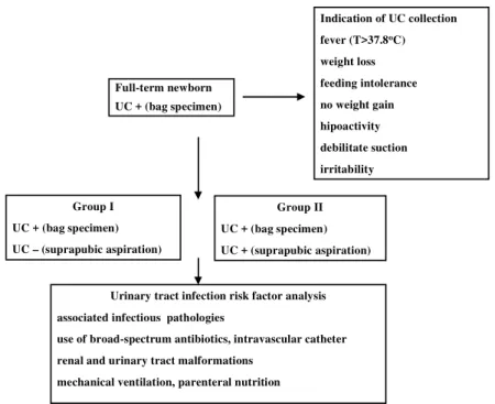
Figure 1 shows the design of the study.
The finding of 100 000 CFU/ml of the same microorganisms defined the positivity of the bag specimen
collec-tion26. A suprapubic aspiration
speci-men was defined as positive at any CFU/ml rate26.
The population of study was char-acterized by gestational age, birth weight, sex, perinatal asphyxia (Apgar score < 6 at 5 minute), and membrane rupture time (<24 hours and ≥ 24 hours). Symptoms were: fever (T>37.8o C), weight loss > 10% of birth
weight or nonspecific symptoms (feed-ing intolerance, failure to thrive, hypoactivity, debilitate suction, or irri-tability).
The technique of bag specimen
col-lection was: antisepsy and sterile plas-tic bag fitted on the genitalia25. If
di-uresis would not occur within 30 min-utes, procedure would have been re-peated until success25.
The suprapubic aspiration
speci-men required a full bladder16,23. The
overlying skin was disinfected, the bladder was punctured above the sym-physis pubis with a 25-gauge needle on a syringe, and about 2 ml of urine was aspirated2,21.
All urine specimens were
trans-ported promptly to the laboratory9.
Urine specimens were processed onto CLED, MacConkey, Thayer-Martin, thioglicolate, and blood agar plates. Culture plates were incubated at 35–
37o C and read at 24–48 hours; if
re-sults were positive, the antibiogram
was determinated9. The absence of
mi-croorganisms within 48 hours charac-terized the culture as negative9.
Data were analyzed through
chi-square, Fischer exact test, Student t
test, and relative risk. Significance was defined as p<0.05.
Figure 1 - Design of the study. UC = urine culture
Full-term newborn UC + (bag specimen)
Indication of UC collection fever (T>37.8oC) weight loss feeding intolerance no weight gain hipoactivity debilitate suction irritability
Urinary tract infection risk factor analysis associated infectious pathologies
use of broad-spectrum antibiotics, intravascular catheter renal and urinary tract malformations
mechanical ventilation, parenteral nutrition Group I
UC + (bag specimen) UC – (suprapubic aspiration)
RESULTS
Sixty-one full-term newborn infants were included in this study, represent-ing 5.1% of the total infants born alive in the study period. The diagnosis was confirmed (positive urine culture by suprapubic aspiration) on 19/61 (31.1%) of full-term infants included in the study (1.6% of the total infants born alive).
The infants were distributed in two groups according to the positivity of urine culture collected by suprapubic aspiration. Group I had 42 infants (68.9%) and Group II had 19 infants (31.1%).
The population of the study is shown in the Table 1 and clinical manifestations are shown in Table 2 and Fig. 2.
No complication occurred from specimen collection by the suprapubic method.
The microbial content of cultures of
urine specimens was: Escherichia coli
(42.9%), Staphylococcus aureus
(10.5%) Staphylococcus coagulase
-negative (26.3%), Enterococcus
faecalis (15.8%) and Klebsiela
pneumoniae (5.3%).
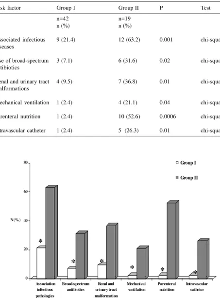
The studied risk factors (associated infectious pathologies, use of broad-spectrum antibiotics, renal and urinary tract malformations, mechanical venti-lation, parenteral nutrition, and intra-vascular catheter) were more frequent in Group II (p<0.05). These results are described in Table 3 and Fig. 3.
Nine newborn infants (21.5%) pre-sented infectious pathologies: ompha-litis (33.3%), pyodermitis (33.3%), bronchopneumonia (22.2%), and sep-sis (11.1%) in the Group I. In Group II this value was increased to 63.2% (meningitis - 8.3%, omphalitis - 25%, bronchopneumonia - 25%, and sepsis - 41.6%).
The urological malformations were as follows: hydronephrosis (27.3% in Group I and 9.1% in Group II), ure-teropelvic junction obstruction (27.3%
Table 1 - Population of the study – Group I and Group II.
Group I Group II P Test
n=42 n=19
Birth weight (g) Average 3399.52 3171.05 0.07 Student t
Variation 2560-4270 2420-4260
SD 418.36 515.08
Sex n (%) n (%)
Male 16 (38.1) 10 (52.6) 0.28 chi-square
Female 26 (61.9) 9 (47.4) 0.28 chi-square
Asphyxia
Apgar 5’ < 6 1 (2.4) 3 (15.8) 0.14 chi-square
Membrane 7(16.7) 5 (26.3) 0.47 chi-square
Rupture time
≥ 24h
Table 2 - Clinical data – Group I and Group II.
Group I Group II P Test
N=42 n=19
n (%) n (%)
Fever (T > 37.8ºC) 38 (90.5) 15 (78.9) 0.31 chi-square
Weight loss > 10% 20 (47.6) 3 (15.8) 0.01 chi-square
of birth weight
Nonspecific 4 (9.5) 10 (52.6) 0.0004 chi-square
symptoms
0 20 40 60 80 100
N (% )
Fever Weight los Non-s pecific s ymptomatology
Group I Group II
*
*
in Group II), posterior uretero valves (27.3% in Group II, and policystic kid-ney (9.1% in Group II).
Correlation between risk factors and urinary tract infection, in both studied groups, using relative risk analysis is shown in Table 4.
Table 5 shows the same analysis, drawing from more than one risk factor.
DISCUSSION
The first description of urinary tract
infection in the neonatal period dates from the beginning of the century, and the observations made are still valid. Due to the absence of specific symp-toms, the confirmation of diagnosis is based on laboratory data4. The clinical
presentation may vary from fever and other signs of sepsis to minimal changes, or the infant may be without signs4.
Until the decade of 1960, studies of urinary tract infection in the neona-tal period were discrepant because they presented different criteria to define
this infection and used divergent meth-odologies17.
Currently, research indicates that urinary tract infection incidence is cor-related to the presence of risk factors. The environment of the studied popu-lation, i.e., the intensive care unit or the nursery, should be taken into consider-ation.
The study population had a urinary tract infection frequency of 1.6%, a rate close to the low incidence limit of
this disease in the neonatal period12.
This finding may be due to the low risk in this study population, because in the absence of risk factors the full-term in-fant rarely develops urinary tract infec-tion.
The population of this study (61 full-term infants) presented nonspecific symptoms (Table 2 – Fig. 2) and posi-tive urine culture collected by bag specimen, which was considered to be the presumptive urinary tract infection diagnosis. Diagnosis (positive urine culture by suprapubic aspiration) was confirmed in 19 full-term newborn in-fants, emphasizing the need for this method to make accurate etiologic di-agnosis.
Another consideration concerning the incidence of the urinary tract infec-tion is its relainfec-tionship to associated in-fectious pathologies; when newborn infants develop sepsis, for instance, there is a higher chance of developing urinary tract infection5,6. The results
showed that in Group II, presence of infection was one of the most prevalent associated risk factors (63.2%), with significant statistical difference as com-pared to Group I (Table 3 – Fig. 3). Therefore, urinary tract infection should be considered in all infants who have signs of sepsis.
Prematurity and male gender are classically called risk factors8. There is
also a proportional relationship with the presence of renal and urinary tract
malformations17. Furthermore, a
pro-longed stay in the nursery may be
con-Table 3 - Risk factors for urinary tract infection.
Risk factor Group I Group II P Test
n=42 n=19
n (%) n (%)
Associated infectious 9 (21.4) 12 (63.2) 0.001 chi-square
diseases
Use of broad-spectrum 3 (7.1) 6 (31.6) 0.02 chi-square
antibiotics
Renal and urinary tract 4 (9.5) 7 (36.8) 0.01 chi-square
malformations
Mechanical ventilation 1 (2.4) 4 (21.1) 0.04 chi-square
Parenteral nutrition 1 (2.4) 10 (52.6) 0.0006 chi-square
Intravascular catheter 1 (2.4) 5 (26.3) 0.01 chi-square
0 20 40 60 80
N (%)
Association infectious pathologies
Broad-spectrum antibiotics
Renal and urinary tract malformation
Mechanical ventilation
Parenteral nutrition
Intravascular catheter
Group I
Group II
*
*
*
*
*
*
sidered as a risk factor, for instance, prematurity, perinatal asphyxia, me-chanical ventilation, and inadequate nutrition. The incidence of neonatal urinary tract infection as a complica-tion of intensive care has risen sharply in recent years.
Risk factors included in this re-search (associated infectious patholo-gies, use of broad-spectrum antibiotics, renal and urinary tract malformations,
mechanical ventilation, and intravascu-lar catheter) were more frequent (p<0.05) in Group II (Table 3 – Fig. 3). Congenital renal and urinary tract defects, mainly the obstructive anoma-lies17,19, exhibit a close relationship
with urinary tract infection, and these were more frequent (p<0.05) in Group II (Table 3 – Fig. 3).
The male presents a high incidence of renal and urinary tract
malforma-tions and sepsis, leading us to believe that these malformations increase the frequency of urinary tract infections. Circumcision seems to reduce the over-all incidence of urinary tract infections, although a few studies have suggested that this procedure may be a predispos-ing factor for urinary tract infection, mainly due to the complications
en-countered10. In this study group, the
absence of circumcision led us to ex-clude this procedure as a risk factor or protector factor of neonatal urinary tract infection. In our study, Group II presented no difference related to gen-der. The low sample size is one of the factors that could explain this finding.
Natural defenses in the urinary tract include antibacterial properties of urine and urinary tract mucosa, anti-adher-ence mechanisms, machanical effects of urinary flow, presence of phagocytic
cells, and immune mechanisms22. In
the neonatal period, these mechanisms may fail, facilitating an urinary tract infection3. Furthermore, in the neonate,
it is frequently difficult to know whether an urinary tract infection is the cause or the result of bacteremia14.
Another factor to be considered is the use of broad-spectrum antibiotics, since use of these antibiotics is neces-sary for treatment of undetermined in-fection. Use of broad spectrum antibi-otics may change neonate natural flora, increasing infections by opportunistic
microorganisms18. Fungal urinary tract
infections, mainly Candida albicans
well exemplify this fact15.
Table 3 and Fig. 3 reinforce that the use of broad-spectrum antibiotics (cefotaxime and vancomycin) can be considered as a factor risk, through analyzed data-frequency 31.6% (p<0.05).
All invasive procedures, even with antisepsy, could lead to a bacterial con-tamination, especially in the neonatal period. Intravascular catheterization, often necessary to deliver prescription drugs or parenteral nutrition, can
facili-Table 4 - Relative risk (RR) analysis of the risk factors for urinary tract infection.
Risk factors RR CI:95% P Test
Associated infectious 3.27 1.51-7.04 0.001 chi-square
pathologies
Use of broad-spectrum 3.03 1.51-6.08 0.012 Fisher exact
antibiotics
Renal and urinary tract 2.97 1.57-5.64 0.007 Fisher exact
malformations
Mechanical ventilation 2.99 1.61-5.53 0.029 Fisher exact
Parenteral nutrition 5.05 2.72-9.39 0.0009 Fisher exact
Intravascular catheter 3.27 1.84-5.83 0.009 Fisher exact
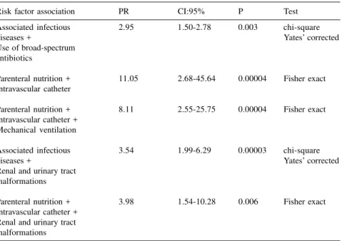
Table 5 - Relative risk (RR) analysis using more than one risk factor for urinary
tract infection.
Risk factor association PR CI:95% P Test
Associated infectious 2.95 1.50-2.78 0.003 chi-square
diseases + Yates’ corrected
Use of broad-spectrum antibiotics
Parenteral nutrition + 11.05 2.68-45.64 0.00004 Fisher exact Intravascular catheter
Parenteral nutrition + 8.11 2.55-25.75 0.00004 Fisher exact Intravascular catheter +
Mechanical ventilation
Associated infectious 3.54 1.99-6.29 0.00003 chi-square
diseases + Yates’ corrected
Renal and urinary tract malformations
Parenteral nutrition + 3.98 1.54-10.28 0.006 Fisher exact
tate a bacteremia and consequently a urinary tract infection. Also, intubation, a necessary mechanical ventilation pro-cedure, can provoke infection.
The frequency of mechanical ven-tilation, intravascular catheterization, and parenteral nutrition (Table 3 – Fig. 3) was higher in Group II.
Finally, all analyzed risk factors in this research (associated infectious pa-thologies, use of broad-spectrum anti-biotics, renal and urinary tract malfor-mations, mechanical ventilation, parenteral nutrition, and intravascular catheter) were present at high frequen-cies in Group II (Table 3 – Fig. 3), with statistical significance (p<0.05). These
findings support the relationship be-tween risk factors and urinary tract in-fection in the neonatal period.
The relative risk of urinary tract in-fection risk factors was, in decreasing importance: parenteral nutrition, intra-vascular catheter, associated infectious diseases, use of broad-spectrum antibi-otics, mechanical ventilation, and renal and urinary tract malformations (Table 4).
Analysis using more than one uri-nary tract infection risk factor (Table 5) revealed an increased relative risk, ris-ing from 2.95 to 11.05. The combina-tion of intravascular catheter and
parenteral nutrition had the highest re-lationship with neonatal urinary tract infection frequency.
In conclusion, the results of this study showed that the presence of pre-disposing factors or risk factors (parenteral nutrition, intravascular catheter, associated infectious patholo-gies, use of broad-spectrum antibiotics, mechanical ventilation, and renal and urinary tract malformations) can con-tribute to increased urinary tract infec-tion frequency in the neonatal period, and in the presence of more than one risk factor, the urinary tract infection occurrence rose up to 11 times.
RESUMO RHCFAP/2997
FALCÃO MC e col. - Infecção urinária em recém-nascido de termo: análise de fatores de risco.
Rev. Hosp. Clín. Fac. Med. S. Paulo 55 (1):9-16, 2000.
Objetivos: Analisar a contribuição
dos fatores de risco para a ocorrência de infecção urinária em recém-nascidos de termo.
Casuística e metodologia: Estudo
retrospectivo (1997), incluindo recém-nascidos de termo com urocultura positiva por saco coletor. A indicação desta coleta foi baseada em:
hipertermia (T>37,8oC), perda de
peso>10% do peso de nascimento,
alterações do estado geral (recusa alimentar, ganho insuficiente de peso e hipoatividade) ou presença de malformações nefro-urológicas. Nesses recém-nascidos foi realizada punção suprapúbica para confirmação diagnóstica. Os recém-nascidos foram divididos em dois grupos, segundo o resultado das uroculturas: Grupo I (diagnóstico presuntivo de infecção urinária) e Grupo II (diagnóstico confirmado de infecção urinária), para avaliação dos fatores de risco pela análise do risco relativo.
Resultados: Foram estudadas 61
crianças (5,1% dos recém-nascidos de termo) - Grupo I n=42 (68,9%) e
Grupo II n=19 (31,1%). Os fatores de risco avaliados (patologias infecciosas associadas, uso prévio de antibióticos, malformações nefro-urológicas, ventilação mecânica, nutrição parenteral e o uso de cateteres) foram mais freqüentes no Grupo II (p<0,05). Analisando-se esses fatores, através de risco relativo, encontrou-se, em ordem decrescente de importância: nutrição parenteral, uso de cateteres, patologias infecciosas associadas, antibiótico prévio, ventilação mecânica e a presença de malformações nefro-urológicas.
Conclusões: Os resultados
parenteral, uso de catéteres e patologias infecciosas associadas contribuiram para aumentar a freqüência da infecção do
trato urinário no período analisado e, na presença de mais de um desses fatores, o risco elevou-se em até 11 vezes.
DESCRITORES: Infecção
uriná-ria. Fatores de risco. Recém-nascido.
REFERENCES
1. BACHUR R & CAPUTO GL - Bacteremia and meningitis among infants with urinary tract infections. Pediatr Emerg Care 1995;11:280-284.
2. BARKEMEYER BM - Suprapubic aspiration in very low birth weight infants. Pediatrics 1993; 89: 457-458.
3. BENADOR D, BENADOR N, SLOSMAN D et al. - Are younger children at highest risk of renal sequelae after pyelonephritis. Lancet1997; 9:349-317.
4. BRION CP, SATLIN LM & EDELMANN CM - Renal disease. In: AVERY GB, FLETCHER MA & MACDONALD MG eds. Neonatology: pathophysiology and management of the newborn. 4th ed. Philadelphia, Lippincott, 199. p. 792-886.
5. CHIU C, LIN T & BULLARD MJ - Identification of febrile neonates unlikely to have bacterial infections. Pediatr Infect Dis J 1997; 16:59-63.
6. CRAIN EF & GERSHEL JC - Urinary tract infection in febrile infants younger than 8 weeks of age. Pediatrics 1990; 86:363-367.
7. HANSSON S, BRANDSTROM P, JODAL U et al. - Low bacterial counts in infants with urinary tract infection. J Pediatr 1998; 2:132:180.
8. HELLERSTEIN S - Urinary tract infections: old and new aspects. Pediatr Clin North Am 1995; 42:1433-1457.
9. HENRY JB, LANZON RB & SCHUMANN GB - Basic examination of urine. In: HENRY JB ed. Clinical Diagnosis and management by laboratory methods. 19th. Philadelphia, Saunders, 1996. p. 411-56.
10. HERZOG LW - Urinary tract infection and circumcision. A case-control study. Am J Dis Child 1989; 143:348-350.
11. HOBERMAN A, WALD ER, PENCHANSKY L et al. - Enhanced urinalysis as a screening test for urinary tract infection. Pediatrics 1993; 89: 1196-1198.
12. HOBERMAN A & WALD ER - Urinary infections in young children. Pediatr Infect Dis J 1997; 16:11-17.
13. HOBERMAN A - Is urine culture necessary to rule out urinary tract infection in young febrile children? Pediatr Infect Dis J 1996; 15:304-309.
14. JAMESELLISON MY, ROBERTS R, VERRIERJONES K et al. -Mucosal immunity in the urinary tract: changes in sIgA, FSC and total IgA with age and in urinary tract infection. Clin Nephrol 1997; 48:69-78.
15. KARLOWICZ G & PHILLIPS JR - Prevalence of Candida species in hospital-acquired urinary tract infections in a neonatal intensive care unit. Pediatr Infect Dis J 1997; 16:190-194.
16. KIERMAN SC, PINCKERT TL & KESLER M - Ultrasound guidance of suprapubic bladder aspiration in neonates. J Pediatr 1993; 123:789-791.
17. KLEIN JO & LONG SS - Bacterial infections of the urinary tract. In: REMINGTON JS & KLEIN JO eds. - Infectious diseases of the fetus and newborn. 4th. Philadelphia, Saunders, 1995. p. 925-34.
18. LOHR JA, DOWNS SM, DUDLEY S et al. - Hospital acquired urinary tract infections in pediatric patient: a prospective study. Pediatr Infect Dis J 1994; 13:8-12.
19. MAN P, CLAESON I, JOHANSON IM et al. - Bacterial attachment as a predictor of renal abnormalities in boys with urinary tract infection. J Pediatr 1989; 115:915-922.
20. MITCHELL CK, FRANCO SM & VOGEL RL - Incidence of urinary tract infection in a inner-city outpatient. J Perinatol 1995; 15:131-134.
21. MORELLL RE, DURITZ G & OLTORF C - Suprapubic aspiration associated with hematoma. Pediatrics 1982; 69:455.
22. PISACANE A, GRAZIANI L, MAZZARELLA G et al. - Breast-feeding and urinary tract infection. J Pediatr 1992; 120:87-89.
24. VOORA S, SNIRIVASAN G, LILIEN LD et al. - Fever in full-term newborns in the first four days of live. Pediatrics 1982; 69:40-44. 25. WOODA GL - Specimen collection and handling for diagnosis of infectious diseases. In: HENRY JB ed. - Clinical Diagnosis and management by laboratory methods. 19th. Philadelphia,
Saunders, 1999. p. 1311-1334.
26. WOODS GL, AYERS LW, WASHINGTON JA et al. - Medical bacteriology. In: HENRY JB ed. – Clinical Diagnosis and management by laboratory methods. 19th. Philadelphia,
Saunders, 1996. p. 1132-1169.