Elevated gamma glutamyl transferase levels are
associated with the location of acute pulmonary
embolism. Cross-sectional evaluation in hospital setting
Níveis elevados de gama glutamil transferase estão associados com a localização de
embolia pulmonar aguda. Estudo transversal em ambiente hospitalar
Ozge Korkmaz
I, Hasan Yucel
II, Ali Zorlu
II, Ocal Berkan
III, Hakki Kaya
II, Sebahattin Goksel
I, Osman Beton
II, Mehmet Birhan Yilmaz
IVDepartment of Cardiology, Cumhuriyet University Medical Faculty, Sivas, Turkey
ABSTRACT
CONTEXT AND OBJECTIVE: The location of embolism is associated with clinical indings and disease severity in cases of acute pulmonary embolism. The level of gamma-glutamyl transferase increases under oxidative stress-related conditions. In this study, we investigated whether gamma-glutamyl transferase levels could predict the location of pulmonary embolism.
DESIGN AND SETTING: Hospital-based cross-sectional study at Cumhuriyet University, Sivas, Turkey.
METHODS: 120 patients who were diagnosed with acute pulmonary embolism through computed tomography-assisted pulmonary angiography were evaluated. They were divided into two main groups (proximally and distally located), and subsequently into subgroups according to thrombus localization as follows: irst group (thrombus in main pulmonary artery; n = 9); second group (thrombus in main pulmo-nary artery branches; n = 71); third group (thrombus in pulmopulmo-nary artery segmental branches; n = 34); and fourth group (thrombus in pulmonary artery subsegmental branches; n = 8).
RESULTS: Gamma-glutamyl transferase levels on admission, heart rate, oxygen saturation, right ventricu-lar dilatation/hypokinesia, pulmonary artery systolic pressure and cardiopulmonary resuscitation require-ment showed prognostic signiicance in univariate analysis. The multivariate logistic regression model showed that gamma-glutamyl transferase level on admission (odds ratio, OR = 1.044; 95% conidence interval, CI: 1.011-1.079; P = 0.009) and pulmonary artery systolic pressure (OR = 1.063; 95% CI: 1.005-1.124; P = 0.033) remained independently associated with proximally localized thrombus in pulmonary artery.
CONCLUSIONS: The indings revealed a signiicant association between increased existing embolism load in the pulmonary artery and increased serum gamma-glutamyl transferase levels.
RESUMO
CONTEXTO E OBJETIVO: A localização da embolia está associada com os resultados clínicos e a gra-vidade da doença do embolismo pulmonar agudo (EPA). O nível de gama-glutamil transferase (GGT) aumenta em condições relacionadas com estresse oxidativo. Investigou-se se os níveis de GGT podem prever a localização do EPA.
TIPO DE ESTUDO E LOCAL: Estudo observacional transversal na Universidade Cumhuriyet, Sivas, Turquia.
MÉTODOS: Avaliamos 120 pacientes diagnosticados com EPA após a realização de angiograia pulmonar assistida por tomograia computadorizada. Eles foram divididos em dois grupos principais (localização proximal e distal) e depois em subgrupos de acordo com a localização do trombo da seguinte forma: pri-meiro grupo (trombo na artéria pulmonar [AP] principal, n = 9); segundo (trombo no ramo da AP principal; n = 71); terceiro grupo (trombo na segmentar da AP; n = 34); quarto grupo (trombo na subsegmentar da AP; n = 8).
RESULTADOS: Na análise univariada, os níveis de GGT tiveram signiicado prognóstico em relação à ad-missão, pulsação arterial, saturação de oxigênio, dilatação do ventrículo direito/hipocinesia, pressão sistó-lica da artéria pulmonar (PSAP) e necessidade de ressuscitação cardiopulmonar. O modelo de regressão logística multivariada demonstrou que o nível de GGT na admissão (razão de possibilidades, OR: 1,044; 95% intervalo de coniança, CI: 1,011-1,079; P = 0,009) e PSAP (OR: 1,063, 95% CI: 1,005-1,124; P = 0,033) permaneceram independentemente associados com trombo localizado proximalmente na AP.
CONCLUSÕES: Os resultados demonstraram associação signiicativa entre aumento da carga existente de embolia da AP e aumento dos níveis séricos da GGT.
IMD. Associate Professor, Department of
Cardiovascular Surgery, Cumhuriyet University Medical Faculty, Sivas, Turkey.
IIMD. Associate Professor, Department of
Cardiology, Cumhuriyet University Medical Faculty, Sivas, Turkey.
IIIMD. Professor and Head of Department of
Cardiovascular Surgery, Cumhuriyet University Medical Faculty, Sivas, Turkey.
IVMD. Professor and Head of Department of
Cardiology, Cumhuriyet University Medical Faculty, Sivas, Turkey.
KEY WORDS:
Gamma-glutamyltransferase. Pulmonary embolism. Biological markers. Pulmonary artery.
Multidetector computed tomography.
PALAVRAS-CHAVE: Gama-glutamiltransferase. Embolia pulmonar. Marcadores biológicos. Artéria pulmonar.
INTRODUCTION
Acute pulmonary embolism (APE) is a common disease result-ing from acute full or partial obstruction of the pulmonary artery bed and can lead to disability or mortality.1,2 In the majority of APE patients, mortality occurs as a result of hemodynamic fail-ure and, for this reason, mortality rates are signiicantly high.3,4 he general rule for treatment is to diagnose patients as early as possible, so as to determine the risk factors and begin treatment.1 Diferent clinical evaluations, radiological examinations (includ-ing echocardiography) and blood biomarkers are analyzed for diagnosis and risk classiication. he location of the embolism is associated with clinical indings and disease severity.5 Cardiac biomarkers, including troponin and natriuretic peptide, are used as mortality predictors for APE.1-6 Although some studies have indicated a correlation between the location of the embolism and blood D-dimer and creatinine levels, others indicate the oppo-site, i.e. suggesting that there is no correlation.7,8
Gamma-glutamyl transferase (GGT) is a membrane protein that functions in the extracellular catabolism of glutathione. It is mostly synthesized in the liver, but also in various other organs. GGT levels become increased especially in cases of hep-atobiliary dysfunction and alcohol consumption, and recent studies have shown that GGT levels increase under oxidative stress-related conditions, including inlammation, carcinogen-esis, ageing, atherosclerosis and reperfusion injury.9-11 he GGT level is independently associated with the prognosis in certain cardiopulmonary diseases, especially acute coronary syndrome, acute myocardial infarction and heart failure, and has also been described as a predictor of early mortality in cases of acute pul-monary embolism.12-14
OBJECTIVES
he objective of this study was to investigate the association between serum GGT levels and the location of APE.
METHODS
he current study is a subgroup analysis on a study previously published.14 In this study, a total of 192 consecutive patients who were admitted to the emergency unit and then hospitalized with suspected APE were considered for enrolment between January 2009 and July 2011. Excluded from the study were 15 patients with cholecystitis, seven patients with chronic liver disease, 4 patients who were receiving dialysis treatment for chronic renal failure, 5 patients with a previous diagnosis of malignancy, 2 patients who were chronic consumers of alcohol, 20 patients whose serum GGT had not been measured and 12 patients in whom the diagnosis of APE was ruled out through scintigra-phy and/or tomograscintigra-phy (alternative diagnoses were conirmed). Also excluded from the current study were patients who were
diagnosed with APE through imaging methods other than com-puted tomography-assisted pulmonary angiography (ventila-tion/perfusion scintigraphy, n = 2) and those with incomplete medical records (n = 3). In the end, evaluations were made on 122 patients (female/male: 63/59) who were admitted to the emergency service, and who were diagnosed with APE following computed tomography-assisted pulmonary angiography.
Ater the results from contrast spiral computed tomogra-phy had been obtained, the patients were divided into two main groups (proximally and distally located), and these two main groups were then divided into subgroups according to thrombus localization as follows: irst group (thrombus in main pulmonary artery; n = 9); second group (thrombus in main pulmonary artery branches; n = 71); third group (throm-bus in pulmonary artery segmental branches; n = 34); and fourth group (thrombus in pulmonary artery subsegmental branches; n = 8). Embolisms localized in the pulmonary trunk, main pul-monary arteries and lower arteries were classiied as “proximally located”, while lobular-segmental-located and subsegmental embolisms were classiied as “distally located”.
All the patients were evaluated in accordance with the clini-cal, laboratory and radiological indings regarding the location of APE. he following information was obtained from the medi-cal records, and was evaluated retrospectively in detail: identity information, demographic data, symptoms, risk factors, comor-bidities (history of coronary artery disease, diabetes mellitus, hypertension and chronic obstructive pulmonary disease), phys-ical examinations, laboratory indings, 12-lead electrocardiogra-phy, echocardiography and computed tomography-assisted pul-monary angiography indings.
Vital signs and blood samples were obtained within 30 min-utes of admission to the emergency unit. GGT activity was mea-sured by means of a Beckman Coulter Synchron LX20 (Brea, CA, USA) autoanalyzer, using original kits. he laboratory reference limits difer signiicantly according to sex, and were set at 9 U/l to 35 U/l for women and 9 U/l to 40 U/l for men, in accordance with the test kit speciications.
evaluated. Furthermore, the duration of hospitalization ater the APE diagnosis was recorded; and pulmonary embolism-associ-ated early mortality events were recorded as pulmonary embo-lism-dependent in-hospital mortality.
Echocardiography was carried out within the irst 24 hours of admission to the emergency room. All examinations were eval-uated via the Vivid 7 system (GE Healthcare, Wauwatosa, WI, USA) in all participating centers using a 2.5 to 5-MHz probe. he modiied Simpson method was used to calculate ejection fractions, with chamber sizes deined in line with recent guide-lines.15 Right ventricular dysfunction was deined as dilatation of the right ventricle (right ventricle dimension > 3.4 cm at basal plane or > 3.8 cm at the midplane), combined with the pres-ence of McConnell’s sign.15,16 Severe tricuspid regurgitation was deined using color low jet Doppler signal intensity, in combina-tion with vena contracta width, in accordance with recommen-dations.17 Pulmonary artery systolic pressure was calculated as previously described.18
First, posteroanterior lung graphs were produced. Patients with suspected pulmonary embolism were evaluated in detail within the irst 24 hours in the radiology clinic, through contrast spiral computerized tomography imaging (imaging performed with 120 kV, 210-250 mAs, slice thickness of 3 mm, table speed of 5 mm/s, reconstruction index of 2 mm, pitch of 2 and 120 ml of intravenous contrast dye). he pulmonary artery, its branches and the lung parenchyma were examined in detail.
Continuous variables that did not have normal distribu-tion were expressed as mean ± standard deviation (SD) or as the interquartile range, while the categorical variables were expressed as percentages. A receiver operator characteristic curve analysis was carried out to identify the optimal cutof point for GGT (at which sensitivity and speciicity would be maximum), in order to predict the proximal localization of the arteries. he area under the curve (AUC) was calculated as a measure of the accuracy of the tests. he researchers compared the AUC using a Z test. Patients with APE were classiied into two main groups: proximal localization of the artery (Group I) and distal localization of the artery (Group II). Comparisons between groups of patients were made using a χ2 test for cat-egorical variables, an independent-sample t test for normally distributed continuous variables and a Mann-Whitney U test when the distribution was skewed. An evaluation of the cor-relation between the variables was made using either Pearson’s or Spearman’s correlation test. We conducted univariate anal-ysis to quantify the associations of variables with proximally localized thrombus in pulmonary artery. he variables found to be statistically signiicant in the univariate analysis and other potential confounders were used in a multiple logistic regression model using the forward stepwise method in order
to determine the independent prognostic factors for proximal artery localization. In the logistic regression analyses, GGT was evaluated as a continuous variable. All statistical analyses were carried out using the SPSS sotware version 14.0 (SPSS Inc., Chicago, IL, USA), and P values < 0.05 were considered to be statistically signiicant.
RESULTS
Enrolled in the study were 122 consecutive patients with conirmed APE, with a mean age of 64 ± 13 years (52% female, 48% male).
he comparisons between the four subgroups of patients with APE, along with their baseline characteristics and hemo-dynamic, electrocardiographic, echocardiographic and labora-tory indings, are summarized in Table 1. he baseline char-acteristics and all previously mentioned electrocardiographic parameters were similar in all groups. Most of the transtho-racic echocardiograms were examined within the irst 24 hours of emergency unit admission (upon admission for most of the patients), while echocardiographic examinations with extended deadlines (n = 19) were not considered in this study due to the potential for altered values. No signiicant diferences were iden-tiied between the groups regarding let ventricle ejection frac-tion levels; however, pulmonary artery systolic pressure levels (an indicator of right ventricle load) and right ventricular dila-tation rates in the right heart cavities increased as the severity of pulmonary artery involvement increased. When the patients’ hemodynamic parameters were evaluated, heart rates were found to increase with increasing pulmonary artery involve-ment, whereas oxygen saturation decreased. No signiicant dif-ference was observed between systolic and diastolic blood pres-sures. An evaluation of the laboratory parameters revealed that GGT levels increased and albumin levels decreased as the severity of pulmonary artery involvement increased (Figure 1). For all of the remaining parameters, no signiicant diference was identiied between the groups (P > 0.05).
GGT levels were found to be correlated with admission heart rate (r = 0.398; P < 0.001), systolic blood pressure (r = -0.251; P: 0.018), diastolic blood pressure (r = -0.316; P = 0.003), oxy-gen saturation (r = -0.454; P < 0.001), presence of right bundle branch block (r = 0.217; P = 0.009), alkaline phosphatase level (r = 0.528; P < 0.001), alanine aminotransferase level (r = 0.508; P < 0.001) and creatinine level (r = 0.256; P = 0.005). here was no signiicant correlation between GGT levels and any other lab-oratory indings (P > 0.05).
requirement were found to have prognostic signiicance in the univariate analysis. he multivariate logistic regression model using a forward stepwise method showed that GGT levels on
admission (odds ratio = 1.044; 95% conidence interval: 1.011-1.079; P = 0.009) and pulmonary artery systolic pressure (odds ratio = 1.063; 95% conidence interval = 1.005-1.124; P = 0.033)
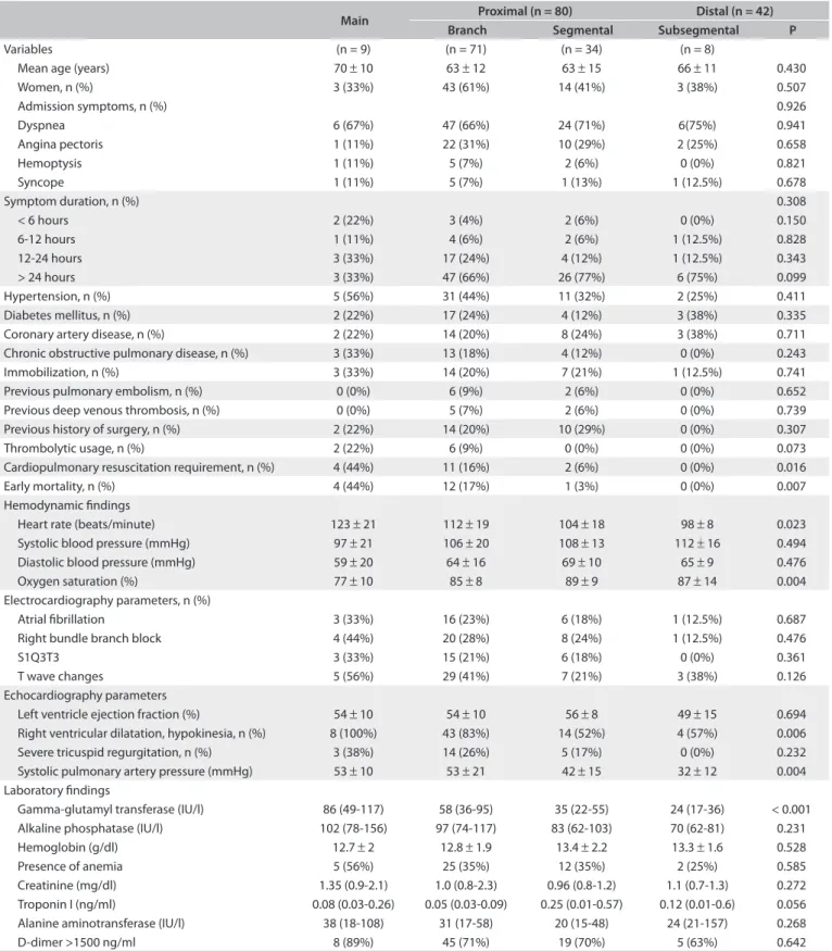
Table 1. Baseline characteristics of study patients with thrombus localization in pulmonary artery (n = 122)
Main Proximal (n = 80) Distal (n = 42) Branch Segmental Subsegmental P
Variables (n = 9) (n = 71) (n = 34) (n = 8)
Mean age (years) 70 ± 10 63 ± 12 63 ± 15 66 ± 11 0.430
Women, n (%) 3 (33%) 43 (61%) 14 (41%) 3 (38%) 0.507
Admission symptoms, n (%) 0.926
Dyspnea 6 (67%) 47 (66%) 24 (71%) 6(75%) 0.941
Angina pectoris 1 (11%) 22 (31%) 10 (29%) 2 (25%) 0.658
Hemoptysis 1 (11%) 5 (7%) 2 (6%) 0 (0%) 0.821
Syncope 1 (11%) 5 (7%) 1 (13%) 1 (12.5%) 0.678
Symptom duration, n (%) 0.308
< 6 hours 2 (22%) 3 (4%) 2 (6%) 0 (0%) 0.150
6-12 hours 1 (11%) 4 (6%) 2 (6%) 1 (12.5%) 0.828
12-24 hours 3 (33%) 17 (24%) 4 (12%) 1 (12.5%) 0.343
> 24 hours 3 (33%) 47 (66%) 26 (77%) 6 (75%) 0.099
Hypertension, n (%) 5 (56%) 31 (44%) 11 (32%) 2 (25%) 0.411
Diabetes mellitus, n (%) 2 (22%) 17 (24%) 4 (12%) 3 (38%) 0.335
Coronary artery disease, n (%) 2 (22%) 14 (20%) 8 (24%) 3 (38%) 0.711
Chronic obstructive pulmonary disease, n (%) 3 (33%) 13 (18%) 4 (12%) 0 (0%) 0.243
Immobilization, n (%) 3 (33%) 14 (20%) 7 (21%) 1 (12.5%) 0.741
Previous pulmonary embolism, n (%) 0 (0%) 6 (9%) 2 (6%) 0 (0%) 0.652
Previous deep venous thrombosis, n (%) 0 (0%) 5 (7%) 2 (6%) 0 (0%) 0.739
Previous history of surgery, n (%) 2 (22%) 14 (20%) 10 (29%) 0 (0%) 0.307
Thrombolytic usage, n (%) 2 (22%) 6 (9%) 0 (0%) 0 (0%) 0.073
Cardiopulmonary resuscitation requirement, n (%) 4 (44%) 11 (16%) 2 (6%) 0 (0%) 0.016
Early mortality, n (%) 4 (44%) 12 (17%) 1 (3%) 0 (0%) 0.007
Hemodynamic indings
Heart rate (beats/minute) 123 ± 21 112 ± 19 104 ± 18 98 ± 8 0.023
Systolic blood pressure (mmHg) 97 ± 21 106 ± 20 108 ± 13 112 ± 16 0.494
Diastolic blood pressure (mmHg) 59 ± 20 64 ± 16 69 ± 10 65 ± 9 0.476
Oxygen saturation (%) 77 ± 10 85 ± 8 89 ± 9 87 ± 14 0.004
Electrocardiography parameters, n (%)
Atrial ibrillation 3 (33%) 16 (23%) 6 (18%) 1 (12.5%) 0.687
Right bundle branch block 4 (44%) 20 (28%) 8 (24%) 1 (12.5%) 0.476
S1Q3T3 3 (33%) 15 (21%) 6 (18%) 0 (0%) 0.361
T wave changes 5 (56%) 29 (41%) 7 (21%) 3 (38%) 0.126
Echocardiography parameters
Left ventricle ejection fraction (%) 54 ± 10 54 ± 10 56 ± 8 49 ± 15 0.694
Right ventricular dilatation, hypokinesia, n (%) 8 (100%) 43 (83%) 14 (52%) 4 (57%) 0.006
Severe tricuspid regurgitation, n (%) 3 (38%) 14 (26%) 5 (17%) 0 (0%) 0.232
Systolic pulmonary artery pressure (mmHg) 53 ± 10 53 ± 21 42 ± 15 32 ± 12 0.004
Laboratory indings
Gamma-glutamyl transferase (IU/l) 86 (49-117) 58 (36-95) 35 (22-55) 24 (17-36) < 0.001
Alkaline phosphatase (IU/l) 102 (78-156) 97 (74-117) 83 (62-103) 70 (62-81) 0.231
Hemoglobin (g/dl) 12.7 ± 2 12.8 ± 1.9 13.4 ± 2.2 13.3 ± 1.6 0.528
Presence of anemia 5 (56%) 25 (35%) 12 (35%) 2 (25%) 0.585
Creatinine (mg/dl) 1.35 (0.9-2.1) 1.0 (0.8-2.3) 0.96 (0.8-1.2) 1.1 (0.7-1.3) 0.272
Troponin I (ng/ml) 0.08 (0.03-0.26) 0.05 (0.03-0.09) 0.25 (0.01-0.57) 0.12 (0.01-0.6) 0.056
Alanine aminotransferase (IU/l) 38 (18-108) 31 (17-58) 20 (15-48) 24 (21-157) 0.268
remained associated with proximal pulmonary artery involve-ment ater adjusting for the signiicant variables in the univariate analysis, and were correlated with the GGT level.
According to the ROC curve analysis, the optimal cutof value for GGT for predicting proximal pulmonary artery involvement was measured as > 40 IU/l, with 73.7% sensitivity, 66.7% spec-iicity, 80.8% positive predictive value and 57.1% negative pre-dictive value (AUC 0.851; 95% conidence interval 0.777-0.908; Figure 2). Furthermore, an admission GGT level of > 139 IU/l [n = 8 (7%)] had speciicity of 95%, sensitivity of 7.5%, positive predictive value of 75% and negative predictive value of 35%. In contrast, an admission GGT level < 20 IU/l [n = 13 (11%)] had
sensitivity of 95%, speciicity of 28.6%, positive predictive value of 72% and negative predictive value of 75%.
DISCUSSION
To the best of our knowledge, this is the irst study to demon-strate the correlation between GGT levels and the degree of pul-monary artery involvement. he indings revealed that there was a signiicant correlation between increased existing embolism load in the pulmonary artery and increased serum GGT levels. his inding may prove to be useful both in treatment planning and as a predictor of mortality.
Pulmonary embolism is a cardiovascular disease with high potential for mortality that can progress into a broad spectrum of clinical manifestations (ranging from silent clinical progress
Table 2. Univariate and multivariate analyses on proximal pulmonary artery involvement
Univariate Multivariate
P OR (95% CI) P OR (95% CI)
Systolic pulmonary artery pressure (mmHg) 0.001 1.052 1.020-1.086 0.033 1.063 1.005-1.124
Right ventricular dilatation/hypokinesia 0.001 5.037 1.895-13.388
Gamma-glutamyl transferase (IU/l), GGT 0.008 1.015 1.004-1.027 0.009 1.044 1.011-1.079
Heart rate (beats/minute) 0.012 1.033 1.007-1.033
Oxygen saturation (%) 0.029 0.942 0.892-0.994
Cardiopulmonary resuscitation requirement 0.050 4.615 1.002-21.252
Systolic blood pressure (mmHg) 0.326 0.988 0.964-1.012
Diastolic blood pressure (mmHg) 0.226 0.981 0.951-1.012
Presence of right bundle branch block 0.313 0.636 0.264-1.532
Alkaline phosphatase (IU/l) 0.084 1.009 0.999-1.019
Alanine aminotransferase (IU/l) 0.741 1.001 0.996-1.005
Creatinine (mg/dl) 0.066 2.273 0.946-5.461
All the variables from Table 1 were examined and only those signiicant at the level P < 0.05 and correlated with GGT level are shown in the univariate analysis. The multivariate logistic regression model with forward stepwise method included all univariate predictors and those with correlated GGT level. CI = conidence interval; OR = odds ratio.
Figure 1. Comparison of gamma-glutamyl transferase (GGT)
levels between four groups. 100 Gamma-glutamyl transferase (IU/I)
90
80
70
60
50
40
30
20
10
0
P < 0.001
Main pulmonary artery
Main pulmonary artery branches
Main pulmonary artery segmental
branches
Main pulmonary artery subsegmental
branches
Figure 2. Receiver operating characteristic (ROC) curve
for gamma-glutamyl transferase (GGT) levels to predict proximal pulmonary artery involvement.
100
Gamma-glutamyl transferase (IU/I)
20 80
40 60
80 40
60 20
100 0
0
100-specificity
S
ensitivit
y
to hemodynamic instability). Although there have been studies indicating a direct correlation between the obstruction level of the pulmonary artery, coagulation load and mortality,5 Mansencal et al. did not ind any discriminant correlation between pulmo-nary hemodynamic data and prognosis.19 Similarly, Araos et al. demonstrated that coagulation load was a weak predictor of mor-tality,20 while Ghuysen et al. stated that pulmonary arterial sys-tolic pressure and the obstruction index of the pulmonary artery were correlated with clinical severity, but were unsuitable for use as mortality predictors.21 In risk classiication, detection of prox-imal extension in pulmonary embolism patients is a prognostic factor with unrecognized value.5
he cardiac biomarkers troponin and natriuretic peptide have emerged as promising tools for evaluating the risk among patients diagnosed with APE. Increased cardiac troponin levels in APE can be explained by the increased right ventricular wall pressure, which leads to compression of the right coronary artery and direct myocardial damage.22 Moreover, the risks of mortality and com-plications have been reported to be higher among patients with elevated cardiac troponin-T during the acute phase of pulmonary embolism.6 Natriuretic peptide is used both as a diagnostic and as a prognostic marker for patients sufering from congestive heart failure, since it is a prohormone that does not accumulate to any signiicant degree in normal ventricular myocytes. In this regard, its levels increase signiicantly a few hours ater acute myocardial contractions.23 Similar to cardiac troponins, brain natriuretic pep-tide and N-terminal pro-brain natriuretic peppep-tide are associated with right ventricle dysfunction in APE.24,25
GGT assaying is a liver function test that is used as a sen-sitive indicator of alcohol consumption and hepatobiliary dys-function.26 GGT is found not only in the liver, but also in the kid-neys and vascular epithelium, as well as in the extracellular luid, where it is attached to albumin carrier molecules and lipopro-teins.27 Previous studies have shown that increased GGT levels are associated with decreased oxygen levels and increased levels of inlammatory markers.28,29 Moreover, the CARDIA study has shown that increased GGT levels are associated with oxidative stress, inlammatory events and diet.28 Wanamethee et al. iden-tiied a correlation between high GGT levels and cardiovascu-lar diseases and stroke in a prospective study on 6,997 patients.12 Furthermore, diferent studies have indicated that serum GGT levels can be used as an early predictor during the development of cardiovascular events and metabolic syndrome.30,31
APE is a complex process involving diferent mechanisms, and GGT levels increase as a result of contributions from all these mechanisms. In the event of a pulmonary artery embolism, embolism severity and involvement levels increase directly pro-portionally to the coagulation load. By extension, hypoxia inten-siies, and increased activity is seen in the neurohormonal and
adrenergic systems. In addition, an increased coagulation load leads to an increase in pulmonary arterial pressure, and thus an increase in hepatic congestion, and the increase in coagula-tion load is further correlated with an increase in GGT levels. As shown in the current study, GGT levels increase with increas-ing numbers of pulmonary artery segments involved, because of the activity of the mechanisms described above. Moreover, Zorlu et al. showed that increased GGT levels could be a predictor of early mortality in cases of APE.14
Our study has some limitations. he most important of these is the single-center nature of the indings. B-type natriuretic pep-tides are established biomarkers for pulmonary embolism, but this was not examined in our study. Selection bias, and the like-lihood that serious cases may have been lost before any tests or evaluations could be conducted, even before admittance to the hospital, may also have inluenced this result, in that patients with more severe proximal involvement are more likely to die acutely. Furthermore, this study was conducted in a tertiary care center: patients with more severe complaints are referred to such cen-ters, while those with milder symptoms are not brought to ter-tiary care centers, but are managed in secondary care hospitals.
CONCLUSION
he present study identiied a signiicant correlation between serum GGT levels and pulmonary artery involvement levels, i.e. the severity of an embolism event.
REFERENCES
1. Torbicki A, Perrier A, Konstantinides S, et al. Guidelines on the
diagnosis and management of acute pulmonary embolism: the
Task Force for the Diagnosis and Management of Acute Pulmonary
Embolism of the European Society of Cardiology (ESC). Eur Heart J.
2008;29(18):2276-315.
2. Goldhaber SZ. Pulmonary embolism. Lancet. 2004;363(9417):1295-305.
3. Dalen JE, Alpert JS. Natural history of pulmonary embolism. Prog
Cardiovasc Dis. 1975;17(4):259-70.
4. Gölbaşı Z. [Chronic thromboembolic pulmonary hypertension:
diagnosis, medical therapy and monitoring]. Anadolu Kardiyol Derq.
2010;10 Suppl 2:56-60.
5. Ghanima W, Abdelnoor M, Holmen LO, Nielssen BE, Sandset PM.
The association between the proximal extension of the clot and
the severity of pulmonary embolism (PE): a proposal for a new
radiological score for PE. J Intern Med. 2007;261(1):74-81.
6. Becattini C, Vedovati MC, Agnelli G. Prognostic value of troponins in acute
pulmonary embolism: a meta-analysis. Circulation. 2007;116(4):427-33.
7. De Monyé W, Sanson BJ, Mac Gillavry MR, et al. Embolus location
afects the sensitivity of rapid quantitative D-dimer assay in the
diagnosis of pulmonary embolism. Am J Respir Crit Care Med.
8. Sen N, Ermis H, Altinkaya N. Pulmonary Embolism in Young and
Elderly Patients: Clinical Characteristics, Laboratory and Instrumental
Findings and Diferences Between Age Groups. Turkish Thoracic
Journal. 2010;11(4):160-6. Available from: http://toraks.dergisi.org/
pdf/pdf_Toraksder_700.pdf. Accessed in 2015 (Jun 30).
9. Gohel MG, Chacko AN. Serum GGT activity and hsCRP level in patients
with type 2 diabetes mellitus with good and poor glycemic control:
An evidence linking oxidative stress, inlammation and glycemic
control. J Diabetes Metab Disord. 2013;12(1):56.
10. Whitield JB. Gamma glutamyl transferase. Crit Rev Clin Lab Sci.
2001;38(4):263-335.
11. Dröge W. Free radicals in the physiological control of cell function.
Physiol Rev. 2002;82(1):47-95.
12. Wannamethee G, Ebrahim S, Shaper AG. Gamma-glutamyltransferase:
determinants and association with mortality from ischemic heart
disease and all causes. Am J Epidemiol. 1995;142(7):699-708.
13. Jiang S, Jiang D, Tao Y. Role of gamma-glutamyltransferase in
cardiovascular diseases. Exp Clin Cardiol. 2013;18(1):53-6.
14. Zorlu A, Yucel H, Bektasoglu G, et al. Increased γ-glutamyl transferase
levels predict early mortality in patients with acute pulmonary
embolism. Am J Emerg Med. 2012;30(6):908-15.
15. Lang RM, Bierig M, Devereux RB, et al. Recommendations for chamber
quantiication. Eur J Echocardiogr. 2006;7(2):79-108.
16. McConnell MV, Solomon SD, Rayan ME, et al. Regional right ventricular
dysfunction detected by echocardiography in acute pulmonary
embolism. Am J Cardiol. 1996;78(4):469-73.
17. Lancellotti P, Moura L, Pierard LA, et al. European Association of
Echocardiography recommendations for the assessment of valvular
regurgitation. Part 2: mitral and tricuspid regurgitation (native valve
disease). Eur J Echocardiogr. 2010;11(4):307-32.
18. Yock PG, Popp RL. Noninvasive estimation of right ventricular
systolic pressure by Doppler ultrasound in patients with tricuspid
regurgitation. Circulation. 1984;70(4):657-62.
19. Mansencal N, Joseph T, Vieillard-Baron A, et al. Comparison of diferent
echocardiographic indexes secondary to right ventricular obstruction
in acute pulmonary embolism. Am J Cardiol. 2003;92(1):116-9.
20. Araoz PA, Gotway MB, Trowbridge RL, et al. Helical CT pulmonary
angiography predictors of in-hospital morbidity and mortality
in patients with acute pulmonary embolism. J Thorac Imaging.
2003;18(4):207-16.
21. Ghuysen A, Ghaye B, Willems V, et al. Computed tomographic
pulmonary angiography and prognostic signiicance in patients with
acute pulmonary embolism. Thorax. 2005;60(11):956-61.
22. Kucher N, Goldhaber SZ. Cardiac biomarkers for risk stratiication
of patients with acute pulmonary embolism. Circulation.
2003;108(18):2191-4.
23. Hama N, Itoh H, Shirakami G, et al. Rapid ventricular induction of
brain natriuretic peptide gene expression in experimental acute
myocardial infarction. Circulation. 1995;92(6):1558-64.
24. Kucher N, Printzen G, Doernhoefer T, et al. Low pro-brain natriuretic
peptide levels predict benign clinical outcome in acute pulmonary
embolism. Circulation. 2003;107(12):1576-8.
25. Tulevski H, Hirsch A, Sanson BJ, et al. Increased brain natriuretic
peptide as a marker for right ventricular dysfunction in acute
pulmonary embolism. Throm Haemost. 2001;86(5):1193-6.
26. Mason JE, Starke RD, Van Kirk JE. Gamma-glutamyl transferase: a
novel cardiovascular risk biomarker. Prev Cardiol. 2010;13(1):36-41.
27. Sacchetti L, Castaldo G, Fortunato G, Salvatore F. Improved procedure
for measuring gamma-glutamyltransferase isoenzymes in serum.
Clin Chem. 1988;34(2):419-22.
28. Lee DH. Stefen LM, Jacobs DR Jr. Association between serum
gamma-glutamyltransferase and dietary factors: the Coronary Artery
Risk Development in Yong Adults (CARDIA) Study. Am J Clin Nutr.
2004;79(4):600-5.
29. Ikeda Y, Fujii J, Tanguchi N, Meister A. Expression of an active
glycosylated human gamma-glutamyl transpeptidase mutant
that lacks a membrane anchor domain. Proc Natl Acad Sci U S A.
1995;92(1):126-30.
30. Emdin M, Passino C, Donato L, Paolicchi A, Pompella A. Serum
gamma-glutamyltransferase as a risk factor of ischemic stroke might
be independent of alcohol consumption. Stroke. 2002;33(4):1163-4.
31. Emdin M, Passino C, Pompella A, Paolicchi A. Serum gamma-glutamyl
transpeptidase: a prognostic marker in cardiovascular diseases.
Biofactors. 2003;17(1-4):199-205.
Sources of funding: None
Conlict of interest: None
Date of irst submission: January 19, 2015
Last received: June 8, 2015
Accepted: June 18, 2015
Address for correspondence:
Hasan Yucel
Department of Cardiology
Cumhuriyet University Medical School, Sivas; Turkey
Tel. +903462581805
Fax. +903462191268