Arq Neuropsiquiatr 2007;65(4-B):1237-1240
1237
BILATERAL CORTICAL ATROPHY AFTER SEVERE
BRAIN TRAUMA AND EXTRADURAL HEMATOMA
Paulo Roberto Louzada
1, Rafael Pereira Vaitsman
2,
Arthur Borges Martins de Souza
3, Pedro de Oliveira Coutinho
4,
Renata Teixeira Lengruber
5, Francisco Weldes Brito das Neves
6,
Herbert Missaka
7, Marco Aurélio Albuquerque Lima
7, José Massoud Salame
8ABSTRACT - We report the case of a severe head injured 43-year old male patient with a large extradural he-matoma, Glasgow Coma Scale 3 and dilated fixed pupils. Patient was promptly submitted to surgical evac-uation of the lesion, but remained in persistent vegetative state in the post-operative time. Head comput-ed tomography scans performcomput-ed before surgery, and at early and late post-operative periods comparative-ly revealed extreme bilateral cortical atrophy. Late consequences of severe head trauma drasticalcomparative-ly affect the prognosis of patients, being its prevention, and neuroprotection against secondary injury still a thera-peutical challenge for neurosurgeons.
KEY WORDS: brain atrophy, extradural hematoma, epidural hematoma, severe head injury, secondary injury.
Atrofia cortical bilateral após traumatismo cranioencefálico grave e hematoma extradural
RESUMO - Relatamos o caso de um paciente de 43 anos, com traumatismo cranioencefálico grave, com gran-de hematoma extradural, Escala gran-de Coma gran-de Glasgow 3 e pupilas fixas e dilatadas. O paciente foi pronta-mente submetido à evacuação cirúrgica da lesão mas permaneceu em estado vegetativo persistente no pe-ríodo pós-operatório. As TC de crânio realizadas antes da cirurgia e nos pepe-ríodos pós-operatórios precoce e tardio revelaram comparativamente extrema atrofia cerebral bilateral. As conseqüências tardias do trau-matismo craniano grave afetam drasticamente o prognóstico dos pacientes, sendo sua prevenção, e a neu-roproteção contra a injúria secundária ainda um desafio terapêutico para os neurocirurgiões.
PALAVRAS-CHAVE: atrofia cerebral, hematoma extradural, hematoma epidural, traumatismo craniano gra-ve, injúria secundária.
1MD, PhD, Neurosurgeon, Serviço de Neurocirurgia, Hospital Municipal Souza Aguiar, Rio de Janeiro RJ, Brazil; 2MD, Neurosurgery
Resident, Serviço de Neurocirurgia, Hospital Municipal Souza Aguiar, Rio de Janeiro RJ, Brazil; 3MD, Neurosurgeon, Serviço de
Neurocirurgia, Instituto Nacional de Câncer (INCA). Rio de Janeiro, RJ, Brazil; 4MD, Neurosurgery Resident, Serviço de
Neurociru-rgia, Hospital Municipal Souza Aguiar, Rio de Janeiro RJ, Brazil; 5Neurology Resident, Serviço de Neurologia, Hospital Universitário
Antônio Pedro, Universidade Federal Fluminense, Niterói RJ, Brazil; 6MD, Neurosurgeon, Serviço de Neurocirurgia, Hospital
Mu-nicipal Souza Aguiar, Rio de Janeiro RJ, Brazil; 7MD, ICU Physician, Unidade de Pacientes Graves (UPG / CTI - Emergência), Hospital
Municipal Souza Aguiar, Rio de Janeiro, RJ, Brazil; 8MD, Neurosurgeons, Serviço de Neurocirurgia, Hospital Municipal Souza Aguiar,
Rio de Janeiro RJ, Brazil. Support: Prefeitura Municipal do Rio de Janeiro. Received 31 May 2007. Accepted 12 September 2007.
Dr. Paulo Roberto Louzada - Serviço de Neurocirurgia / Hospital Municipal Souza Aguiar - Praça da República 111 / 4º andar - 20211-350 Rio de Janeiro RJ - Brasil. E-mail: [email protected]
Extraaxial hematomas, extradural hematoma (EH) and subdural hematoma (SH), stand among the most common neurosurgical emergencies in general hos-pitals worldwide. Prognosis and mortality rates of pa-tients with extra-axial hematomas are clearly depen-dent on neurological status on admittance1. Literature
reports a mortality rate of 4.7% for patients with ex-tradural hematoma operated at Glasgow Coma Scale (GCS) 13-15, with 66.7% of good recovery, ranging to a mortality rate of 43%, and only 29% of good recov-ery for patients admitted at GCS 3-81. In this line,
Kot-wica and Jakubowski studied brain injured patients admitted in GCS 3, reporting an overall mortality of
89%2. In the same study, better outcomes for GCS 3
patients were reached for those with no abnormal-ities on CT scan or for patients with extradural he-matoma surgically treated2. Better prognosis is
ob-tained when surgical evacuation is performed ear-ly after trauma, as classicalear-ly described for SH, when surgery is performed within the fi rst four hours af-ter trauma3.
post-trau-Arq Neuropsiquiatr 2007;65(4-B)
1238
Brain trauma: extradural hematoma, bilateral cortical atrophy Louzada et al.
matic brain atrophy is an uncommon event, with few descriptions in the literature and no effi cient treat-ment currently available to preclude such condition. We discuss the possible relation of the poor post-op-erative outcome with the radiological findings of brain atrophy highlighting the consequences of sec-ondary brain injury.
CASE
A 43-year old man, probably victim of aggression, was admitted to the emergency room with a history of rapid neurological deterioration from GCS 6 (at the ambulance) to GCS 3 within the past 30 minutes. Patient was intubated and no respiratory drive was detected. Pupils were dilated
andfi xed. CT scan performed 10 minutes after admission
revealed a huge (4.2 cm) left extradural temporo-parietal hematoma with significant (1.6 cm) midline shift (Fig 1A and 1D). The patient was promptly conducted to operating room. A large fronto-temporo-parietal incision was made to access the entire lesion. A skull fracture was identifi ed crossing a branch of middle meningeal artery, being impli-cated as the cause of the hemorrhagic lesion. A temporo-pa-rietal craniectomy was performed with complete evacuation of the lesion in less than 1 hour after patient admittance in
the hospital. In the immediate post-operative period, pa-tient mildly ameliorate its neurological status from bilater-al midriasis to anisocoria (left > right), being conducted to the intensive care unit for post-operative support.
A CT scan performed 48 hours after surgery reveals en-tire evacuation of the hematoma, improvement of midline shift and signs of left parietal ischemic injury by compres-sion of underlying neural tissue (Fig 1B and 1E). Pupillary re-activity became normal after several weeks, when patient was awake but unresponsive to any stimuli, thus character-izing a persistent vegetative state.
Four months and eleven days after surgery, a new CT scan were made to access any late complication of the head trauma responsible for the patient unresponsiveness. Inter-estingly, remarkable bilateral brain atrophy (Fig 1C and 1F) was observed. Enlarged sulci were more predominant in the fronto-temporal lobes than in right parietal and occipi-tal lobes. Patient has no history of alcoholism or other pre-disposing condition to cortical atrophy before head trau-ma. Encefalomalacia and ventricular occipital horn ectasia could be observed at left hemisphere, consequent to isch-emic insult already observed in the fi rst post-op exam. Pa-tient remained in persistent vegetative state and died near six months after surgery from pulmonary sepsis and respi-ratory failure.
Arq Neuropsiquiatr 2007;65(4-B)
1239
Brain trauma: extradural hematoma, bilateral cortical atrophy Louzada et al.
DISCUSSION
Bilateral cerebral atrophy secondary to brain trau-ma are an uncommon fi nding, poorly reported in the literature. Levin et al. report two cases of children with reversible cerebral atrophy years after a SH4.
Tomita et al. showed a large series of delayed hemi-spheric brain atrophy after EH and SH5. In this study,
authors reported that from 42 patients victims of neu-rotrauma, only 9 (21%) presented enlarged ventricles and cerebral atrophy5. More recently, Mackenzie et
al. presented a retrospective study of patients with closed, mild to moderate head injury that evolved to whole brain atrophy at an average time of 11 months6.
We demonstrated a case of severe head injured patient evolved to secondary bilateral brain atrophy about four months after trauma. Brain atrophy is de-fi ned as enlarged lateral ventricles with prominent cerebral sulci7,8. In our study, the size of the
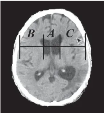
later-al ventricles was measured using the the bicaudate cerebroventricular index (BCVI) on the CT scan taken in the plane parallel to orbitomeatal line as demon-strated by Tomita et al.5. The BCVI was measured by
the ratio between the width of the ventricular fron-tal horns and the tofron-tal width of the two brain hemi-spheres measured at the head the caudate nucleus level5. Figure 2, indicates the measures A, B and C,
to obtain BCVI by the formula (A / A + B + C) x 100.
The normal reported BCVI in adults is 15±3%. Values higher than 18%, in the presence of enlarged sulci, are considered to be indicative of cerebral atrophy. In the series of Tomita et al. the higher BCVI obtained in a patient victim of EH was 24.6%5. In the case
re-ported here, we obtained a BCVI of 25.5%, confi rm-ing the larger bilateral cerebral atrophy caused by an EH reported in the literature.
Secondary brain injury, the sequence of pathophys-iological and intracellular events that follows primary mechanical damage to the neural tissue, has been extensively studied, however the precise mecha-nisms underlying secondary brain injury remains still unclear. Delayed neural injury may results from rochemical imbalances (mainly excitotoxicity), neu-roinfl ammation, oxidative stress etc9. Apoptotic cell
death and ischemia, two biological events related to secondary brain injury, among others, was already reported to be involved in posttraumatic neurode-generation and neuroatrophy5,6. In the case described
here, neuronal loss occurs bilaterally, mainly at the cerebral cortex but also at the brainstem and cerebel-lum (not shown), therefore away from the site of the lesion (left temporal-parietal lobes), thus, could not be explained simply by the effects of direct neural tissue compression, being probably due to secondary and late toxic neurochemical and neuroinfl ammatory phenomena affecting the whole brain.
Brain atrophy could be, in part, responsible for the devastating neurological sequelae presented by the patient reported in this case, once delayed neuroana-tomical changes were more predominant in the bi-frontal and bitemporal cortices, leading to a compro-mising of cognition and motor function. Moreover, the intensity if neuronal loss observed (BCVI 25.5%) could be closely related to the severity of neurological status presented by the patient on admission (GCS 3, dilated and fi xed pupils).
Encouraging therapeutical strategies against sec-ondary brain injury has been investigated in clinical trials2,10. However, currently, there is no treatment
able to block secondary brain injury and the delayed cerebral atrophy. Once the full mechanisms of neural damage after primary insult can be elucidated, neu-roprotective strategies must to be developed to im-prove outcome of severe brain damaged patients.
REFERENCES
1. Cheung PS, Lam JM, Yeung JH, Graham CA, Rainer TH. Outcome of traumatic extradural haematoma in Hong Kong. Injury 2007;38:76-80. 2. Kotwica Z, Jakubowski JK. Head-injured adult patients with GCS of 3
on admission--who have a chance to survive ? Acta Neurochir (Wien) 1995;133:56-59.
Arq Neuropsiquiatr 2007;65(4-B)
1240
Brain trauma: extradural hematoma, bilateral cortical atrophy Louzada et al.
3. Seelig JM, Becker DP, Miller JD, Greenberg RP, Ward JD, Choi SC. Trau-matic acute subdural hematoma: major mortality reduction in comatose patients treated within four hours. N Engl J Med 1981;304:1511-1518. 4. Levin HS, Mendelsohn D, Bruce D, Harward H, Culhane KA, Eisenberg
HM. Reversibility of cerebral atrophy aĞ er head injury in children. Neu-rosurgery 1992;31:1117-1121.
5. Tomita H, Tone O, Ito U. Hemispheric cerebral atrophy aĞ er traumatic ex-tra-axial hematoma in adults. Neurol Med Chir (Tokyo) 1997; 37:819-824. 6. MacKenzie JD, Siddiqi F, Babb JS, et al. Brain atrophy in mild or
mod-erate traumatic brain injury: a longitudinal quantitative analysis. Am J Neuroradiol 2002;23:1509-1515.
7. Kishore PR, Lipper MH, Miller JD, Girevendulis AK, Becker DP, Vines FS. Post-traumatic hydrocephalus in patients with severe head injury. Neuroradiology 1978;16:261-265.
8. Synek V, Reuben JR, Du Boulay GH. Comparing Evans’ index and com-puterized axial tomography in assessing relationship of ventricular size to brain size. Neurology 1976;26:231-233.
9. McIntosh TK, Smith DH, Garde E. Therapeutic approaches for the pre-vention of secondary brain injury. Eur J Anaesthesiol 1996;13:291-309. 10. Clausen T, Bullock R. Medical treatment and neuroprotection in